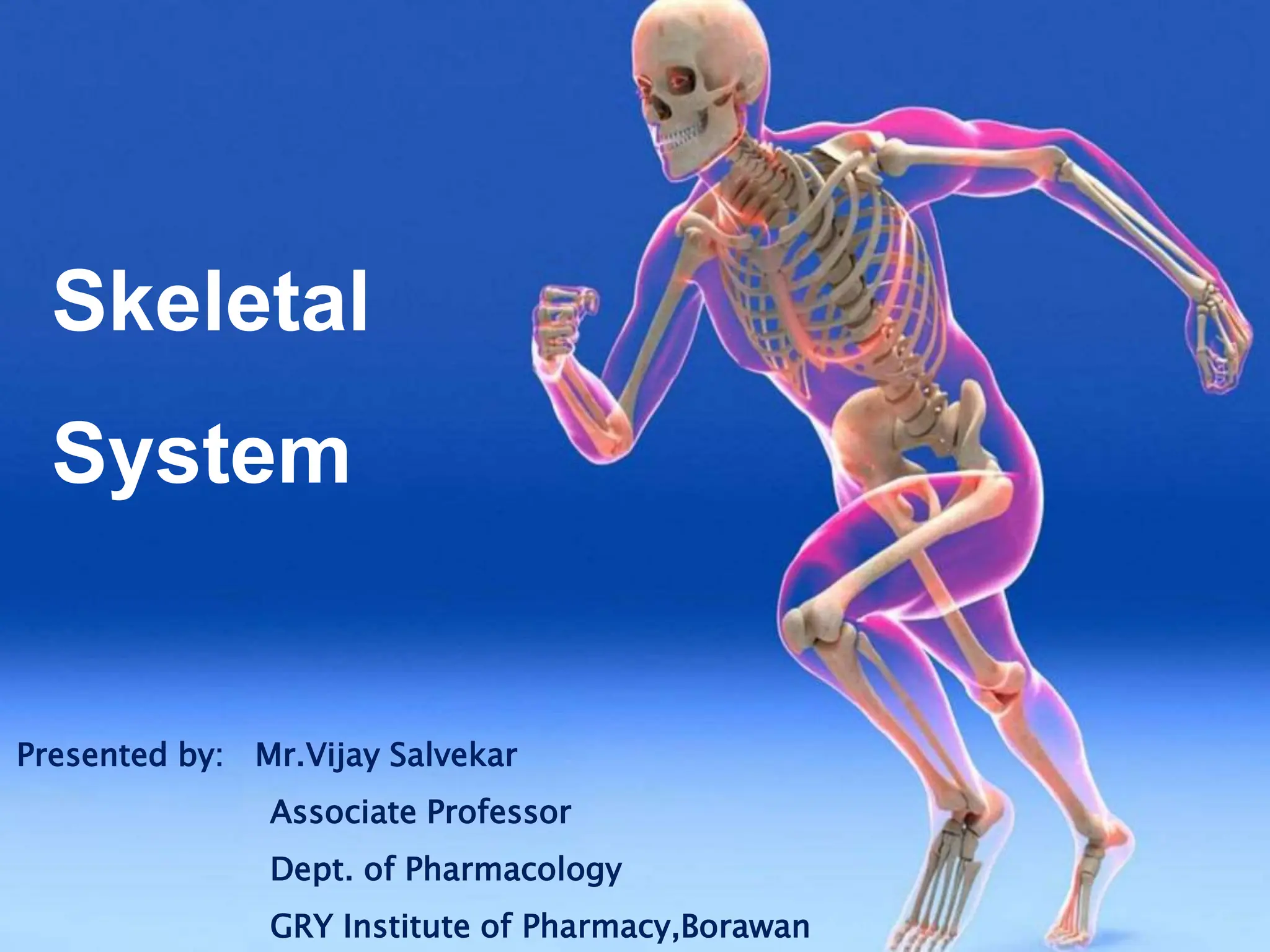
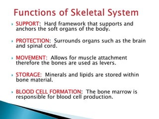
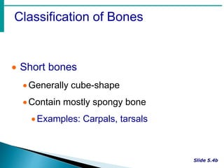
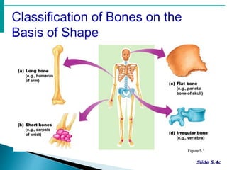
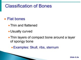
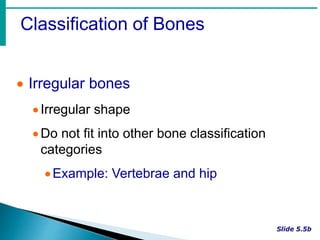
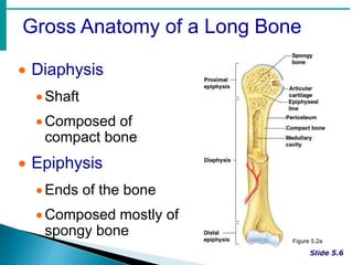
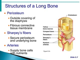
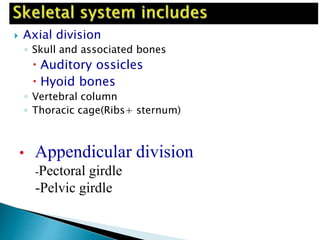
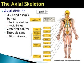
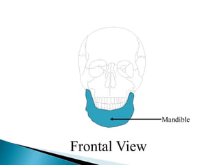
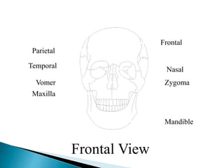
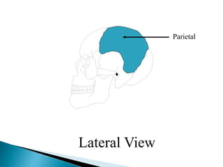
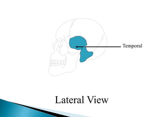
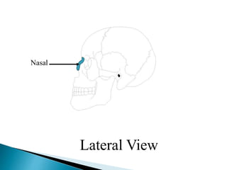
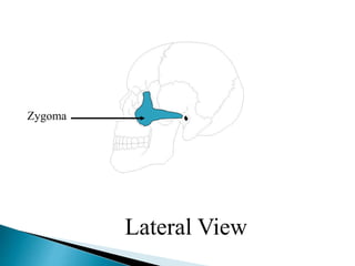
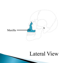
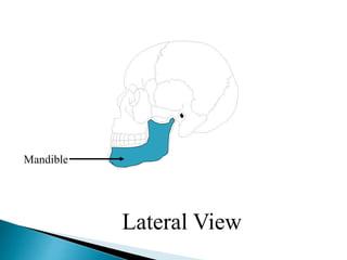
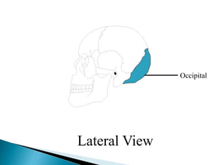
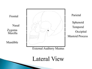
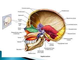
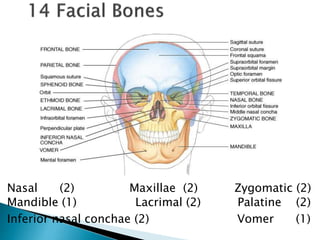
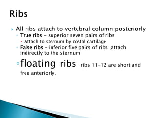
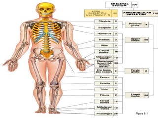
SlideShare a Scribd company logo
1 of 130
Download to read offline
Skeletal
System
Presented by: Mr.Vijay Salvekar
Associate Professor
Dept. of Pharmacology
GRY Institute of Pharmacy,Borawan
 COMPOSED OF:
-Bones
-Cartilage
-Joints
-Ligaments
 SUPPORT: Hard framework that supports and
anchors the soft organs of the body.
 PROTECTION: Surrounds organs such as the brain
and spinal cord.
 MOVEMENT: Allows for muscle attachment
therefore the bones are used as levers.
 STORAGE: Minerals and lipids are stored within
bone material.
 BLOOD CELL FORMATION: The bone marrow is
responsible for blood cell production.
Bone Markings
 Bone Surface is not smooth, but shows:
 Bone markings which reveal where:
-muscles, tendons, and ligaments attatched
-nerves and blood vessels pass
*bone marking may be:
1-projections or processes or
2-depressions or cavities
 Compact bone
◦ Outer layer of bone, very hard and dense.
◦ Organized in structural units called Haversian
systems.
◦ Matrix is composed of Ca salts (Ca carbonate and
Ca phosphate)
◦ Osteocytes – living bone cells that live in matrix.
 Porous (Spongy) bone
◦ Located in the ends of long bones.
◦ Many spaces that are filled with red bone marrow
which produces bone cells.
 Spongy bone
◦ Trabeculae – needle-like threads of spongy bone
that surround the spaces. Add strength to this
portion of the bone.
 Cartilage
◦ Matrix is a firm gel with chondrocytes suspended in
the matrix.
Classification of Bones
Copyright © 2003 Pearson Education, Inc. publishing as Benjamin Cummings
 Long bones
Typically longer than wide
Have a shaft with heads at both ends
Contain mostly compact bone
• Examples: Femur, humerus
Classification of Bones
 Short bones
Generally cube-shape
Contain mostly spongy bone
Examples: Carpals, tarsals
Classification of Bones on the
Basis of Shape
Figure 5.1
Classification of Bones
 Flat bones
Thin and flattened
Usually curved
Thin layers of compact bone around a layer
of spongy bone
Examples: Skull, ribs, sternum
Classification of Bones
 Irregular bones
Irregular shape
Do not fit into other bone classification
categories
Example: Vertebrae and hip
Gross Anatomy of a Long Bone
 Diaphysis
Shaft
Composed of
compact bone
 Epiphysis
Ends of the bone
Composed mostly of
spongy bone
Figure 5.2a
Structures of a Long Bone
 Periosteum
 Outside covering of
the diaphysis
 Fibrous connective
tissue membrane
 Sharpey’s fibers
 Secure periosteum
and underlying bone
 Arteries
 Supply bone cells
with nutrients
Figure 5.2c
Structures of a Long Bone
 Articular cartilage
Covers the
external surface of
the epiphyses
Made of hyaline
cartilage
Decreases friction
at joint surfaces Figure 5.2a
Structures of a Long Bone
 Medullary cavity
Cavity of the shaft
Contains yellow
marrow (mostly fat)
in adults
Contains red marrow
(for blood cell
formation) Figure 5.2a
Microscopic Anatomy of Bone
 Osteon (Haversian System)
 A unit of bone
 Central (Haversian) canal
 Opening in the center of an osteon
 Carries blood vessels and nerves
 Perforating (Volkman’s) canal
 Canal perpendicular to the central canal
 Carries blood vessels and nerves
Microscopic Anatomy of Bone
Figure 5.3
Microscopic Anatomy of Bone
 Lacunae
 Cavities containing
bone cells
(osteocytes)
 Arranged in
concentric rings
 Lamellae
 Rings around the
central canal
 Sites of lacunae Figure 5.3
Microscopic Anatomy of Bone
 Canaliculi
Tiny canals
Radiate from the
central canal to
lacunae
Form a transport
system
Figure 5.3
Changes in the Human Skeleton
 In embryos, the skeleton is primarily hyaline
cartilage
 During development, much of this cartilage
is replaced by bone
 Cartilage remains in isolated areas
 Bridge of the nose
 Parts of ribs
 Joints
Bone Growth
Copyright © 2003 Pearson Education, Inc. publishing as Benjamin Cummings
 Epiphyseal plates allow for growth of
long bone during childhood
New cartilage is continuously formed
Older cartilage becomes ossified
Cartilage is broken down
Bone replaces cartilage
Bone Growth
 Bones are remodeled and lengthened
until growth stops
Bones change shape by gravity &muscle
pull
Bones grow in width through periostium
Long Bone Formation and Growth
Figure 5.4a
Types of Bone Cells
 Osteocytes
 Mature bone cells
 Osteoblasts
 Bone-forming cells
 Osteoclasts
 Bone-destroying cells
 Break down bone matrix for remodeling and
release of calcium
 Bone remodeling is a process by both
osteoblasts and osteoclasts
 Closed fracture (simple): skin is intact
 Open fracture (compound): skin is open
 Fracture reduction :
1-closed reduction ,no surgery is needed
2-open reduction ,surgery is needed
 Healing time for simple fracture is 6-8 weeks
(longer in elderly people)
 It occurs in FOUR major events
 1-hematoma formation
 2-fibrocartilage callus formation
 3-bony callus formation
 4-bone remodelling
 Axial division
◦ Skull and associated bones
 Auditory ossicles
 Hyoid bones
◦ Vertebral column
◦ Thoracic cage(Ribs+ sternum)
• Appendicular division
-Pectoral girdle
-Pelvic girdle
 Axial division
◦ Skull and associated
bones:
 Auditory ossicles
 Hyoid bones
◦ Vertebral column
◦ Thoracic cage
 Ribs + sternum
 Sutures – Immovable joints that join skull
bones together
 Form boundaries between skull bones
 Four sutures:
◦ Coronal – between parietal and frontal
◦ Sagittal– between parietal bones
◦ Lambdoid – between the parietal and occipital
◦ Squamous – between the parietal and temporal
Fontanels – usually ossify by 2 years of age
Frontal
(Coronal)
Sagittal
Squamous
Lambdoid
Sutures
•skull = 22 bones
•cranium = 8 bones: frontal, occipital, 2 temporals, 2 parietals, sphenoid and
ethmoid
•facial bones = 14 bones: nasals, maxillae, zygomatics, mandible, lacrimals,
palatines, inferior nasal conchae, vomer.
•skull forms a larger cranial cavity
-also forms the nasal cavity, the orbits, paranasal sinuses
mandible and auditory ossicles are the only movable skull bones
•cranial bones also: attach to membranes called meninges
-stabilize positions of the brain, blood vessels
-outer surface provides large areas for muscle attachment that
move the head or provide facial expressions
The Adult Skull
Bones of the Cranium
Frontal View
Frontal
Frontal View
Parietal
Frontal View
Temporal
Frontal View
Nasal
Frontal View
Vomer
Frontal View
Zygoma
Frontal View
Maxilla
Frontal View
Mandible
Frontal View
Frontal
Parietal
Temporal
Zygoma
Nasal
Vomer
Maxilla
Mandible
Frontal View
Lateral View
Frontal
Lateral View
Parietal
Lateral View
Temporal
Lateral View
Nasal
Lateral View
Zygoma
Lateral View
Maxilla
Lateral View
Mandible
Lateral View
Occipital
Lateral View
Mastoid Process
Lateral View
External Auditory Meatus
Lateral View
Frontal
Nasal
Zygoma
Maxilla
Mandible
Parietal
Sphenoid
Temporal
Occipital
External Auditory Meatus
Mastoid Process
Lateral View
Nasal (2) Maxillae (2) Zygomatic (2)
Mandible (1) Lacrimal (2) Palatine (2)
Inferior nasal conchae (2) Vomer (1)
 part of the nasal complex
 Paired cavities in ethmoid,
sphenoid, frontal and
maxillary
 Lined with mucous
membranes and open into
nasal cavity though
openings called ostia
 Resonating chambers for
voice, lighten the skull
 Sinusitis is inflammation
of the membrane (allergy)
 infection can easily spread
from one sinus to the
other through the nasal
cavity
 can also spread to other
tissues
 frontal sinuses
 sphenoid sinuses
 ethmoid sinuses
 maxillary
http://www.wisc-online.com/objects/index.asp?objID=AP12104
 26 vertebrae
◦ 24 individual vertebrae
◦ Sacrum
◦ Coccyx
 Seven cervical vertebrae
 Twelve thoracic vertebrae
 Five lumbar vertebrae
 Sacrum and coccyx are
 Fused together.
 Body
◦ weight bearing
 Vertebral arch
◦ pedicles
◦ laminae
 Vertebral foramen
 Seven processes
◦ 2 transverse
◦ 1 spinous
◦ 4 articular
 Smaller bodies
 Larger spinal canal
 1st and 2nd cervical
vertebrae are unique
◦ atlas & axis
 All articulate with
ribs
 Have heart-
shaped bodies
 Each side of the
body bears
demifacets for
articulation with
ribs
◦Allows rotation and prevents
flexion and extension
 Bodies are thick
and strong
 Allows flexion
and extension –
rotation
prevented
 Forms the posterior wall of pelvis
 Formed from 5 fused vertebrae
 Superior surface articulates with L5
 Inferiorly articulates with coccyx
Figure 7.18a, b
 Is the “tailbone”
 Formed from 3 – 5 fused vertebrae
 Offers only slight support to pelvic organs
 Forms the framework of the chest
 Components of the bony thorax
◦ Thoracic vertebrae – posteriorly
◦ Ribs – laterally
◦ Sternum and costal cartilage – anteriorly
 Protects thoracic organs
 Supports shoulder girdle and upper limbs
 Provides attachment sites for muscles
Figure 7.19a
 Formed from three parts :
◦ Manubrium – superior part
 Articulates with medial end of clavicles
◦ Body – bulk of sternum
 Sides are articulate for costal cartilage of ribs 2–7
◦ Xiphoid process – inferior end of sternum
 Ossifies around age 40
 All ribs attach to vertebral column posteriorly
◦ True ribs - superior seven pairs of ribs
 Attach to sternum by costal cartilage
◦ False ribs – inferior five pairs of ribs ,attach
indirectly to the sternum
◦floating ribs ribs 11–12 are short and
free anteriorly.
 Abnormal spinal curvatures
◦ Scoliosis – an abnormal lateral curvature
◦ Kyphosis – an exaggerated thoracic curvature
◦ Lordosis – an inward lumbar curvature – “swayback”
 Stenosis of the lumbar spine
◦ A narrowing of the vertebral canal
 Allows us to move and manipulate
objects
 Includes all bones other than axial
skeleton, it includes:
◦ the limbs (upper & lower limbs)
◦ the supportive girdles (pectoral
&pelvic girdles)
Figure 8–1
 Also called the shoulder girdle
 Connects the arms to the body
 Positions the shoulders
 Provides a base for arm movement
Figure 8–2a
 Consists of:
◦ 2 clavicles
◦ 2 scapulae
 Connects with the axial skeleton only at the
manubrium(claviculosternal joint)
Figure 8–2b, c
 Also called collarbones
 Long, S-shaped bones
 Originate at the manubrium (sternal end)
 Articulate with the scapulae (acromial end)
 Also called shoulder blades
 Broad, flat and triangular
 Articulate with arms and collarbone
Figure 8–3a
 Arms, forearms, wrists, and hands
Note: arm (brachium) = 1 bone, the
humerus
Figure 8–4
 Also called the arm
 The long, upper armbone
 Articulates with the pectoral girdle
Figure 8–5
Also called the
antebrachium
Consists of 2 long bones:
◦ulna (medial)
◦radius (lateral)
Figure 8–6
◦8 carpal bones:
4 proximal carpal bones
4 distal carpal bones
allow wrist to bend and
twist
The 5 long bones of the
hand
Numbered I–V from lateral
(thumb) to medial
Articulate with proximal
phalanges
Thumb:
◦2 phalanges (proximal,
distal)
Fingers:
◦3 phalanges (proximal,
middle, distal)
Consists of 2 ossa coxae,
the sacrum, and the
coccyx
Stabilized by ligaments of
pelvic girdle, sacrum, and
lumbar vertebrae
Figure 8–7
 Also called hipbones
 Strong to bear body weight
&stress of movement
 Each is made up of 3 fused bones:
◦ ilium (articulates with sacrum)
◦ ischium
◦ pubis
Also called the hip socket
Is the meeting point of the
ilium, ischium, and pubis
Articulates with head of the
femur (Hip joint))
Figure 8–8
Figure 8–9
Female pelvis:
◦smoother
◦lighter
◦less prominent muscle and
ligament attachments
Figure 8–10
 Enlarged pelvic outlet
 Broad pubic angle (> 100°)
 Less curvature of sacrum and coccyx
 Wide, circular pelvic inlet
 Broad, low pelvis
 Ilia project laterally, not upwards
Functions:
◦weight bearing
◦motion
Note: leg = lower leg; thigh =
upper leg
 Femur (thigh)
 Patella (kneecap)
 Tibia and fibula (leg)
 Tarsals (ankle)
 Metatarsals (foot)
 Phalanges (toes)
Figure 8–11
 Also called the kneecap
 Formed within tendon of quadriceps femoris
Figure 8–13
 Also called the shinbone
 Supports body weight
 Larger than fibula
 Medial to fibula
The Fibula
• Attaches muscles of feet and toes
• Smaller than tibia
• Lateral to tibia
 Talus:
 Calcaneus (heel bone):
◦ transfers weight to ground
◦ attaches Achilles tendon
 Also called the tarsus:
◦ consists of 7 tarsal bones
Figure 8–14a
 5 long bones of foot
 Numbered I–V, medial to lateral
 Articulate with toes
 Phalanges:
◦ bones of the toes
 Hallux:
◦ big toe, 2 phalanges (distal, proximal)
 Other 4 toes:
◦ 3 phalanges (distal, medial, proximal)
 Arches transfer weight from 1 part of the foot
to another
Figure 8–14b
 Bones are arranged to form THREE strong
arches, 2 longitudinal (medial & lateral)&
1 transverse
 Ligaments & tendons help to hold the bones
firmly in the arched position but still allow a
certain amount of spriginess
 Week arches are referred to as flat foot
Articulations (Joints)
 Holds bones together
 Allows bones to move
 All bones articulate with at least one other
bone except the hyoid.
 Functional classification: focuses on the
amount of movement (synarthrosis,
amphiarthrosis and diarthrosis)
 Structural classification:based on whether
Fibrous, Cartilage or a joint cavity separates
the bony regions at the joint.
 As a general rule, fibrous joints are
immovable and synovial joints are freely
movable .
 Synarthroses
 No movements
◦ Primarily axial
skeleton
◦ Bones connected
with fibrous
tissue ligament
◦ Examples: Skull
sutures and
distal
Tibia/Fibula
 Amphiarthrose
s
◦ Slightly
movable
◦ Axial skeleton
◦ Connected by
cartilage
◦ Intervertebral
joints, pubic
symphysis
 Diarthroses – freely movable
◦ Also called synovial (fluid filled joint cavity)
◦ Primarily found in the limbs
◦ Plane of movement depends on the joint
1. Articular cartilage: hyaline
2. Joint Cavity: space filled with lubricating
fluid
3. Fibrous Capsule: fibrous CT lined with a
smooth synovial membrane
4. Reinforcing Ligament: can be inside or
outside the joint capsule
5. Synovial Fluid: viscous and lubricating
6. Tendons sheath an elongated bursa that
rapes around a tendon subjected to
friction.
7. Menisci: cartilaginous discs
 Dislocation: Bone is forced out of its
position, Reduction is done by experts only
 Sprain: excessive stretch on a ligament
 Arthritis: inflammation of joints, may be
-Acute: usually bacterial
-Chronic: Rheumatoid ,Osteoarthritis and
Gouty arthritis

More Related Content

Similar to skeletal-system-220930065657-a14b470a (1).pdf (20)

Skeletal system
Skeletal systemSkeletal system
Skeletal system
 
Skeletal System Notes
Skeletal System NotesSkeletal System Notes
Skeletal System Notes
 
Lecture bone structure & markings
Lecture bone structure & markingsLecture bone structure & markings
Lecture bone structure & markings
 
skeletal
skeletalskeletal
skeletal
 
Skeletal_System.ppt
Skeletal_System.pptSkeletal_System.ppt
Skeletal_System.ppt
 
Skeletal system
Skeletal systemSkeletal system
Skeletal system
 
Hap osseous system
Hap osseous systemHap osseous system
Hap osseous system
 
Anatomy, bones & Joints
Anatomy, bones & JointsAnatomy, bones & Joints
Anatomy, bones & Joints
 
Bone function and_formation_2009
Bone function and_formation_2009Bone function and_formation_2009
Bone function and_formation_2009
 
B11introbone
B11introboneB11introbone
B11introbone
 
The skeletal system anatomy
The skeletal system anatomyThe skeletal system anatomy
The skeletal system anatomy
 
Medical Surgical Nursing - Musculoskeletal Disorders
Medical Surgical Nursing - Musculoskeletal DisordersMedical Surgical Nursing - Musculoskeletal Disorders
Medical Surgical Nursing - Musculoskeletal Disorders
 
Musculoskeletal disorders notes
Musculoskeletal disorders notesMusculoskeletal disorders notes
Musculoskeletal disorders notes
 
6. The Skeletal System
6. The Skeletal System6. The Skeletal System
6. The Skeletal System
 
Anatomy, bones & joints
Anatomy, bones & jointsAnatomy, bones & joints
Anatomy, bones & joints
 
intro bones
intro bonesintro bones
intro bones
 
Anatomy, bones & joints
Anatomy, bones & jointsAnatomy, bones & joints
Anatomy, bones & joints
 
Introduction to Human Skeletal System
Introduction to Human Skeletal SystemIntroduction to Human Skeletal System
Introduction to Human Skeletal System
 
Axial skeleton
Axial skeletonAxial skeleton
Axial skeleton
 
1.3 human movement system
1.3 human movement system1.3 human movement system
1.3 human movement system
 

Recently uploaded

Temporomandibular joint Muscles of Mastication
Temporomandibular joint Muscles of MasticationTemporomandibular joint Muscles of Mastication
Temporomandibular joint Muscles of Masticationvidulajaib
 
Gas_Laws_powerpoint_notes.ppt for grade 10
Gas_Laws_powerpoint_notes.ppt for grade 10Gas_Laws_powerpoint_notes.ppt for grade 10
Gas_Laws_powerpoint_notes.ppt for grade 10ROLANARIBATO3
 
RESPIRATORY ADAPTATIONS TO HYPOXIA IN HUMNAS.pptx
RESPIRATORY ADAPTATIONS TO HYPOXIA IN HUMNAS.pptxRESPIRATORY ADAPTATIONS TO HYPOXIA IN HUMNAS.pptx
RESPIRATORY ADAPTATIONS TO HYPOXIA IN HUMNAS.pptxFarihaAbdulRasheed
 
Welcome to GFDL for Take Your Child To Work Day
Welcome to GFDL for Take Your Child To Work DayWelcome to GFDL for Take Your Child To Work Day
Welcome to GFDL for Take Your Child To Work DayZachary Labe
 
Dashanga agada a formulation of Agada tantra dealt in 3 Rd year bams agada tanta
Dashanga agada a formulation of Agada tantra dealt in 3 Rd year bams agada tantaDashanga agada a formulation of Agada tantra dealt in 3 Rd year bams agada tanta
Dashanga agada a formulation of Agada tantra dealt in 3 Rd year bams agada tantaPraksha3
 
Call Girls in Mayapuri Delhi 💯Call Us 🔝9953322196🔝 💯Escort.
Call Girls in Mayapuri Delhi 💯Call Us 🔝9953322196🔝 💯Escort.Call Girls in Mayapuri Delhi 💯Call Us 🔝9953322196🔝 💯Escort.
Call Girls in Mayapuri Delhi 💯Call Us 🔝9953322196🔝 💯Escort.aasikanpl
 
Call Girls in Munirka Delhi 💯Call Us 🔝8264348440🔝
Call Girls in Munirka Delhi 💯Call Us 🔝8264348440🔝Call Girls in Munirka Delhi 💯Call Us 🔝8264348440🔝
Call Girls in Munirka Delhi 💯Call Us 🔝8264348440🔝soniya singh
 
Solution chemistry, Moral and Normal solutions
Solution chemistry, Moral and Normal solutionsSolution chemistry, Moral and Normal solutions
Solution chemistry, Moral and Normal solutionsHajira Mahmood
 
‏‏VIRUS - 123455555555555555555555555555555555555555
‏‏VIRUS -  123455555555555555555555555555555555555555‏‏VIRUS -  123455555555555555555555555555555555555555
‏‏VIRUS - 123455555555555555555555555555555555555555kikilily0909
 
Vision and reflection on Mining Software Repositories research in 2024
Vision and reflection on Mining Software Repositories research in 2024Vision and reflection on Mining Software Repositories research in 2024
Vision and reflection on Mining Software Repositories research in 2024AyushiRastogi48
 
Best Call Girls In Sector 29 Gurgaon❤️8860477959 EscorTs Service In 24/7 Delh...
Best Call Girls In Sector 29 Gurgaon❤️8860477959 EscorTs Service In 24/7 Delh...Best Call Girls In Sector 29 Gurgaon❤️8860477959 EscorTs Service In 24/7 Delh...
Best Call Girls In Sector 29 Gurgaon❤️8860477959 EscorTs Service In 24/7 Delh...lizamodels9
 
LIGHT-PHENOMENA-BY-CABUALDIONALDOPANOGANCADIENTE-CONDEZA (1).pptx
LIGHT-PHENOMENA-BY-CABUALDIONALDOPANOGANCADIENTE-CONDEZA (1).pptxLIGHT-PHENOMENA-BY-CABUALDIONALDOPANOGANCADIENTE-CONDEZA (1).pptx
LIGHT-PHENOMENA-BY-CABUALDIONALDOPANOGANCADIENTE-CONDEZA (1).pptxmalonesandreagweneth
 
Bentham & Hooker's Classification. along with the merits and demerits of the ...
Bentham & Hooker's Classification. along with the merits and demerits of the ...Bentham & Hooker's Classification. along with the merits and demerits of the ...
Bentham & Hooker's Classification. along with the merits and demerits of the ...Nistarini College, Purulia (W.B) India
 
Analytical Profile of Coleus Forskohlii | Forskolin .pptx
Analytical Profile of Coleus Forskohlii | Forskolin .pptxAnalytical Profile of Coleus Forskohlii | Forskolin .pptx
Analytical Profile of Coleus Forskohlii | Forskolin .pptxSwapnil Therkar
 
Neurodevelopmental disorders according to the dsm 5 tr
Neurodevelopmental disorders according to the dsm 5 trNeurodevelopmental disorders according to the dsm 5 tr
Neurodevelopmental disorders according to the dsm 5 trssuser06f238
 
Module 4: Mendelian Genetics and Punnett Square
Module 4:  Mendelian Genetics and Punnett SquareModule 4:  Mendelian Genetics and Punnett Square
Module 4: Mendelian Genetics and Punnett SquareIsiahStephanRadaza
 
Artificial Intelligence In Microbiology by Dr. Prince C P
Artificial Intelligence In Microbiology by Dr. Prince C PArtificial Intelligence In Microbiology by Dr. Prince C P
Artificial Intelligence In Microbiology by Dr. Prince C PPRINCE C P
 
Manassas R - Parkside Middle School 🌎🏫
Manassas R - Parkside Middle School 🌎🏫Manassas R - Parkside Middle School 🌎🏫
Manassas R - Parkside Middle School 🌎🏫qfactory1
 

Recently uploaded (20)

Temporomandibular joint Muscles of Mastication
Temporomandibular joint Muscles of MasticationTemporomandibular joint Muscles of Mastication
Temporomandibular joint Muscles of Mastication
 
Gas_Laws_powerpoint_notes.ppt for grade 10
Gas_Laws_powerpoint_notes.ppt for grade 10Gas_Laws_powerpoint_notes.ppt for grade 10
Gas_Laws_powerpoint_notes.ppt for grade 10
 
RESPIRATORY ADAPTATIONS TO HYPOXIA IN HUMNAS.pptx
RESPIRATORY ADAPTATIONS TO HYPOXIA IN HUMNAS.pptxRESPIRATORY ADAPTATIONS TO HYPOXIA IN HUMNAS.pptx
RESPIRATORY ADAPTATIONS TO HYPOXIA IN HUMNAS.pptx
 
Volatile Oils Pharmacognosy And Phytochemistry -I
Volatile Oils Pharmacognosy And Phytochemistry -IVolatile Oils Pharmacognosy And Phytochemistry -I
Volatile Oils Pharmacognosy And Phytochemistry -I
 
Engler and Prantl system of classification in plant taxonomy
Engler and Prantl system of classification in plant taxonomyEngler and Prantl system of classification in plant taxonomy
Engler and Prantl system of classification in plant taxonomy
 
Welcome to GFDL for Take Your Child To Work Day
Welcome to GFDL for Take Your Child To Work DayWelcome to GFDL for Take Your Child To Work Day
Welcome to GFDL for Take Your Child To Work Day
 
Dashanga agada a formulation of Agada tantra dealt in 3 Rd year bams agada tanta
Dashanga agada a formulation of Agada tantra dealt in 3 Rd year bams agada tantaDashanga agada a formulation of Agada tantra dealt in 3 Rd year bams agada tanta
Dashanga agada a formulation of Agada tantra dealt in 3 Rd year bams agada tanta
 
Call Girls in Mayapuri Delhi 💯Call Us 🔝9953322196🔝 💯Escort.
Call Girls in Mayapuri Delhi 💯Call Us 🔝9953322196🔝 💯Escort.Call Girls in Mayapuri Delhi 💯Call Us 🔝9953322196🔝 💯Escort.
Call Girls in Mayapuri Delhi 💯Call Us 🔝9953322196🔝 💯Escort.
 
Call Girls in Munirka Delhi 💯Call Us 🔝8264348440🔝
Call Girls in Munirka Delhi 💯Call Us 🔝8264348440🔝Call Girls in Munirka Delhi 💯Call Us 🔝8264348440🔝
Call Girls in Munirka Delhi 💯Call Us 🔝8264348440🔝
 
Solution chemistry, Moral and Normal solutions
Solution chemistry, Moral and Normal solutionsSolution chemistry, Moral and Normal solutions
Solution chemistry, Moral and Normal solutions
 
‏‏VIRUS - 123455555555555555555555555555555555555555
‏‏VIRUS -  123455555555555555555555555555555555555555‏‏VIRUS -  123455555555555555555555555555555555555555
‏‏VIRUS - 123455555555555555555555555555555555555555
 
Vision and reflection on Mining Software Repositories research in 2024
Vision and reflection on Mining Software Repositories research in 2024Vision and reflection on Mining Software Repositories research in 2024
Vision and reflection on Mining Software Repositories research in 2024
 
Best Call Girls In Sector 29 Gurgaon❤️8860477959 EscorTs Service In 24/7 Delh...
Best Call Girls In Sector 29 Gurgaon❤️8860477959 EscorTs Service In 24/7 Delh...Best Call Girls In Sector 29 Gurgaon❤️8860477959 EscorTs Service In 24/7 Delh...
Best Call Girls In Sector 29 Gurgaon❤️8860477959 EscorTs Service In 24/7 Delh...
 
LIGHT-PHENOMENA-BY-CABUALDIONALDOPANOGANCADIENTE-CONDEZA (1).pptx
LIGHT-PHENOMENA-BY-CABUALDIONALDOPANOGANCADIENTE-CONDEZA (1).pptxLIGHT-PHENOMENA-BY-CABUALDIONALDOPANOGANCADIENTE-CONDEZA (1).pptx
LIGHT-PHENOMENA-BY-CABUALDIONALDOPANOGANCADIENTE-CONDEZA (1).pptx
 
Bentham & Hooker's Classification. along with the merits and demerits of the ...
Bentham & Hooker's Classification. along with the merits and demerits of the ...Bentham & Hooker's Classification. along with the merits and demerits of the ...
Bentham & Hooker's Classification. along with the merits and demerits of the ...
 
Analytical Profile of Coleus Forskohlii | Forskolin .pptx
Analytical Profile of Coleus Forskohlii | Forskolin .pptxAnalytical Profile of Coleus Forskohlii | Forskolin .pptx
Analytical Profile of Coleus Forskohlii | Forskolin .pptx
 
Neurodevelopmental disorders according to the dsm 5 tr
Neurodevelopmental disorders according to the dsm 5 trNeurodevelopmental disorders according to the dsm 5 tr
Neurodevelopmental disorders according to the dsm 5 tr
 
Module 4: Mendelian Genetics and Punnett Square
Module 4:  Mendelian Genetics and Punnett SquareModule 4:  Mendelian Genetics and Punnett Square
Module 4: Mendelian Genetics and Punnett Square
 
Artificial Intelligence In Microbiology by Dr. Prince C P
Artificial Intelligence In Microbiology by Dr. Prince C PArtificial Intelligence In Microbiology by Dr. Prince C P
Artificial Intelligence In Microbiology by Dr. Prince C P
 
Manassas R - Parkside Middle School 🌎🏫
Manassas R - Parkside Middle School 🌎🏫Manassas R - Parkside Middle School 🌎🏫
Manassas R - Parkside Middle School 🌎🏫
 

skeletal-system-220930065657-a14b470a (1).pdf

  • 1. Skeletal System Presented by: Mr.Vijay Salvekar Associate Professor Dept. of Pharmacology GRY Institute of Pharmacy,Borawan
  • 3.  SUPPORT: Hard framework that supports and anchors the soft organs of the body.  PROTECTION: Surrounds organs such as the brain and spinal cord.  MOVEMENT: Allows for muscle attachment therefore the bones are used as levers.  STORAGE: Minerals and lipids are stored within bone material.  BLOOD CELL FORMATION: The bone marrow is responsible for blood cell production.
  • 4. Bone Markings  Bone Surface is not smooth, but shows:  Bone markings which reveal where: -muscles, tendons, and ligaments attatched -nerves and blood vessels pass *bone marking may be: 1-projections or processes or 2-depressions or cavities
  • 5.  Compact bone ◦ Outer layer of bone, very hard and dense. ◦ Organized in structural units called Haversian systems. ◦ Matrix is composed of Ca salts (Ca carbonate and Ca phosphate) ◦ Osteocytes – living bone cells that live in matrix.  Porous (Spongy) bone ◦ Located in the ends of long bones. ◦ Many spaces that are filled with red bone marrow which produces bone cells.
  • 6.  Spongy bone ◦ Trabeculae – needle-like threads of spongy bone that surround the spaces. Add strength to this portion of the bone.  Cartilage ◦ Matrix is a firm gel with chondrocytes suspended in the matrix.
  • 7. Classification of Bones Copyright © 2003 Pearson Education, Inc. publishing as Benjamin Cummings  Long bones Typically longer than wide Have a shaft with heads at both ends Contain mostly compact bone • Examples: Femur, humerus
  • 8. Classification of Bones  Short bones Generally cube-shape Contain mostly spongy bone Examples: Carpals, tarsals
  • 9. Classification of Bones on the Basis of Shape Figure 5.1
  • 10. Classification of Bones  Flat bones Thin and flattened Usually curved Thin layers of compact bone around a layer of spongy bone Examples: Skull, ribs, sternum
  • 11. Classification of Bones  Irregular bones Irregular shape Do not fit into other bone classification categories Example: Vertebrae and hip
  • 12. Gross Anatomy of a Long Bone  Diaphysis Shaft Composed of compact bone  Epiphysis Ends of the bone Composed mostly of spongy bone Figure 5.2a
  • 13. Structures of a Long Bone  Periosteum  Outside covering of the diaphysis  Fibrous connective tissue membrane  Sharpey’s fibers  Secure periosteum and underlying bone  Arteries  Supply bone cells with nutrients Figure 5.2c
  • 14. Structures of a Long Bone  Articular cartilage Covers the external surface of the epiphyses Made of hyaline cartilage Decreases friction at joint surfaces Figure 5.2a
  • 15. Structures of a Long Bone  Medullary cavity Cavity of the shaft Contains yellow marrow (mostly fat) in adults Contains red marrow (for blood cell formation) Figure 5.2a
  • 16. Microscopic Anatomy of Bone  Osteon (Haversian System)  A unit of bone  Central (Haversian) canal  Opening in the center of an osteon  Carries blood vessels and nerves  Perforating (Volkman’s) canal  Canal perpendicular to the central canal  Carries blood vessels and nerves
  • 17. Microscopic Anatomy of Bone Figure 5.3
  • 18. Microscopic Anatomy of Bone  Lacunae  Cavities containing bone cells (osteocytes)  Arranged in concentric rings  Lamellae  Rings around the central canal  Sites of lacunae Figure 5.3
  • 19. Microscopic Anatomy of Bone  Canaliculi Tiny canals Radiate from the central canal to lacunae Form a transport system Figure 5.3
  • 20. Changes in the Human Skeleton  In embryos, the skeleton is primarily hyaline cartilage  During development, much of this cartilage is replaced by bone  Cartilage remains in isolated areas  Bridge of the nose  Parts of ribs  Joints
  • 21. Bone Growth Copyright © 2003 Pearson Education, Inc. publishing as Benjamin Cummings  Epiphyseal plates allow for growth of long bone during childhood New cartilage is continuously formed Older cartilage becomes ossified Cartilage is broken down Bone replaces cartilage
  • 22. Bone Growth  Bones are remodeled and lengthened until growth stops Bones change shape by gravity &muscle pull Bones grow in width through periostium
  • 23. Long Bone Formation and Growth Figure 5.4a
  • 24. Types of Bone Cells  Osteocytes  Mature bone cells  Osteoblasts  Bone-forming cells  Osteoclasts  Bone-destroying cells  Break down bone matrix for remodeling and release of calcium  Bone remodeling is a process by both osteoblasts and osteoclasts
  • 25.  Closed fracture (simple): skin is intact  Open fracture (compound): skin is open  Fracture reduction : 1-closed reduction ,no surgery is needed 2-open reduction ,surgery is needed
  • 26.  Healing time for simple fracture is 6-8 weeks (longer in elderly people)  It occurs in FOUR major events  1-hematoma formation  2-fibrocartilage callus formation  3-bony callus formation  4-bone remodelling
  • 27.  Axial division ◦ Skull and associated bones  Auditory ossicles  Hyoid bones ◦ Vertebral column ◦ Thoracic cage(Ribs+ sternum) • Appendicular division -Pectoral girdle -Pelvic girdle
  • 28.  Axial division ◦ Skull and associated bones:  Auditory ossicles  Hyoid bones ◦ Vertebral column ◦ Thoracic cage  Ribs + sternum
  • 29.
  • 30.
  • 31.  Sutures – Immovable joints that join skull bones together  Form boundaries between skull bones  Four sutures: ◦ Coronal – between parietal and frontal ◦ Sagittal– between parietal bones ◦ Lambdoid – between the parietal and occipital ◦ Squamous – between the parietal and temporal Fontanels – usually ossify by 2 years of age
  • 33. •skull = 22 bones •cranium = 8 bones: frontal, occipital, 2 temporals, 2 parietals, sphenoid and ethmoid •facial bones = 14 bones: nasals, maxillae, zygomatics, mandible, lacrimals, palatines, inferior nasal conchae, vomer. •skull forms a larger cranial cavity -also forms the nasal cavity, the orbits, paranasal sinuses mandible and auditory ossicles are the only movable skull bones •cranial bones also: attach to membranes called meninges -stabilize positions of the brain, blood vessels -outer surface provides large areas for muscle attachment that move the head or provide facial expressions The Adult Skull
  • 34. Bones of the Cranium
  • 57.
  • 58.
  • 59.
  • 60.
  • 61. Nasal (2) Maxillae (2) Zygomatic (2) Mandible (1) Lacrimal (2) Palatine (2) Inferior nasal conchae (2) Vomer (1)
  • 62.  part of the nasal complex  Paired cavities in ethmoid, sphenoid, frontal and maxillary  Lined with mucous membranes and open into nasal cavity though openings called ostia  Resonating chambers for voice, lighten the skull  Sinusitis is inflammation of the membrane (allergy)  infection can easily spread from one sinus to the other through the nasal cavity  can also spread to other tissues  frontal sinuses  sphenoid sinuses  ethmoid sinuses  maxillary
  • 64.  26 vertebrae ◦ 24 individual vertebrae ◦ Sacrum ◦ Coccyx  Seven cervical vertebrae  Twelve thoracic vertebrae  Five lumbar vertebrae  Sacrum and coccyx are  Fused together.
  • 65.  Body ◦ weight bearing  Vertebral arch ◦ pedicles ◦ laminae  Vertebral foramen  Seven processes ◦ 2 transverse ◦ 1 spinous ◦ 4 articular
  • 66.  Smaller bodies  Larger spinal canal  1st and 2nd cervical vertebrae are unique ◦ atlas & axis
  • 67.  All articulate with ribs  Have heart- shaped bodies  Each side of the body bears demifacets for articulation with ribs
  • 68. ◦Allows rotation and prevents flexion and extension
  • 69.  Bodies are thick and strong  Allows flexion and extension – rotation prevented
  • 70.  Forms the posterior wall of pelvis  Formed from 5 fused vertebrae  Superior surface articulates with L5  Inferiorly articulates with coccyx
  • 72.  Is the “tailbone”  Formed from 3 – 5 fused vertebrae  Offers only slight support to pelvic organs
  • 73.  Forms the framework of the chest  Components of the bony thorax ◦ Thoracic vertebrae – posteriorly ◦ Ribs – laterally ◦ Sternum and costal cartilage – anteriorly  Protects thoracic organs  Supports shoulder girdle and upper limbs  Provides attachment sites for muscles
  • 75.  Formed from three parts : ◦ Manubrium – superior part  Articulates with medial end of clavicles ◦ Body – bulk of sternum  Sides are articulate for costal cartilage of ribs 2–7 ◦ Xiphoid process – inferior end of sternum  Ossifies around age 40
  • 76.  All ribs attach to vertebral column posteriorly ◦ True ribs - superior seven pairs of ribs  Attach to sternum by costal cartilage ◦ False ribs – inferior five pairs of ribs ,attach indirectly to the sternum ◦floating ribs ribs 11–12 are short and free anteriorly.
  • 77.  Abnormal spinal curvatures ◦ Scoliosis – an abnormal lateral curvature ◦ Kyphosis – an exaggerated thoracic curvature ◦ Lordosis – an inward lumbar curvature – “swayback”  Stenosis of the lumbar spine ◦ A narrowing of the vertebral canal
  • 78.  Allows us to move and manipulate objects  Includes all bones other than axial skeleton, it includes: ◦ the limbs (upper & lower limbs) ◦ the supportive girdles (pectoral &pelvic girdles)
  • 80.
  • 81.  Also called the shoulder girdle  Connects the arms to the body  Positions the shoulders  Provides a base for arm movement
  • 83.  Consists of: ◦ 2 clavicles ◦ 2 scapulae  Connects with the axial skeleton only at the manubrium(claviculosternal joint)
  • 85.  Also called collarbones  Long, S-shaped bones  Originate at the manubrium (sternal end)  Articulate with the scapulae (acromial end)
  • 86.  Also called shoulder blades  Broad, flat and triangular  Articulate with arms and collarbone
  • 88.
  • 89.  Arms, forearms, wrists, and hands Note: arm (brachium) = 1 bone, the humerus
  • 91.  Also called the arm  The long, upper armbone  Articulates with the pectoral girdle
  • 93. Also called the antebrachium Consists of 2 long bones: ◦ulna (medial) ◦radius (lateral)
  • 95. ◦8 carpal bones: 4 proximal carpal bones 4 distal carpal bones allow wrist to bend and twist
  • 96. The 5 long bones of the hand Numbered I–V from lateral (thumb) to medial Articulate with proximal phalanges
  • 97. Thumb: ◦2 phalanges (proximal, distal) Fingers: ◦3 phalanges (proximal, middle, distal)
  • 98.
  • 99. Consists of 2 ossa coxae, the sacrum, and the coccyx Stabilized by ligaments of pelvic girdle, sacrum, and lumbar vertebrae
  • 101.  Also called hipbones  Strong to bear body weight &stress of movement  Each is made up of 3 fused bones: ◦ ilium (articulates with sacrum) ◦ ischium ◦ pubis
  • 102. Also called the hip socket Is the meeting point of the ilium, ischium, and pubis Articulates with head of the femur (Hip joint))
  • 105.
  • 108.  Enlarged pelvic outlet  Broad pubic angle (> 100°)  Less curvature of sacrum and coccyx  Wide, circular pelvic inlet  Broad, low pelvis  Ilia project laterally, not upwards
  • 109.
  • 110. Functions: ◦weight bearing ◦motion Note: leg = lower leg; thigh = upper leg
  • 111.  Femur (thigh)  Patella (kneecap)  Tibia and fibula (leg)  Tarsals (ankle)  Metatarsals (foot)  Phalanges (toes)
  • 113.  Also called the kneecap  Formed within tendon of quadriceps femoris
  • 115.  Also called the shinbone  Supports body weight  Larger than fibula  Medial to fibula The Fibula • Attaches muscles of feet and toes • Smaller than tibia • Lateral to tibia
  • 116.  Talus:  Calcaneus (heel bone): ◦ transfers weight to ground ◦ attaches Achilles tendon
  • 117.  Also called the tarsus: ◦ consists of 7 tarsal bones Figure 8–14a
  • 118.  5 long bones of foot  Numbered I–V, medial to lateral  Articulate with toes
  • 119.  Phalanges: ◦ bones of the toes  Hallux: ◦ big toe, 2 phalanges (distal, proximal)  Other 4 toes: ◦ 3 phalanges (distal, medial, proximal)
  • 120.  Arches transfer weight from 1 part of the foot to another Figure 8–14b
  • 121.  Bones are arranged to form THREE strong arches, 2 longitudinal (medial & lateral)& 1 transverse  Ligaments & tendons help to hold the bones firmly in the arched position but still allow a certain amount of spriginess  Week arches are referred to as flat foot
  • 123.  Holds bones together  Allows bones to move  All bones articulate with at least one other bone except the hyoid.
  • 124.  Functional classification: focuses on the amount of movement (synarthrosis, amphiarthrosis and diarthrosis)  Structural classification:based on whether Fibrous, Cartilage or a joint cavity separates the bony regions at the joint.  As a general rule, fibrous joints are immovable and synovial joints are freely movable .
  • 125.  Synarthroses  No movements ◦ Primarily axial skeleton ◦ Bones connected with fibrous tissue ligament ◦ Examples: Skull sutures and distal Tibia/Fibula
  • 126.  Amphiarthrose s ◦ Slightly movable ◦ Axial skeleton ◦ Connected by cartilage ◦ Intervertebral joints, pubic symphysis
  • 127.  Diarthroses – freely movable ◦ Also called synovial (fluid filled joint cavity) ◦ Primarily found in the limbs ◦ Plane of movement depends on the joint
  • 128. 1. Articular cartilage: hyaline 2. Joint Cavity: space filled with lubricating fluid 3. Fibrous Capsule: fibrous CT lined with a smooth synovial membrane 4. Reinforcing Ligament: can be inside or outside the joint capsule 5. Synovial Fluid: viscous and lubricating 6. Tendons sheath an elongated bursa that rapes around a tendon subjected to friction. 7. Menisci: cartilaginous discs
  • 129.
  • 130.  Dislocation: Bone is forced out of its position, Reduction is done by experts only  Sprain: excessive stretch on a ligament  Arthritis: inflammation of joints, may be -Acute: usually bacterial -Chronic: Rheumatoid ,Osteoarthritis and Gouty arthritis