Downloaded 716 times
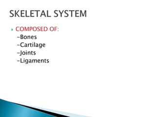
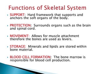
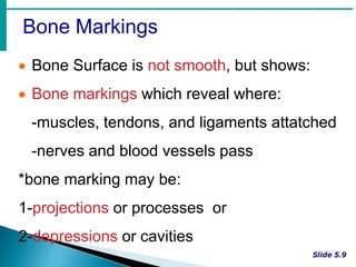
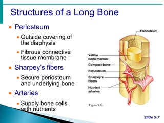
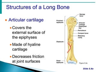
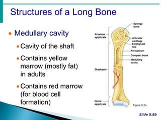
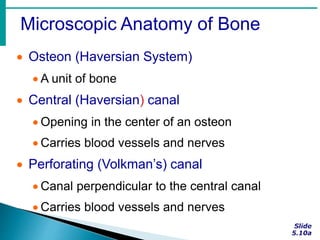


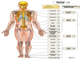
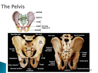
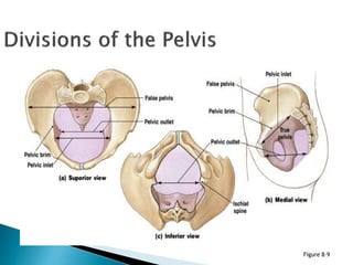

The document provides an overview of the skeletal system, its composition, functions, and classifications of bones. It details the anatomy of long bones, types of bone cells, processes of bone growth, and the structure of the spine and pelvis. Additionally, the document explains joint classifications, types of fractures, and conditions related to joint health.


