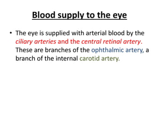
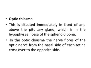
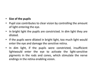
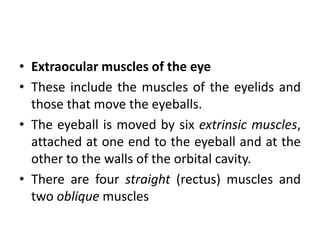
Downloaded 377 times


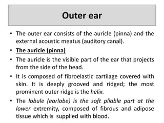


This document provides an overview of the anatomy and structure of the sensory systems, including the eye, ear, nose, and tongue. It describes in detail: - The three parts of the ear: outer, middle, and inner ear. The outer ear collects sounds and directs them to the middle ear, which transfers sounds to the inner ear for conversion into nerve impulses. - The structures of the eye, including the sclera, choroid, iris, lens, retina, and extraocular muscles. The eye focuses light through the cornea and lens onto the light-sensitive retina to initiate vision. - The nose contains olfactory receptors that detect smells and transmit signals to the brain.


![Skin anatomy chc training 2012 [compatibility mode] [repaired]](https://cdn.slidesharecdn.com/ss_thumbnails/skinanatomychctraining2012compatibilitymoderepaired-131009014235-phpapp01-thumbnail.jpg?width=640&height=640&fit=bounds)


