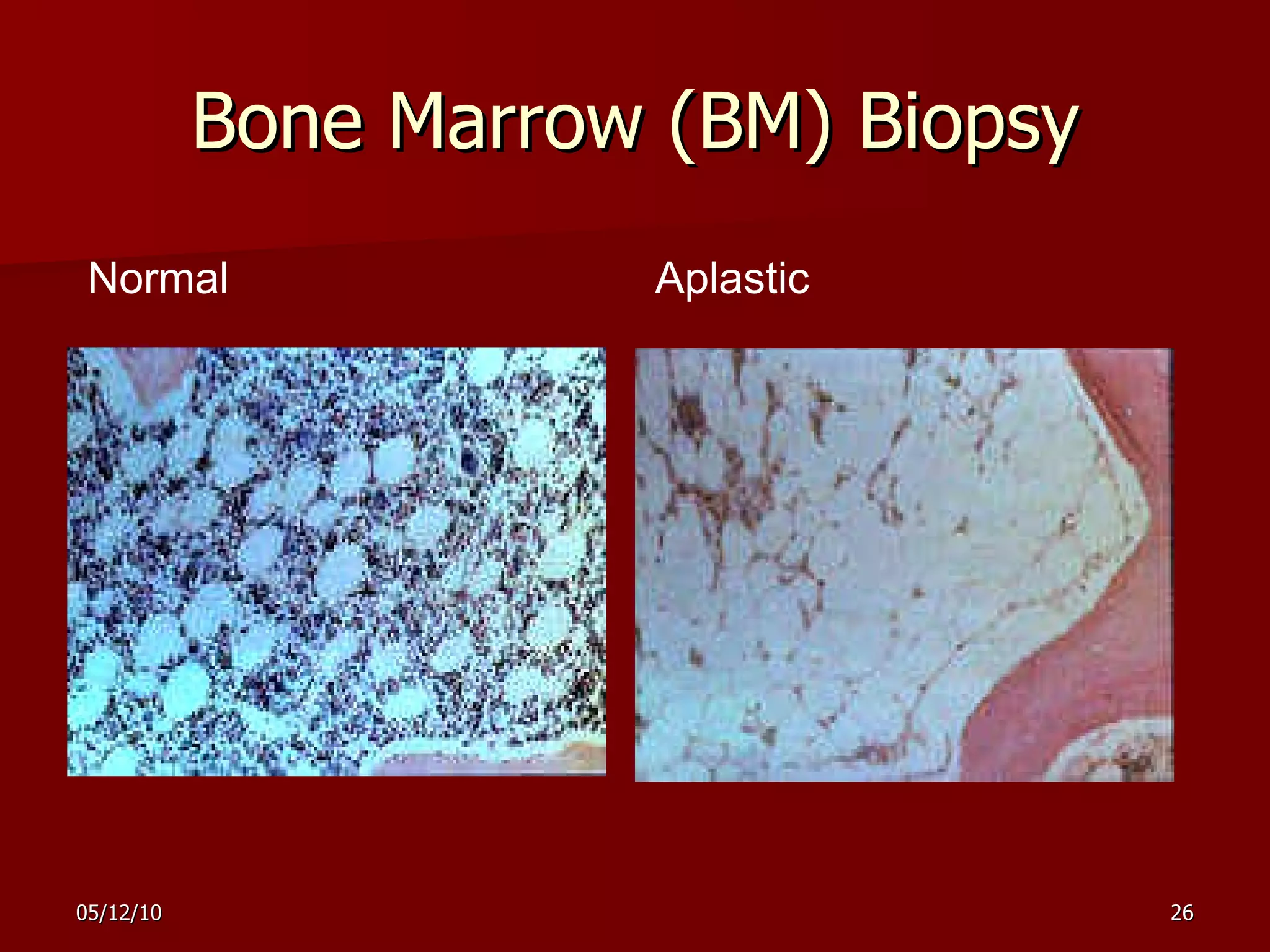
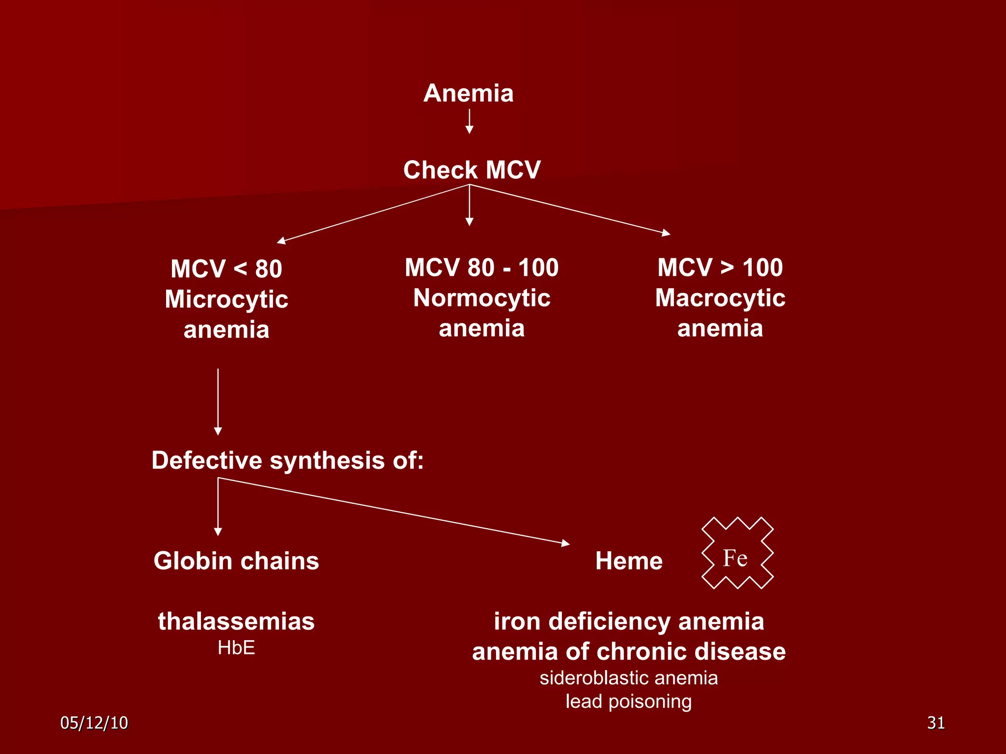
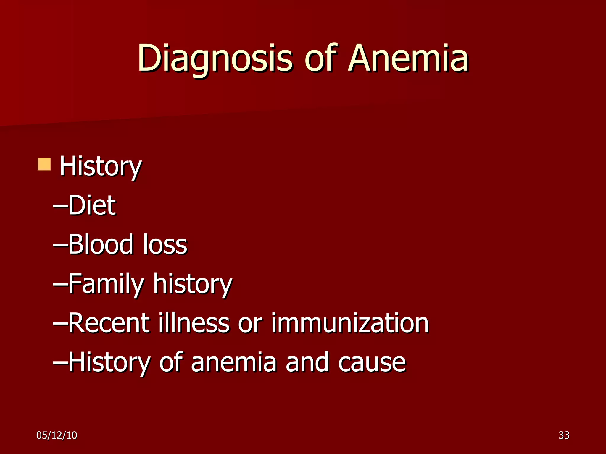
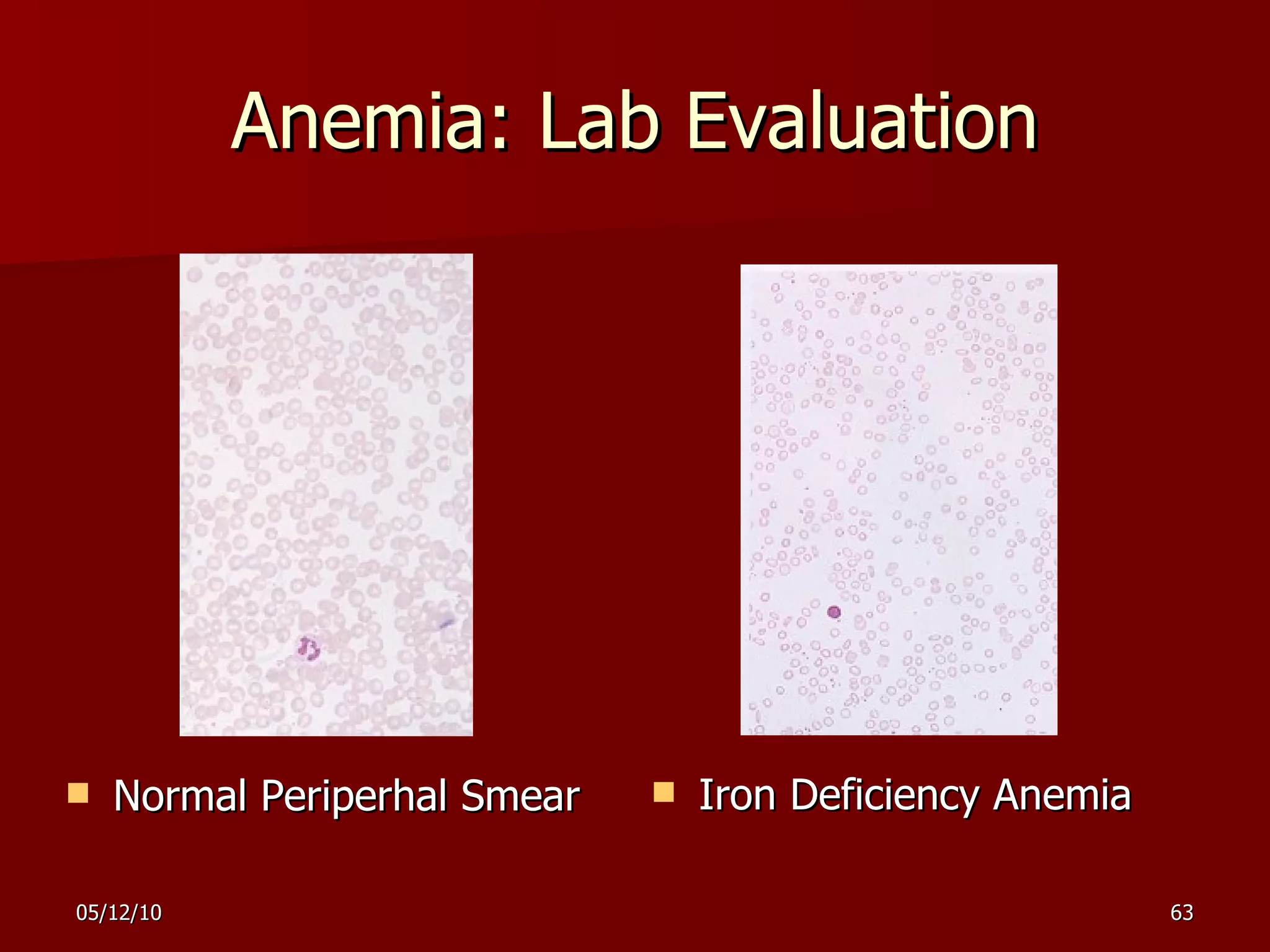
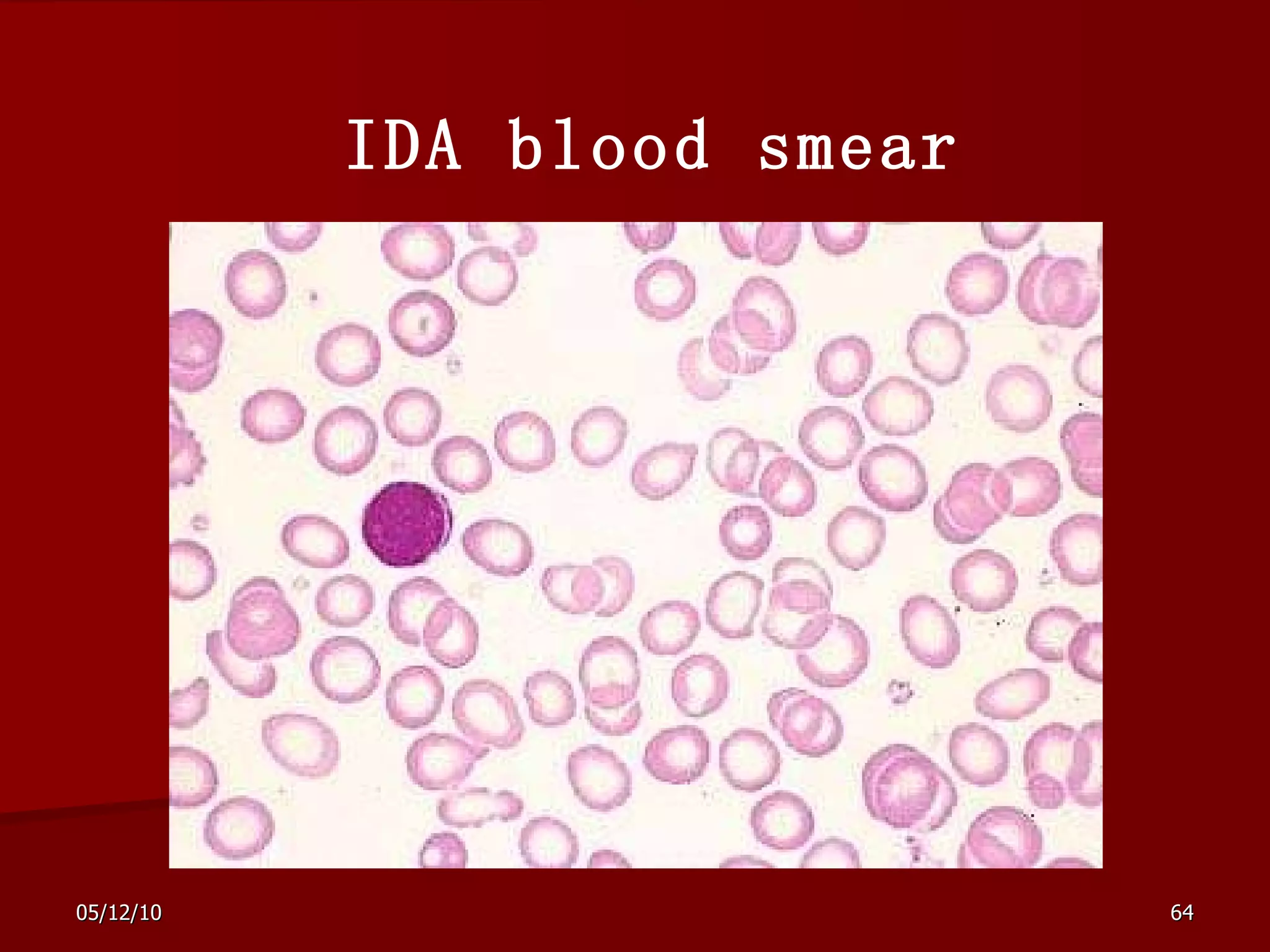
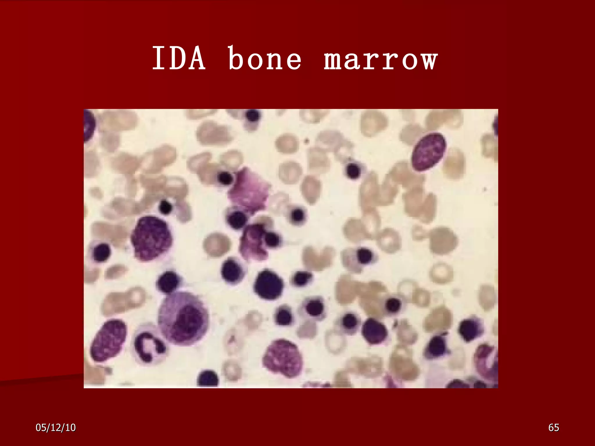
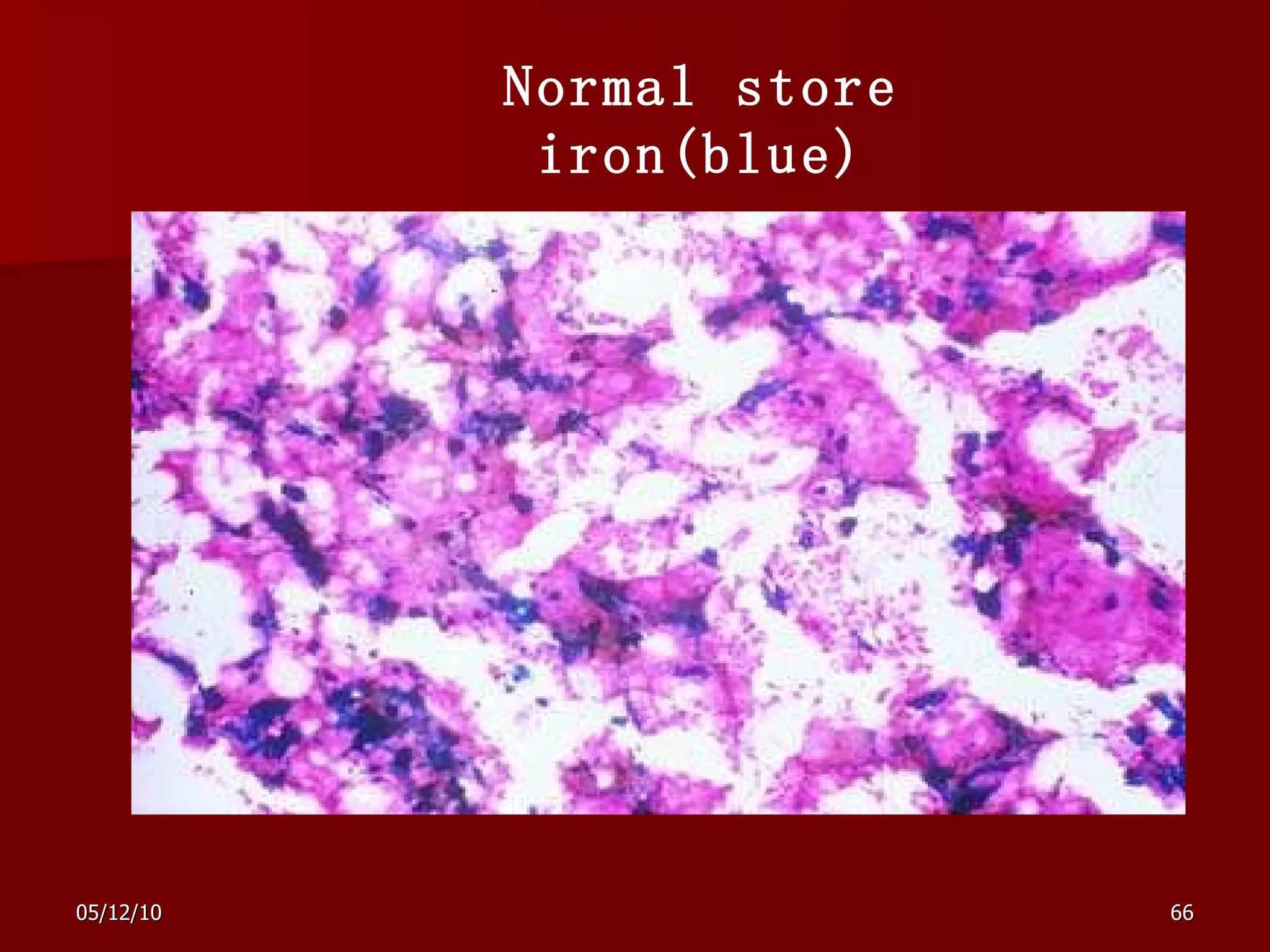
Anemia Overview Ruozhi Xiao The Third Hospital of Sun Yat-sen University [email_address]
Erythrocytes parameters RBC = red blood cells Hgb= hemoglobin Mean corpuscular volume (MCV) N: 80-100 fl RDW(Red cell Distr i bution Width)  Mean corpuscular hemoglobin (MCH) N: 27-34 pg Mean corpuscular hemoglobin concentration (MCHC) N: 310 – 370 g/lRBC (31-37 g/dl)
Survival and Production of RBC Formed in bone marrow  Life span is 120 days (+/-20 days) Cleared in spleen  Reticulocytes are newly formed RBC  in circulation  If no new production, Hgb drops 1 gm/week
 
ERYTHROPOIESIS In developing from the stem cell, the RBC has to undergo the most changes, which can be categorized into several morphological/stainable stages  Proerythroblast RBC * -blast is the common suffix for an immature form of a cell Early erythroblast Intermediate erythroblast Late erythroblast Reticulocyte … … … … Stem cell
Hematocrits Normal, Hemorrhage, IDA, Leukemia, Hemolysis, B12, P Vera Plasma White cells Red cells
Definitinon “ Low blood” Anemia is simply a hemoglobin level lower than the normal range for a particular age and sex of the patient.  Most common hematologic disorder by far
The normal range for Hb and RBC   Hb  RBC  Males :  120 - 160g/L  (4.0-5.5)x  10 12 /L Females :  110 - 150g/L  (3.5-5.0)x  10 12 /L Neonates : 170 - 200g/L  (6.0-7.0)x  10 12 /L
Anemia classification Based on general mechanisms morphological classification
Anemia morphological classification Microcytic Normocytic Macrocytic
morphological classification Type  MCV   fl  MCH  pg   MCHC %   Macrocytic anemia  > 100  > 32  32-35 Normocytic  anemia  80-100  27-32  32-35 Microcytic anemia   < 80  < 27  < 32
Normal Red Blood Cells - Peripheral Blood Smear
Normal Red Blood Cells
 
Microcytic anemia
Thalassemia Thalassa  = the sea Defective globin synthesis Normal   1     thalassemia
b-Thalassemia
 
  Macrocytic anemia
General mechanisms of anemia 1. RBC Loss without RBC destruction 2. Deficient RBC production: Marrow failure 3. Increased RBC destruction over production: Hemolysis
ANEMIA Causes - Cytoplasmic Protein Production Decreased hemoglobin synthesis Disorders of globin synthesis Disorders of heme synthesis Heme synthesis Decreased Iron Iron not in utilizable form Decreased heme synthesis
Pathophysiology Decreased RBC production Iron deficiency  Folic acid deficiency Aplastic anemia Increased RBC loss or destruction  sickle cell anemia blood loss infection
The third hospital of Sun yat-sen university  :Leukemia : trauma,surgery : cancer and ulcer,menstrual periods :Renal disease :  Malaria :Lead poisoning : SLE :PNH
Bone Marrow Disorders Aplastic anemia Myelodysplastic Syndromes Acute Leukemia
Aplastic Anemia Blood Bone Marrow Biopsy
Bone Marrow (BM) Biopsy Normal Aplastic
Definitions Aplastic Anemia (AA) Pancytopenia Hypocellular bone marrow Myelodysplastic Syndrome (MDS) Cytopenias with hypercellular bone marrow Acute Leukemia (AL) Malignant proliferation of immature cells
Aplastic Anemia: Signs and Symptoms Anemia (low Hb, Hct) fatigue, lassitude, dyspnea Thrombocytopenia (low platelets) bruises, petechiae serious bleeding Neutropenia (low neutrophils, a type of white cell) infections
Acquired Aplastic Anemia Drugs Chemicals Viruses Immune diseases Paroxysmal nocturnal hemoglobinuria (PNH) Pregnancy IDIOPATHIC
Myelodysplastic Syndromes (MDS) Clonal diseases Neoplastic Refractory anemias Potential for acute myeloid leukemia (AML)
Anemia  Check MCV MCV < 80 Microcytic anemia MCV 80 - 100 Normocytic anemia MCV > 100 Macrocytic anemia Defective synthesis of: Heme iron deficiency anemia anemia of chronic disease sideroblastic anemia lead poisoning Globin chains thalassemias HbE Fe
Clinical features Mild:Mild dyspnea on exertion, palpitation Moderate: As with MILD ANEMIA, may also have excessive dyspnea  Severe:Anemia:Dyspnea at rest, tachycardia with pounding pulse, weakness, dizziness,  headache, insomnia
Diagnosis of Anemia History –Diet  –Blood loss –Family history  –Recent illness or immunization  –History of anemia and cause
Diagnosis of Anemia Physical Examination – Evaluate conjunctiva and mucous membranes for paleness – Cardiovascular system for murmur  – Liver  – Spleen  – Nodes – Look for jaundice or purpura
Diagnosis of Anemia Labs – Complete blood count with differential and platelets  – Evaluation of smear with red cell indices – Reticulocyte count
Diagnosis of Anemia Other tests – Serum bilirubin, LDH, urinary hemosiderin, hgb electrophoresis, quantitative hgbA2 and F
Common treatment to All “Anemias” Support Hematopoietic growth factors Blood transfusions, blood substitutes Iron  Cure Stem cell transplant Gene therapy
IRON DEFICIENCY ANEMIA
Terms Fe = iron  TIBC = total iron binding capacity  RDW = red cell distribution width
CASE 1 A  50  year old man comes to see you because of fatigue and a change in bowel habit.  He is found to have a hemoglobin of 105 g/L (normal 120-170) and MCV of 78 fL (80-100).  Peripheral blood film shows microcytes and hypochromia.  He previously had a hemoglobin of 165 g/L three years ago, with a normal MCV.
Case 1 – Question 1 What is your approach to the history and physical examination?
Case 1 - Question 1 Discussion iron deficiency most likely symptoms suspicious for lower GI tract malignancy. still ask about chronic inflammatory diseases ask about melena, hematochezia, weight loss, family history of colon ca rectal exam indicated
CASE 1  -  Question 2 What other investigations are appropriate?
Investigations serum ferritin  12 ug/L  (30-400) iron  8 umol/L  (10-28) TIBC  80 umol/L (38-76) transferrin sat.  10 %  (20-55) Conclusion:  Iron deficiency anemia
IRON DEFICIENCY ANEMIA Prevalence
IRON Functions as electron transporter; vital for life Must be in ferrous (Fe +2 ) state for activity Ferric (Fe +3 ) ions cannot transport electrons or O 2
IRON DEFICIENCY ANEMIA IRON METABOLISM ABSORPTION IN DUODENUM TRANSFERRIN TRANSPORTS IRON TO THE CELLS FERRITIN AND HEMOS I DERIN STORE IRON  10% of daily iron is absorbed
Iron Metabolism Heme Iron Hemoglobin and myoglobin Non-heme Iron Breast milk Cow milk All supplements
Iron Absorption Heme Iron Well absorbed Not dependent on Iron deficiency status Not limited by diet
Iron Absorption Non-heme Iron Absorption is sporadic, generally poor Improved absorption Iron deficient status Heme iron (ie red meat, fish, chicken) Vitamin C Worsened absorption Cow’s milk, cheese Cereal Tea
IRON Body Compartments - 75 kg man 3 mg Absorption < 1 mg/day Excretion < 1 mg/day Stores 1000mg Tissue 500 mg Red Cells 2300 mg
Iron deficiency anemia Causes: inadequate dietary iron intake Malabsorption: gastrectomy, chronic diarrhea,  increased iron needs: pregnancy and lactation chronic occult blood loss: bleeding ulcers, GI inflammation, hemorrhoids, cancer, chronic hemoglobinuria Menstrual blood loss
Most body iron is present in hemoglobin in circulating red cells The macrophages of the reticuloendothelial system store iron released from hemoglobin as ferritin and hemosiderin S mall loss of iron each day in urine, faeces ,  skin and nails and in menstrua ting  females as blood (1-2 mg daily)
IRON DEFICIENCY ANEMIA ETIOLOGY: CHRONIC BLEEDING  MENORRHAGIA  PEPTIC ULCER STOMACH CANCER ULCERATIVE COLITIS INTESTINAL CANCER HAEMORRHOIDS DECREASED IRON INTAKE INCREASED IRON REQUIRMENT  (JUVENILE AGE, PREGNANCY, LACTATION)
IRON DEFICENCY - STAGES Prelatent  reduction in iron stores without reduced serum iron levels Hb (N), MCV (N), iron absorption (  ), transferin saturation (N), serum ferritin (  ), marrow iron (  ) Latent iron stores are exhausted, but the blood hemoglobin level remains normal Hb (N), MCV (N), TIBC (  ), serum ferritin (  ), transfe r rin saturation (  ), marrow iron (absent) Iron deficiency anemia blood hemoglobin concentration falls below the lower limit of normal Hb (  ), MCV (  ), TIBC (  ), serum ferritin (  ), transfer r in saturation (  ), marrow iron (absent)
IRON DEFICIENCY ANEMIA GENERAL ANEMIA’S SYMPTOMS: FATIGABILITY DIZZENES S HEADACHE IRRITABILITY  ROARING PALPITATION CHD, CHF
CHARACTERISTICS SYMPTOMS GLOSSITIS, STOMATITIS DYSPHAGIA (  Plummer-Vinson syndrome ) ATROPHIC GASTRITIS DRY, PALE SKIN SPOON SHAPED NAILS, KOILONYCHIA,  BLUE SCLERAE HAIR LOSS PICA (APETITE FOR NON FOOD SUBSTANCES SUCH AS AN ICE, CLAY) SPLENOMEGALY (10%) INCREASED PLATELET COUNT
KOILONYCHIA      
Smooth tongue
IRON DEFICIENCY ANEMIA MCV MCH  MCHC  N Fe TIBC TRANSFERIN SATURATION FERRITIN
BLOOD ROUTINE
BLOOD AND  BONE MARROW SMEAR BLOOD: microcytosis, hipochromia,  anisocytosis   poikilocytosis BONE MARROW high cellularity  mild to moderate erythroid hyperplasia ( 25-35%; N 16 – 18% )  polychromatic and pyknotic cytoplasm  of  erythroblasts is vacuolated and irregular in outline   ( micronormoblastic   erythropoiesis ) absence of stainable iron
Anemia: Lab Evaluation Normal Periperhal Smear Iron Deficiency Anemia
IDA blood smear
IDA bone marrow
Normal store iron(blue)
IDA
Reticulocytes up
Using special stains such as methylene blue  or brilliant cresyl blue, reticulocytes stain with  dark blue granules whereas mature  erythrocytes evenly stain pale blue.
Management History and physical examination is sufficient to exclude serious disease (e.g pregnant or lactating women, adolescents)  - CURE ANEMIA
Management History and/or physical examination is insufficient  (e.g old men, postmenopausal women)  - FIND ETIOLOGY OF ANEMIA AND CURE (CAUSAL TREATMENT) Benzidine test  Gastroscopy Colonoscopy Gynaecological examination
IRON DEFICIENCY ANEMIA CURE ORAL 3 00 mg of iron daily  after  meal  How long?  3-6 months  to restore iron  reserve Absorption  is enhanced: vit   C, meat, orange juice, fish is inhibited:  tea, milk
IRON DEFICIENCY ANEMIA CURE PARENTERAL IRON SUBSTITUTION Bad oral iron tolerance  (nausea, diarrhoea) Negative oral iron absorption test Necessity of quick management (CHD, CHF)
Remember: Iron deficiency anemia is a manifestation of an underlying process. Look for and treat the cause of the iron deficiency.
Look for the cause colonoscopy reveals colon carcinoma which is subsequently resected.
Iron Deficiency anemia Diagnostics: Iron levels Total iron-binding capacity (TIBC) Serum Ferritin Medications: Iron supplements, oral or parenteral Vit. C
The End

Anemia

  • 1.
    Anemia Overview RuozhiXiao The Third Hospital of Sun Yat-sen University [email_address]
  • 2.
    Erythrocytes parameters RBC= red blood cells Hgb= hemoglobin Mean corpuscular volume (MCV) N: 80-100 fl RDW(Red cell Distr i bution Width) Mean corpuscular hemoglobin (MCH) N: 27-34 pg Mean corpuscular hemoglobin concentration (MCHC) N: 310 – 370 g/lRBC (31-37 g/dl)
  • 3.
    Survival and Productionof RBC Formed in bone marrow Life span is 120 days (+/-20 days) Cleared in spleen Reticulocytes are newly formed RBC in circulation If no new production, Hgb drops 1 gm/week
  • 4.
  • 5.
    ERYTHROPOIESIS In developingfrom the stem cell, the RBC has to undergo the most changes, which can be categorized into several morphological/stainable stages Proerythroblast RBC * -blast is the common suffix for an immature form of a cell Early erythroblast Intermediate erythroblast Late erythroblast Reticulocyte … … … … Stem cell
  • 6.
    Hematocrits Normal, Hemorrhage,IDA, Leukemia, Hemolysis, B12, P Vera Plasma White cells Red cells
  • 7.
    Definitinon “ Lowblood” Anemia is simply a hemoglobin level lower than the normal range for a particular age and sex of the patient. Most common hematologic disorder by far
  • 8.
    The normal rangefor Hb and RBC Hb RBC Males : 120 - 160g/L (4.0-5.5)x 10 12 /L Females : 110 - 150g/L (3.5-5.0)x 10 12 /L Neonates : 170 - 200g/L (6.0-7.0)x 10 12 /L
  • 9.
    Anemia classification Basedon general mechanisms morphological classification
  • 10.
    Anemia morphological classificationMicrocytic Normocytic Macrocytic
  • 11.
    morphological classification Type MCV fl MCH pg MCHC % Macrocytic anemia > 100 > 32 32-35 Normocytic anemia 80-100 27-32 32-35 Microcytic anemia < 80 < 27 < 32
  • 12.
    Normal Red BloodCells - Peripheral Blood Smear
  • 13.
  • 14.
  • 15.
  • 16.
    Thalassemia Thalassa = the sea Defective globin synthesis Normal  1   thalassemia
  • 17.
  • 18.
  • 19.
  • 20.
    General mechanisms ofanemia 1. RBC Loss without RBC destruction 2. Deficient RBC production: Marrow failure 3. Increased RBC destruction over production: Hemolysis
  • 21.
    ANEMIA Causes -Cytoplasmic Protein Production Decreased hemoglobin synthesis Disorders of globin synthesis Disorders of heme synthesis Heme synthesis Decreased Iron Iron not in utilizable form Decreased heme synthesis
  • 22.
    Pathophysiology Decreased RBCproduction Iron deficiency Folic acid deficiency Aplastic anemia Increased RBC loss or destruction sickle cell anemia blood loss infection
  • 23.
    The third hospitalof Sun yat-sen university :Leukemia : trauma,surgery : cancer and ulcer,menstrual periods :Renal disease : Malaria :Lead poisoning : SLE :PNH
  • 24.
    Bone Marrow DisordersAplastic anemia Myelodysplastic Syndromes Acute Leukemia
  • 25.
    Aplastic Anemia BloodBone Marrow Biopsy
  • 26.
    Bone Marrow (BM)Biopsy Normal Aplastic
  • 27.
    Definitions Aplastic Anemia(AA) Pancytopenia Hypocellular bone marrow Myelodysplastic Syndrome (MDS) Cytopenias with hypercellular bone marrow Acute Leukemia (AL) Malignant proliferation of immature cells
  • 28.
    Aplastic Anemia: Signsand Symptoms Anemia (low Hb, Hct) fatigue, lassitude, dyspnea Thrombocytopenia (low platelets) bruises, petechiae serious bleeding Neutropenia (low neutrophils, a type of white cell) infections
  • 29.
    Acquired Aplastic AnemiaDrugs Chemicals Viruses Immune diseases Paroxysmal nocturnal hemoglobinuria (PNH) Pregnancy IDIOPATHIC
  • 30.
    Myelodysplastic Syndromes (MDS)Clonal diseases Neoplastic Refractory anemias Potential for acute myeloid leukemia (AML)
  • 31.
    Anemia CheckMCV MCV < 80 Microcytic anemia MCV 80 - 100 Normocytic anemia MCV > 100 Macrocytic anemia Defective synthesis of: Heme iron deficiency anemia anemia of chronic disease sideroblastic anemia lead poisoning Globin chains thalassemias HbE Fe
  • 32.
    Clinical features Mild:Milddyspnea on exertion, palpitation Moderate: As with MILD ANEMIA, may also have excessive dyspnea Severe:Anemia:Dyspnea at rest, tachycardia with pounding pulse, weakness, dizziness, headache, insomnia
  • 33.
    Diagnosis of AnemiaHistory –Diet –Blood loss –Family history –Recent illness or immunization –History of anemia and cause
  • 34.
    Diagnosis of AnemiaPhysical Examination – Evaluate conjunctiva and mucous membranes for paleness – Cardiovascular system for murmur – Liver – Spleen – Nodes – Look for jaundice or purpura
  • 35.
    Diagnosis of AnemiaLabs – Complete blood count with differential and platelets – Evaluation of smear with red cell indices – Reticulocyte count
  • 36.
    Diagnosis of AnemiaOther tests – Serum bilirubin, LDH, urinary hemosiderin, hgb electrophoresis, quantitative hgbA2 and F
  • 37.
    Common treatment toAll “Anemias” Support Hematopoietic growth factors Blood transfusions, blood substitutes Iron Cure Stem cell transplant Gene therapy
  • 38.
  • 39.
    Terms Fe =iron TIBC = total iron binding capacity RDW = red cell distribution width
  • 40.
    CASE 1 A 50 year old man comes to see you because of fatigue and a change in bowel habit. He is found to have a hemoglobin of 105 g/L (normal 120-170) and MCV of 78 fL (80-100). Peripheral blood film shows microcytes and hypochromia. He previously had a hemoglobin of 165 g/L three years ago, with a normal MCV.
  • 41.
    Case 1 –Question 1 What is your approach to the history and physical examination?
  • 42.
    Case 1 -Question 1 Discussion iron deficiency most likely symptoms suspicious for lower GI tract malignancy. still ask about chronic inflammatory diseases ask about melena, hematochezia, weight loss, family history of colon ca rectal exam indicated
  • 43.
    CASE 1 - Question 2 What other investigations are appropriate?
  • 44.
    Investigations serum ferritin 12 ug/L (30-400) iron 8 umol/L (10-28) TIBC 80 umol/L (38-76) transferrin sat. 10 % (20-55) Conclusion: Iron deficiency anemia
  • 45.
  • 46.
    IRON Functions aselectron transporter; vital for life Must be in ferrous (Fe +2 ) state for activity Ferric (Fe +3 ) ions cannot transport electrons or O 2
  • 47.
    IRON DEFICIENCY ANEMIAIRON METABOLISM ABSORPTION IN DUODENUM TRANSFERRIN TRANSPORTS IRON TO THE CELLS FERRITIN AND HEMOS I DERIN STORE IRON 10% of daily iron is absorbed
  • 48.
    Iron Metabolism HemeIron Hemoglobin and myoglobin Non-heme Iron Breast milk Cow milk All supplements
  • 49.
    Iron Absorption HemeIron Well absorbed Not dependent on Iron deficiency status Not limited by diet
  • 50.
    Iron Absorption Non-hemeIron Absorption is sporadic, generally poor Improved absorption Iron deficient status Heme iron (ie red meat, fish, chicken) Vitamin C Worsened absorption Cow’s milk, cheese Cereal Tea
  • 51.
    IRON Body Compartments- 75 kg man 3 mg Absorption < 1 mg/day Excretion < 1 mg/day Stores 1000mg Tissue 500 mg Red Cells 2300 mg
  • 52.
    Iron deficiency anemiaCauses: inadequate dietary iron intake Malabsorption: gastrectomy, chronic diarrhea, increased iron needs: pregnancy and lactation chronic occult blood loss: bleeding ulcers, GI inflammation, hemorrhoids, cancer, chronic hemoglobinuria Menstrual blood loss
  • 53.
    Most body ironis present in hemoglobin in circulating red cells The macrophages of the reticuloendothelial system store iron released from hemoglobin as ferritin and hemosiderin S mall loss of iron each day in urine, faeces , skin and nails and in menstrua ting females as blood (1-2 mg daily)
  • 54.
    IRON DEFICIENCY ANEMIAETIOLOGY: CHRONIC BLEEDING MENORRHAGIA PEPTIC ULCER STOMACH CANCER ULCERATIVE COLITIS INTESTINAL CANCER HAEMORRHOIDS DECREASED IRON INTAKE INCREASED IRON REQUIRMENT (JUVENILE AGE, PREGNANCY, LACTATION)
  • 55.
    IRON DEFICENCY -STAGES Prelatent reduction in iron stores without reduced serum iron levels Hb (N), MCV (N), iron absorption (  ), transferin saturation (N), serum ferritin (  ), marrow iron (  ) Latent iron stores are exhausted, but the blood hemoglobin level remains normal Hb (N), MCV (N), TIBC (  ), serum ferritin (  ), transfe r rin saturation (  ), marrow iron (absent) Iron deficiency anemia blood hemoglobin concentration falls below the lower limit of normal Hb (  ), MCV (  ), TIBC (  ), serum ferritin (  ), transfer r in saturation (  ), marrow iron (absent)
  • 56.
    IRON DEFICIENCY ANEMIAGENERAL ANEMIA’S SYMPTOMS: FATIGABILITY DIZZENES S HEADACHE IRRITABILITY ROARING PALPITATION CHD, CHF
  • 57.
    CHARACTERISTICS SYMPTOMS GLOSSITIS,STOMATITIS DYSPHAGIA ( Plummer-Vinson syndrome ) ATROPHIC GASTRITIS DRY, PALE SKIN SPOON SHAPED NAILS, KOILONYCHIA, BLUE SCLERAE HAIR LOSS PICA (APETITE FOR NON FOOD SUBSTANCES SUCH AS AN ICE, CLAY) SPLENOMEGALY (10%) INCREASED PLATELET COUNT
  • 58.
  • 59.
  • 60.
    IRON DEFICIENCY ANEMIAMCV MCH MCHC N Fe TIBC TRANSFERIN SATURATION FERRITIN
  • 61.
  • 62.
    BLOOD AND BONE MARROW SMEAR BLOOD: microcytosis, hipochromia, anisocytosis poikilocytosis BONE MARROW high cellularity mild to moderate erythroid hyperplasia ( 25-35%; N 16 – 18% ) polychromatic and pyknotic cytoplasm of erythroblasts is vacuolated and irregular in outline ( micronormoblastic erythropoiesis ) absence of stainable iron
  • 63.
    Anemia: Lab EvaluationNormal Periperhal Smear Iron Deficiency Anemia
  • 64.
  • 65.
  • 66.
  • 67.
  • 68.
  • 69.
    Using special stainssuch as methylene blue or brilliant cresyl blue, reticulocytes stain with dark blue granules whereas mature erythrocytes evenly stain pale blue.
  • 70.
    Management History andphysical examination is sufficient to exclude serious disease (e.g pregnant or lactating women, adolescents) - CURE ANEMIA
  • 71.
    Management History and/orphysical examination is insufficient (e.g old men, postmenopausal women) - FIND ETIOLOGY OF ANEMIA AND CURE (CAUSAL TREATMENT) Benzidine test Gastroscopy Colonoscopy Gynaecological examination
  • 72.
    IRON DEFICIENCY ANEMIACURE ORAL 3 00 mg of iron daily after meal How long? 3-6 months to restore iron reserve Absorption is enhanced: vit C, meat, orange juice, fish is inhibited: tea, milk
  • 73.
    IRON DEFICIENCY ANEMIACURE PARENTERAL IRON SUBSTITUTION Bad oral iron tolerance (nausea, diarrhoea) Negative oral iron absorption test Necessity of quick management (CHD, CHF)
  • 74.
    Remember: Iron deficiencyanemia is a manifestation of an underlying process. Look for and treat the cause of the iron deficiency.
  • 75.
    Look for thecause colonoscopy reveals colon carcinoma which is subsequently resected.
  • 76.
    Iron Deficiency anemiaDiagnostics: Iron levels Total iron-binding capacity (TIBC) Serum Ferritin Medications: Iron supplements, oral or parenteral Vit. C
  • 77.