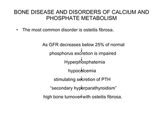
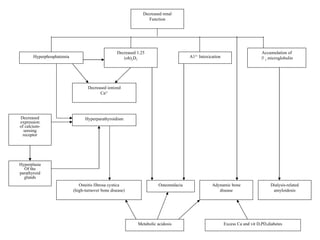
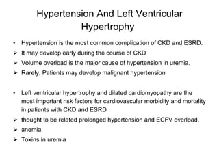
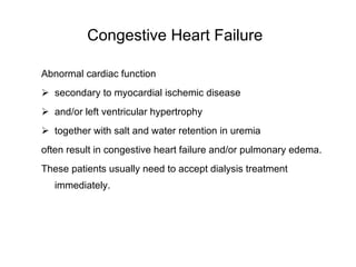
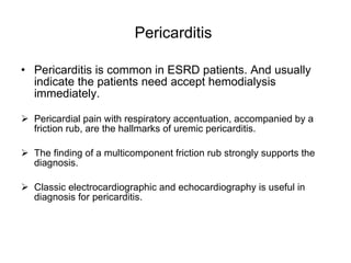
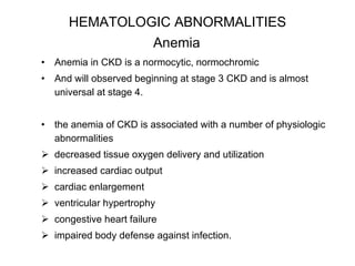
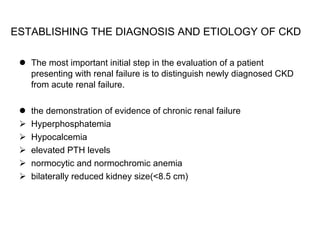
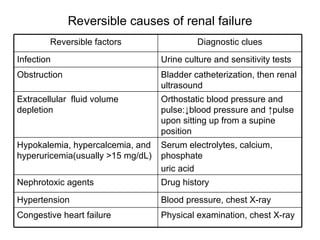
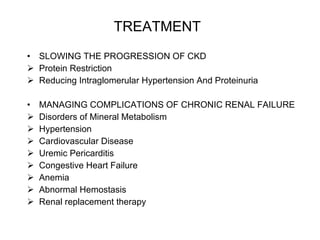
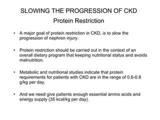
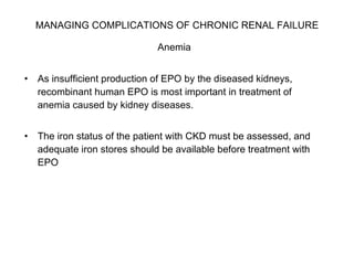
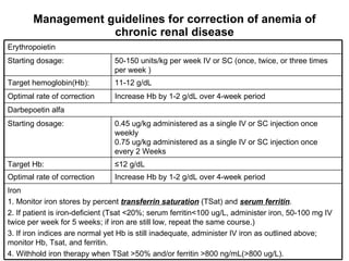
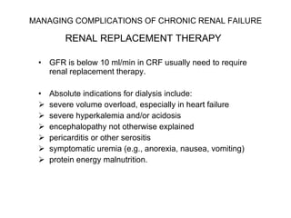
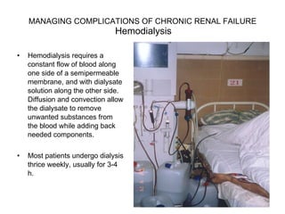
Chronic Renal Failure  Department of Nephrology, The First Affiliated Hospital of Sun Yat-Sen University  Jiang Zongpei
CONTANT DEFINITIONS ETIOLOGY PATHOPHYSIOLOGY OF CKD PATHOPHYSIOLOGY AND BIOCHEMISTRY OF UREMIA CLINICAL MANIFESTATIONS OF CHRONIC RENAL FAILURE EVALUATION AND MANAGEMENT OF PATIENTS WITH CKD TREATMENT
DEFINITIONS  Chronic Kidney disease (CKD) : CKD is a pathophysiologic process with multiple etiologies, resulting in the irreversible attrition of nephron number and function, frequently leading to end stage renal disease (ESRD).  ESRD  represents a clinical state or condition in which there has been an irreversible loss of renal function, and these patients usually need to accept renal replacement therapy (dialysis or transplantation) in order to avoid life-threatening uremia.  Uremia  Uremia is the clinical and laboratory syndrome, reflecting dysfunction of all organ systems as a result of untreated or undertreated acute or chronic renal failure.
Major causes of chronic renal failure   Glomerulopathies Primary glomerular diseases: 1. Focal and segmental glomerulosclerosis(FSGS) 2. Membranoproliferative  glomerulonephritis(MPGN) 3. IgA nephropathy(IgAN) 4. Membranous nephropathy(MN)   Secondary glomerular diseases: 1.Diabetic nephropathy 2.Lupus nephropathy 3. Post-infectious glomerulonephritis 4. Amyloidosis 5.HIV-associated nephropathy 6.Collagen-vascular diseases 7.Sickle cell nephropathy 8. HIV associated membranoproliferative glomerulonephritis Tubulointerstitial nephritis Drug hypersensitivity Heavy metals Analgesic nephropathy Reflux/chronic pyelonephritis Idiopathic Hereditary diseases Polycystic kidney disease Medullary cystic disease Alport’s syndrome Obstructive nephropathies Prostatic disease Nephrolithiasis Retroperitoneal fibrosis/tumor Congenital Vascular diseases Hypertensive nephrosclerosis Renal artery stenosis
ETIOLOGY  The leading underlying etiologies of both CKD and ESRD in China. Glomerulonephritis  60% Diabetic nephropathy  20% Lupus nephropahty  5-10% Hypertensive nephrosclerosis  5-10% Others  5-10%
PATHOPHYSIOLOGY OF CKD   1 The pathophysiology that why CKD progress to ESRD is unclearly now. vasoactive molecules Cytokines growth factors Renin-angiotensin axis glomerular  hyperperfusion, Hypertension hyperfiltration glomerular hypertrophy sclerosis nephron population decrease and reduction of renal mass.  Such reduction of renal mass causes structural and functional hypertrophy of surviving nephrons
Renal pathologic change in CRF Normal Sclerosis
Stages of chronic kidney disease: A clinical action plan Replacement (if uremia is present) Preparation for kidney replacement therapy. Evaluating and treating complications Estimating progression Diagnosis and treatment.  Treatment of basic diseases.  Slowing of progression. Cardiovascular disease risk reduction. Action Uremic symptoms become prominent and usually the patients need to accept renal replacement therapy <15 Kidney failure 5 15-29 Severely↓GFR 4 clinical and laboratory complications of CKD become progressive 30-59 Moderately↓GFR 3 60-89 Kidney damage with mildly↓GFR 2 patients often remain free of symptom ≥ 90 Kidney damage with normal of ↑GFR 1 symptom GFR Description Stage
PATHOPHYSIOLOGY AND BIOCHEMISTRY OF UREMIA   Which toxin is responsible for uremic symptoms remain unclear.  Urea  may contribute to some of the clinical abnormalities, including anorexia, malaise, vomiting, and headache.  Nitrogenous excretory products  (guanido compounds, urates and so on) and  nitrogenous compounds  (so-called  middle molecules toxins ) are associated with neuromuscular abnormalities and believed to contribute to morbidity and mortality in uremic subjects.
PATHOPHYSIOLOGY AND BIOCHEMISTRY OF UREMIA A host of metabolic and endocrine functions normally subserved by the kidney are also impaired, resulting in anemia; malnutrition; impaired metabolism of carbohydrates, fats, and proteins and metabolic bone disease.  The endocrine hormone involved in ESRD patients are PTH, insulin, erythropoietin (EPO) and so on.
CLINICAL MANIFESTATIONS OF CHRONIC RENAL FAILURE   Uremia leads to disturbances in the function of every organ system.  The symptoms of chronic renal failure often develop slowly and are nonspecific.  Individuals can remain asymptomatic until renal failure is far-advanced (GFR<10-15 ml/min).
CLINICAL MANIFESTATIONS OF CHRONIC RENAL FAILURE FLUID, ELECTROLYTE, AND ACID-BASE DISORDERS BONE DISEASE AND DISORDERS OF CALCIUM AND PHOSPHATE METABOLISM CARDIOVASCULAR ABNORMALITIES HEMATOLOGIC ABNORMALITIES NEUROMUSCULAR ABNORMALITIES GASTROINTESTINAL AND NUTRITIONAL ABNORMALITIES ENDOCRINE METABOLIC DISTURBANCES
FLUID, ELECTROLYTE, AND ACID-BASE DISORDERS Sodium And Water Homeostasis   In most patients with stable CKD, the total body contents of Na+ and H 2 O are increased modestly, and will cause edema and hypertention.  Glomerulo-tubular balance impaired and promote Na+ retention Na+ retention may lead to cumulative positive Na+ balance and cause extracellular fluid volume (ECFV) expansion.  Patients with CKD also have impaired renal mechanisms for conserving Na+ and H 2 O. And  these patients are easy to appear volume depletion.
FLUID, ELECTROLYTE, AND ACID-BASE DISORDERS Potassium Homeostasis In CKD hyperkalemia is common in CKD patients dietary intake, protein catabolism, hemorrhage, transfusion of stored red blood cells, metabolic acidosis ACE inhibitors and angiotensin receptor blockers(ARB), and some diuretics Hypokalemia is uncommon in CKD  usually caused by markedly reduced dietary K+ intake Or association with excessive diuretic therapy or gastrointestinal losses.  And also be seen in tubulointerstitial diseases, such as Fanconi’s syndrome, renal tubular acidosis
FLUID, ELECTROLYTE, AND ACID-BASE DISORDERS Metabolic Acidosis  is a common disturbance during the advanced stages of CKD Reduced ability to produce ammonia. Hyperkalemia further depresses urinary ammonium excretion.
BONE DISEASE AND DISORDERS OF CALCIUM AND PHOSPHATE METABOLISM The disorders of calcium, phosphorus, and bone diseases is common in CKD.  The major disorders of bone disease can be classified into: high bone turnover with high PTH levels (including osteitis fibrosa) low bone turnover with low or normal PTH levels (osteomalacia and adynamic bone disease).
BONE DISEASE AND DISORDERS OF CALCIUM AND PHOSPHATE METABOLISM The most common disorder is osteitis fibrosa.  As GFR decreases below 25% of normal  phosphorus excretion is impaired Hyperphosphatemia  hypocalcemia  stimulating secretion of PTH  “ secondary hyperparathyroidism”  high bone turnover with osteitis fibrosa.
Decreased renal Function Decreased 1.25 (oh) 2 D 3 A1 3+  Intoxication Accumulation of β 2  microglobulin Hyperphosphatemia Decreased ionized Ca 2+ Hyperparathyroidism Decreased expression of calcium- sensing receptor Hyperplasia Of the parathyroid glands Osteitis fibrosa cystica (high-turnover bone disease) Osteomalacia Adynamic bone disease Dialysis-related amyloidosis Metabolic acidosis Excess Ca and vit D,PD,diabetes
CARDIOVASCULAR ABNORMALITIES   Cardiovascular disease is the leading cause of morbidity and mortality in patients with CKD at all stages. Ischemic Cardiovascular Disease Congestive Heart Failure Hypertension And Left Ventricular Hypertrophy Pericarditis
Ischemic Cardiovascular Disease   CKD is a major risk factor for ischemic cardiovascular disease, including coronary heart, cerebrovascular, and peripheral vascular diseases Ischemic cardiovascular disease in CKD caused by both traditional risk factors and CKD-related risk factors The traditional risk factors include hypertension, hypervolemia, dyslipidemia, and so on.  The CKD-related risks include anemia, hyperphosphatemia, high PTH level, and a state of “microinflammation” that can be found at all stages of CKD.
Hypertension And Left Ventricular Hypertrophy   Hypertension is the most common complication of CKD and ESRD. It may develop early during the course of CKD  Volume overload is the major cause of hypertension in uremia. Rarely, Patients may develop malignant hypertension Left ventricular hypertrophy and dilated cardiomyopathy are the most important risk factors for cardiovascular morbidity and mortality in patients with CKD and ESRD  thought to be related prolonged hypertension and ECFV overload.  anemia  Toxins in uremia
Congestive Heart Failure   Abnormal cardiac function  secondary to myocardial ischemic disease  and/or left ventricular hypertrophy together with salt and water retention in uremia often result in congestive heart failure and/or pulmonary edema.  These patients usually need to accept dialysis treatment immediately.
Pericarditis  Pericarditis is common in ESRD patients. And usually indicate the patients need accept hemodialysis immediately. Pericardial pain with respiratory accentuation, accompanied by a friction rub, are the hallmarks of uremic pericarditis.  The finding of a multicomponent friction rub strongly supports the diagnosis.  Classic electrocardiographic and echocardiography is useful in diagnosis for pericarditis.
HEMATOLOGIC ABNORMALITIES  Anemia   Anemia in CKD is a normocytic, normochromic  And will observed beginning at stage 3 CKD and is almost universal at stage 4.  the anemia of CKD is associated with a number of physiologic abnormalities decreased tissue oxygen delivery and utilization increased cardiac output cardiac enlargement ventricular hypertrophy congestive heart failure impaired body defense against infection.
HEMATOLOGIC ABNORMALITIES  Anemia The primary cause of anemia in patients with CKD  insufficient production of EPO by the diseased kidneys.  iron and folate deficiency Severe PTH acute and chronic inflammation aluminum toxicity  shortened red cell survival.
NEUROMUSCULAR ABNORMALITIES   Central, peripheral, and autonomic neuropathy, as well as abnormalities in muscle composition and function, are all common complications in CKD.  Retained nitrogenous metabolites and middle molecules toxins as well as PTH ,  all contribute to the pathophysiology of neuromuscular abnormalities.  The “restless legs syndrome ” is characterized by illdefined sensations of discomfort in the legs and legs and feet requiring frequently  movement.
GASTROINTESTINAL AND NUTRITIONAL ABNORMALITIES   Gastrointestinal symptoms (abdominal pain, nausea, vomiting) usually may the first symptom for ESRD patients. Urimic toxins is associated with an unpleasant metallic taste sensation in ESRD patient.  Gastritis, peptic disease are common in ESRD patient. Protein restriction is useful in diminishing the symptoms. But it is sometimes easy to cause malnutrition.
ENDOCRINE-METABOLIC DISTURBANCES   Disturbances in parathyroid function Glucose metabolism impaired in CKD Because the kidney contributes significantly to insulin removal from the circulation, plasma levels of insulin are slightly to moderately elevated in most uremic subjects.  Many hypoglycemic drugs require dose reduction in renal failure The growth and sex hormone   secretion is impaired in CKD , Sexual maturation is often in children with CKD.
EVALUATION AND MANAGEMENT OF PATIENTS WITH CKD   History Hypertension Diabetes systemic infectious Inflammatory metabolic diseases exposure to drugs and toxins a family history of renal and urologic disease. In evaluating the uremic syndrome questions about appetite ; diet ; nausea ; vomiting ; edema ; weight change Physical examination blood pressure  examination of the abdomen for renal masses, examination for edema neurologic examination
EVALUATION AND MANAGEMENT OF PATIENTS WITH CKD   Laboratory Investigations   Laboratory investigation should focus on to find evidence to underlying disease process and its continued activity.  immunologic tests (SLE and vasculitis) Serum and uriary protein electrophoresis. Urinalysis, a 24h urine collection for protein excretion measurements of plasma creatinine and estimation of GFR. to assess metabolic bone disease  hemoglobin.
EVALUATION AND MANAGEMENT OF PATIENTS WITH CKD   Imaging Studies   The most useful imaging study is renal ultrasonography.  Provide an estimate of kidney size and obstructive uropathy.  The small kidneys usually supports the diagnosis of progressive CKD is irreversible Normal kidney size usually suggests the possibility of an acute rather than chronic process.  a vascular imaging procedure, such as  doppler sonography of the renal arteries,  radionuclide scintigraphy,  magnetic resonance angiography (MRA)
EVALUATION AND MANAGEMENT OF PATIENTS WITH CKD   Renal Biopsy   Renal biopsy should be reserved for patients with near normal kidney size, in whom a definitive diagnosis cannot be made yet.  Contraindications to renal biopsy include: bilateral small kidneys  polycystic kidney disease uncontrolled hypertension urinary tract or perinephric infection bleeding diathesis respiratory distress morbid obesity.
ESTABLISHING THE DIAGNOSIS AND ETIOLOGY OF CKD   The most important initial step in the evaluation of a patient presenting with renal failure is to distinguish newly diagnosed CKD from acute renal failure.  the demonstration of evidence of chronic renal failure  Hyperphosphatemia Hypocalcemia elevated PTH levels normocytic and normochromic anemia bilaterally reduced kidney size(<8.5 cm)
Reversible causes of renal failure Physical examination, chest X-ray Congestive heart failure Blood pressure, chest X-ray Hypertension Drug history Nephrotoxic agents Serum electrolytes, calcium, phosphate uric acid Hypokalemia, hypercalcemia, and hy peruricemia(usually >15 mg/dL) Orthostatic blood pressure and pulse:↓blood pressure and ↑pulse upon sitting up from a supine position Extracellular  fluid volume depletion  Bladder catheterization, then renal ultrasound Obstruction Urine culture and sensitivity tests Infection Diagnostic clues Reversible factors
TREATMENT   SLOWING THE PROGRESSION OF CKD Protein Restriction Reducing Intraglomerular Hypertension And Proteinuria MANAGING COMPLICATIONS OF CHRONIC RENAL FAILURE Disorders of Mineral Metabolism Hypertension Cardiovascular Disease Uremic Pericarditis Congestive Heart Failure Anemia Abnormal Hemostasis Renal replacement therapy
SLOWING THE PROGRESSION OF CKD   Protein Restriction   A major goal of protein restriction in CKD, is to slow the progression of nephron injury.  Protein restriction should be carried out in the context of an overall dietary program that keeping nutritional status and avoids malnutrition.  Metabolic and nutritional studies indicate that protein requirements for patients with CKD are in the range of 0.6-0.8 g/kg per day.  And we need give patients enough essential amino acids and energy supply (35 kcal/kg per day).
SLOWING THE PROGRESSION OF CKD   Reducing Intraglomerular Hypertension And Proteinuria   Progressive renal injury in CKD appears to be most closely related to the height of intraglomerular pressure and/or the extent of glomerular hypertrophy. Antihypertensive therapy in patients with CKD also aims to slow the progression of nephron injury, by reduce intraglomerular hypertension and hypertrophy. ACE inhibitors(ACEI) and angiotensin receptor blockers (ARB) are now clearly established as effective, antiproteinuric and anti-intraglomerular hypertension agents.  If patients present side effects of the use of ACEI/ARBthese (e.g. cough, hyperkalemia) . We may choice of calcium channel blockers (CCB) as a second line medicine.
MANAGING COMPLICATIONS OF CHRONIC RENAL FAILURE   Disorders of Mineral Metabolism   Treatment should begin with dietary phosphorus restriction to <1000 mg/d. Oral phosphorus binding agents.  Vitamin D or vitamin D analogs should be given when PTH level is more than two to three times to normal It is particularly important to maintain the calcium-phosphate product in the normal range to avoid metastatic calcification.
MANAGING COMPLICATIONS OF CHRONIC RENAL FAILURE   Hypertension  Volume control with salt restriction is the essential of therapy.  The choice of antihypertensive agent is similar to that in the general population, ACE inhibitior, ARB, CCB, or combination.  In all patients with CKD, blood pressure should be controlled to at least the level of 130/80 to 85mmHg.  In CKD patients with diabetes or proteinuria > 1g per 24 h, blood pressure should be further reduced to 125/75 mmHg.
MANAGING COMPLICATIONS OF CHRONIC RENAL FAILURE   Cardiovascular Disease Dialysis immediately Uremic Pericarditis Water and salt intake  Diuretics  Digoxin  ACE inhibitors ARB  Dialysis immediately Congestive Heart Failure Life-style changes Hyperlipidemia Hypertension CKD related risk factor Risk factor
MANAGING COMPLICATIONS OF CHRONIC RENAL FAILURE   Anemia   As insufficient production of EPO by the diseased kidneys, recombinant human EPO is most important in treatment of anemia caused by kidney diseases.  The iron status of the patient with CKD must be assessed, and adequate iron stores should be available before treatment with EPO
Management guidelines for correction of anemia of chronic renal disease Iron 1. Monitor iron stores by percent  transferrin saturation  (TSat) and  serum ferritin . 2. If patient is iron-deficient (Tsat <20%; serum ferritin<100 ug/L, administer iron, 50-100 mg IV twice per week for 5 weeks; if iron are still low, repeat the same course.) 3. If iron indices are normal yet Hb is still inadequate, administer IV iron as outlined above; monitor Hb, Tsat, and ferritin.  4. Withhold iron therapy when TSat >50% and/or ferritin >800 ng/mL(>800 ug/L). Increase Hb by 1-2 g/dL over 4-week period Optimal rate of correction ≤ 12 g/dL Target Hb: 0.45 ug/kg administered as a single IV or SC injection once weekly  0.75 ug/kg administered as a single IV or SC injection once every 2 Weeks  Starting dosage: Darbepoetin alfa Increase Hb by 1-2 g/dL over 4-week period Optimal rate of correction 11-12 g/dL Target hemoglobin(Hb): 50-150 units/kg per week IV or SC (once, twice, or three times per week ) Starting dosage: Erythropoietin
MANAGING COMPLICATIONS OF CHRONIC RENAL FAILURE   RENAL REPLACEMENT THERAPY   GFR is below 10 ml/min in CRF usually need to require renal replacement therapy.  Absolute indications for dialysis include:  severe volume overload, especially in heart failure severe hyperkalemia and/or acidosis encephalopathy not otherwise explained pericarditis or other serositis symptomatic uremia (e.g., anorexia, nausea, vomiting) protein energy malnutrition.
MANAGING COMPLICATIONS OF CHRONIC RENAL FAILURE    Hemodialysis  Hemodialysis requires a constant flow of blood along one side of a semipermeable membrane, and with dialysate solution along the other side. Diffusion and convection allow the dialysate to remove unwanted substances from the blood while adding back needed components.  Most patients undergo dialysis thrice weekly, usually for 3-4 h.
Peritoneal dialysis is through a peritoneal catheter that allows infusion of a dialysate solution into the abdominal cavity, which allows transfer of solutes across the peritoneal membrane.  Patients generally have the choice of performing their own exchanges (2-3 L of dialysate, 4-5 times during daytime hours) or using an automated device at night. The most common complication of peritoneal dialysis is peritonitis. MANAGING COMPLICATIONS OF CHRONIC RENAL FAILURE Peritoneal dialysis
Up to 50% of all patients with ESRD are suitable for kidney transplantation.  The most common method for kidney transplantantion is put the graft in right side plevic cavity. Two-thirds of kidney transplants come from deceased donors, and the others from living related or unrelated donors.  Immunosuppressive drugs developed very fast in these years. (Cyclosporine, MMF,  tacrolimus and rapamycin.) MANAGING COMPLICATIONS OF CHRONIC RENAL FAILURE kidney transplantation.
Conclusion DEFINITIONS ETIOLOGY PATHOPHYSIOLOGY OF CKD PATHOPHYSIOLOGY AND BIOCHEMISTRY OF UREMIA CLINICAL MANIFESTATIONS OF CHRONIC RENAL FAILURE EVALUATION AND MANAGEMENT OF PATIENTS WITH CKD TREATMENT
Thanks for your attention!

Chronic renal failure(2010505)

  • 1.
    Chronic Renal Failure Department of Nephrology, The First Affiliated Hospital of Sun Yat-Sen University Jiang Zongpei
  • 2.
    CONTANT DEFINITIONS ETIOLOGYPATHOPHYSIOLOGY OF CKD PATHOPHYSIOLOGY AND BIOCHEMISTRY OF UREMIA CLINICAL MANIFESTATIONS OF CHRONIC RENAL FAILURE EVALUATION AND MANAGEMENT OF PATIENTS WITH CKD TREATMENT
  • 3.
    DEFINITIONS ChronicKidney disease (CKD) : CKD is a pathophysiologic process with multiple etiologies, resulting in the irreversible attrition of nephron number and function, frequently leading to end stage renal disease (ESRD). ESRD represents a clinical state or condition in which there has been an irreversible loss of renal function, and these patients usually need to accept renal replacement therapy (dialysis or transplantation) in order to avoid life-threatening uremia. Uremia Uremia is the clinical and laboratory syndrome, reflecting dysfunction of all organ systems as a result of untreated or undertreated acute or chronic renal failure.
  • 4.
    Major causes ofchronic renal failure Glomerulopathies Primary glomerular diseases: 1. Focal and segmental glomerulosclerosis(FSGS) 2. Membranoproliferative glomerulonephritis(MPGN) 3. IgA nephropathy(IgAN) 4. Membranous nephropathy(MN) Secondary glomerular diseases: 1.Diabetic nephropathy 2.Lupus nephropathy 3. Post-infectious glomerulonephritis 4. Amyloidosis 5.HIV-associated nephropathy 6.Collagen-vascular diseases 7.Sickle cell nephropathy 8. HIV associated membranoproliferative glomerulonephritis Tubulointerstitial nephritis Drug hypersensitivity Heavy metals Analgesic nephropathy Reflux/chronic pyelonephritis Idiopathic Hereditary diseases Polycystic kidney disease Medullary cystic disease Alport’s syndrome Obstructive nephropathies Prostatic disease Nephrolithiasis Retroperitoneal fibrosis/tumor Congenital Vascular diseases Hypertensive nephrosclerosis Renal artery stenosis
  • 5.
    ETIOLOGY Theleading underlying etiologies of both CKD and ESRD in China. Glomerulonephritis 60% Diabetic nephropathy 20% Lupus nephropahty 5-10% Hypertensive nephrosclerosis 5-10% Others 5-10%
  • 6.
    PATHOPHYSIOLOGY OF CKD 1 The pathophysiology that why CKD progress to ESRD is unclearly now. vasoactive molecules Cytokines growth factors Renin-angiotensin axis glomerular hyperperfusion, Hypertension hyperfiltration glomerular hypertrophy sclerosis nephron population decrease and reduction of renal mass. Such reduction of renal mass causes structural and functional hypertrophy of surviving nephrons
  • 7.
    Renal pathologic changein CRF Normal Sclerosis
  • 8.
    Stages of chronickidney disease: A clinical action plan Replacement (if uremia is present) Preparation for kidney replacement therapy. Evaluating and treating complications Estimating progression Diagnosis and treatment. Treatment of basic diseases. Slowing of progression. Cardiovascular disease risk reduction. Action Uremic symptoms become prominent and usually the patients need to accept renal replacement therapy <15 Kidney failure 5 15-29 Severely↓GFR 4 clinical and laboratory complications of CKD become progressive 30-59 Moderately↓GFR 3 60-89 Kidney damage with mildly↓GFR 2 patients often remain free of symptom ≥ 90 Kidney damage with normal of ↑GFR 1 symptom GFR Description Stage
  • 9.
    PATHOPHYSIOLOGY AND BIOCHEMISTRYOF UREMIA Which toxin is responsible for uremic symptoms remain unclear. Urea may contribute to some of the clinical abnormalities, including anorexia, malaise, vomiting, and headache. Nitrogenous excretory products (guanido compounds, urates and so on) and nitrogenous compounds (so-called middle molecules toxins ) are associated with neuromuscular abnormalities and believed to contribute to morbidity and mortality in uremic subjects.
  • 10.
    PATHOPHYSIOLOGY AND BIOCHEMISTRYOF UREMIA A host of metabolic and endocrine functions normally subserved by the kidney are also impaired, resulting in anemia; malnutrition; impaired metabolism of carbohydrates, fats, and proteins and metabolic bone disease. The endocrine hormone involved in ESRD patients are PTH, insulin, erythropoietin (EPO) and so on.
  • 11.
    CLINICAL MANIFESTATIONS OFCHRONIC RENAL FAILURE Uremia leads to disturbances in the function of every organ system. The symptoms of chronic renal failure often develop slowly and are nonspecific. Individuals can remain asymptomatic until renal failure is far-advanced (GFR<10-15 ml/min).
  • 12.
    CLINICAL MANIFESTATIONS OFCHRONIC RENAL FAILURE FLUID, ELECTROLYTE, AND ACID-BASE DISORDERS BONE DISEASE AND DISORDERS OF CALCIUM AND PHOSPHATE METABOLISM CARDIOVASCULAR ABNORMALITIES HEMATOLOGIC ABNORMALITIES NEUROMUSCULAR ABNORMALITIES GASTROINTESTINAL AND NUTRITIONAL ABNORMALITIES ENDOCRINE METABOLIC DISTURBANCES
  • 13.
    FLUID, ELECTROLYTE, ANDACID-BASE DISORDERS Sodium And Water Homeostasis In most patients with stable CKD, the total body contents of Na+ and H 2 O are increased modestly, and will cause edema and hypertention. Glomerulo-tubular balance impaired and promote Na+ retention Na+ retention may lead to cumulative positive Na+ balance and cause extracellular fluid volume (ECFV) expansion. Patients with CKD also have impaired renal mechanisms for conserving Na+ and H 2 O. And these patients are easy to appear volume depletion.
  • 14.
    FLUID, ELECTROLYTE, ANDACID-BASE DISORDERS Potassium Homeostasis In CKD hyperkalemia is common in CKD patients dietary intake, protein catabolism, hemorrhage, transfusion of stored red blood cells, metabolic acidosis ACE inhibitors and angiotensin receptor blockers(ARB), and some diuretics Hypokalemia is uncommon in CKD usually caused by markedly reduced dietary K+ intake Or association with excessive diuretic therapy or gastrointestinal losses. And also be seen in tubulointerstitial diseases, such as Fanconi’s syndrome, renal tubular acidosis
  • 15.
    FLUID, ELECTROLYTE, ANDACID-BASE DISORDERS Metabolic Acidosis is a common disturbance during the advanced stages of CKD Reduced ability to produce ammonia. Hyperkalemia further depresses urinary ammonium excretion.
  • 16.
    BONE DISEASE ANDDISORDERS OF CALCIUM AND PHOSPHATE METABOLISM The disorders of calcium, phosphorus, and bone diseases is common in CKD. The major disorders of bone disease can be classified into: high bone turnover with high PTH levels (including osteitis fibrosa) low bone turnover with low or normal PTH levels (osteomalacia and adynamic bone disease).
  • 17.
    BONE DISEASE ANDDISORDERS OF CALCIUM AND PHOSPHATE METABOLISM The most common disorder is osteitis fibrosa. As GFR decreases below 25% of normal phosphorus excretion is impaired Hyperphosphatemia hypocalcemia stimulating secretion of PTH “ secondary hyperparathyroidism” high bone turnover with osteitis fibrosa.
  • 18.
    Decreased renal FunctionDecreased 1.25 (oh) 2 D 3 A1 3+ Intoxication Accumulation of β 2 microglobulin Hyperphosphatemia Decreased ionized Ca 2+ Hyperparathyroidism Decreased expression of calcium- sensing receptor Hyperplasia Of the parathyroid glands Osteitis fibrosa cystica (high-turnover bone disease) Osteomalacia Adynamic bone disease Dialysis-related amyloidosis Metabolic acidosis Excess Ca and vit D,PD,diabetes
  • 19.
    CARDIOVASCULAR ABNORMALITIES Cardiovascular disease is the leading cause of morbidity and mortality in patients with CKD at all stages. Ischemic Cardiovascular Disease Congestive Heart Failure Hypertension And Left Ventricular Hypertrophy Pericarditis
  • 20.
    Ischemic Cardiovascular Disease CKD is a major risk factor for ischemic cardiovascular disease, including coronary heart, cerebrovascular, and peripheral vascular diseases Ischemic cardiovascular disease in CKD caused by both traditional risk factors and CKD-related risk factors The traditional risk factors include hypertension, hypervolemia, dyslipidemia, and so on. The CKD-related risks include anemia, hyperphosphatemia, high PTH level, and a state of “microinflammation” that can be found at all stages of CKD.
  • 21.
    Hypertension And LeftVentricular Hypertrophy Hypertension is the most common complication of CKD and ESRD. It may develop early during the course of CKD Volume overload is the major cause of hypertension in uremia. Rarely, Patients may develop malignant hypertension Left ventricular hypertrophy and dilated cardiomyopathy are the most important risk factors for cardiovascular morbidity and mortality in patients with CKD and ESRD thought to be related prolonged hypertension and ECFV overload. anemia Toxins in uremia
  • 22.
    Congestive Heart Failure Abnormal cardiac function secondary to myocardial ischemic disease and/or left ventricular hypertrophy together with salt and water retention in uremia often result in congestive heart failure and/or pulmonary edema. These patients usually need to accept dialysis treatment immediately.
  • 23.
    Pericarditis Pericarditisis common in ESRD patients. And usually indicate the patients need accept hemodialysis immediately. Pericardial pain with respiratory accentuation, accompanied by a friction rub, are the hallmarks of uremic pericarditis. The finding of a multicomponent friction rub strongly supports the diagnosis. Classic electrocardiographic and echocardiography is useful in diagnosis for pericarditis.
  • 24.
    HEMATOLOGIC ABNORMALITIES Anemia Anemia in CKD is a normocytic, normochromic And will observed beginning at stage 3 CKD and is almost universal at stage 4. the anemia of CKD is associated with a number of physiologic abnormalities decreased tissue oxygen delivery and utilization increased cardiac output cardiac enlargement ventricular hypertrophy congestive heart failure impaired body defense against infection.
  • 25.
    HEMATOLOGIC ABNORMALITIES Anemia The primary cause of anemia in patients with CKD insufficient production of EPO by the diseased kidneys. iron and folate deficiency Severe PTH acute and chronic inflammation aluminum toxicity shortened red cell survival.
  • 26.
    NEUROMUSCULAR ABNORMALITIES Central, peripheral, and autonomic neuropathy, as well as abnormalities in muscle composition and function, are all common complications in CKD. Retained nitrogenous metabolites and middle molecules toxins as well as PTH , all contribute to the pathophysiology of neuromuscular abnormalities. The “restless legs syndrome ” is characterized by illdefined sensations of discomfort in the legs and legs and feet requiring frequently movement.
  • 27.
    GASTROINTESTINAL AND NUTRITIONALABNORMALITIES Gastrointestinal symptoms (abdominal pain, nausea, vomiting) usually may the first symptom for ESRD patients. Urimic toxins is associated with an unpleasant metallic taste sensation in ESRD patient. Gastritis, peptic disease are common in ESRD patient. Protein restriction is useful in diminishing the symptoms. But it is sometimes easy to cause malnutrition.
  • 28.
    ENDOCRINE-METABOLIC DISTURBANCES Disturbances in parathyroid function Glucose metabolism impaired in CKD Because the kidney contributes significantly to insulin removal from the circulation, plasma levels of insulin are slightly to moderately elevated in most uremic subjects. Many hypoglycemic drugs require dose reduction in renal failure The growth and sex hormone secretion is impaired in CKD , Sexual maturation is often in children with CKD.
  • 29.
    EVALUATION AND MANAGEMENTOF PATIENTS WITH CKD History Hypertension Diabetes systemic infectious Inflammatory metabolic diseases exposure to drugs and toxins a family history of renal and urologic disease. In evaluating the uremic syndrome questions about appetite ; diet ; nausea ; vomiting ; edema ; weight change Physical examination blood pressure examination of the abdomen for renal masses, examination for edema neurologic examination
  • 30.
    EVALUATION AND MANAGEMENTOF PATIENTS WITH CKD Laboratory Investigations Laboratory investigation should focus on to find evidence to underlying disease process and its continued activity. immunologic tests (SLE and vasculitis) Serum and uriary protein electrophoresis. Urinalysis, a 24h urine collection for protein excretion measurements of plasma creatinine and estimation of GFR. to assess metabolic bone disease hemoglobin.
  • 31.
    EVALUATION AND MANAGEMENTOF PATIENTS WITH CKD Imaging Studies The most useful imaging study is renal ultrasonography. Provide an estimate of kidney size and obstructive uropathy. The small kidneys usually supports the diagnosis of progressive CKD is irreversible Normal kidney size usually suggests the possibility of an acute rather than chronic process. a vascular imaging procedure, such as doppler sonography of the renal arteries, radionuclide scintigraphy, magnetic resonance angiography (MRA)
  • 32.
    EVALUATION AND MANAGEMENTOF PATIENTS WITH CKD Renal Biopsy Renal biopsy should be reserved for patients with near normal kidney size, in whom a definitive diagnosis cannot be made yet. Contraindications to renal biopsy include: bilateral small kidneys polycystic kidney disease uncontrolled hypertension urinary tract or perinephric infection bleeding diathesis respiratory distress morbid obesity.
  • 33.
    ESTABLISHING THE DIAGNOSISAND ETIOLOGY OF CKD The most important initial step in the evaluation of a patient presenting with renal failure is to distinguish newly diagnosed CKD from acute renal failure. the demonstration of evidence of chronic renal failure Hyperphosphatemia Hypocalcemia elevated PTH levels normocytic and normochromic anemia bilaterally reduced kidney size(<8.5 cm)
  • 34.
    Reversible causes ofrenal failure Physical examination, chest X-ray Congestive heart failure Blood pressure, chest X-ray Hypertension Drug history Nephrotoxic agents Serum electrolytes, calcium, phosphate uric acid Hypokalemia, hypercalcemia, and hy peruricemia(usually >15 mg/dL) Orthostatic blood pressure and pulse:↓blood pressure and ↑pulse upon sitting up from a supine position Extracellular fluid volume depletion Bladder catheterization, then renal ultrasound Obstruction Urine culture and sensitivity tests Infection Diagnostic clues Reversible factors
  • 35.
    TREATMENT SLOWING THE PROGRESSION OF CKD Protein Restriction Reducing Intraglomerular Hypertension And Proteinuria MANAGING COMPLICATIONS OF CHRONIC RENAL FAILURE Disorders of Mineral Metabolism Hypertension Cardiovascular Disease Uremic Pericarditis Congestive Heart Failure Anemia Abnormal Hemostasis Renal replacement therapy
  • 36.
    SLOWING THE PROGRESSIONOF CKD Protein Restriction A major goal of protein restriction in CKD, is to slow the progression of nephron injury. Protein restriction should be carried out in the context of an overall dietary program that keeping nutritional status and avoids malnutrition. Metabolic and nutritional studies indicate that protein requirements for patients with CKD are in the range of 0.6-0.8 g/kg per day. And we need give patients enough essential amino acids and energy supply (35 kcal/kg per day).
  • 37.
    SLOWING THE PROGRESSIONOF CKD Reducing Intraglomerular Hypertension And Proteinuria Progressive renal injury in CKD appears to be most closely related to the height of intraglomerular pressure and/or the extent of glomerular hypertrophy. Antihypertensive therapy in patients with CKD also aims to slow the progression of nephron injury, by reduce intraglomerular hypertension and hypertrophy. ACE inhibitors(ACEI) and angiotensin receptor blockers (ARB) are now clearly established as effective, antiproteinuric and anti-intraglomerular hypertension agents. If patients present side effects of the use of ACEI/ARBthese (e.g. cough, hyperkalemia) . We may choice of calcium channel blockers (CCB) as a second line medicine.
  • 38.
    MANAGING COMPLICATIONS OFCHRONIC RENAL FAILURE Disorders of Mineral Metabolism Treatment should begin with dietary phosphorus restriction to <1000 mg/d. Oral phosphorus binding agents. Vitamin D or vitamin D analogs should be given when PTH level is more than two to three times to normal It is particularly important to maintain the calcium-phosphate product in the normal range to avoid metastatic calcification.
  • 39.
    MANAGING COMPLICATIONS OFCHRONIC RENAL FAILURE Hypertension Volume control with salt restriction is the essential of therapy. The choice of antihypertensive agent is similar to that in the general population, ACE inhibitior, ARB, CCB, or combination. In all patients with CKD, blood pressure should be controlled to at least the level of 130/80 to 85mmHg. In CKD patients with diabetes or proteinuria > 1g per 24 h, blood pressure should be further reduced to 125/75 mmHg.
  • 40.
    MANAGING COMPLICATIONS OFCHRONIC RENAL FAILURE Cardiovascular Disease Dialysis immediately Uremic Pericarditis Water and salt intake Diuretics Digoxin ACE inhibitors ARB Dialysis immediately Congestive Heart Failure Life-style changes Hyperlipidemia Hypertension CKD related risk factor Risk factor
  • 41.
    MANAGING COMPLICATIONS OFCHRONIC RENAL FAILURE Anemia As insufficient production of EPO by the diseased kidneys, recombinant human EPO is most important in treatment of anemia caused by kidney diseases. The iron status of the patient with CKD must be assessed, and adequate iron stores should be available before treatment with EPO
  • 42.
    Management guidelines forcorrection of anemia of chronic renal disease Iron 1. Monitor iron stores by percent transferrin saturation (TSat) and serum ferritin . 2. If patient is iron-deficient (Tsat <20%; serum ferritin<100 ug/L, administer iron, 50-100 mg IV twice per week for 5 weeks; if iron are still low, repeat the same course.) 3. If iron indices are normal yet Hb is still inadequate, administer IV iron as outlined above; monitor Hb, Tsat, and ferritin. 4. Withhold iron therapy when TSat >50% and/or ferritin >800 ng/mL(>800 ug/L). Increase Hb by 1-2 g/dL over 4-week period Optimal rate of correction ≤ 12 g/dL Target Hb: 0.45 ug/kg administered as a single IV or SC injection once weekly 0.75 ug/kg administered as a single IV or SC injection once every 2 Weeks Starting dosage: Darbepoetin alfa Increase Hb by 1-2 g/dL over 4-week period Optimal rate of correction 11-12 g/dL Target hemoglobin(Hb): 50-150 units/kg per week IV or SC (once, twice, or three times per week ) Starting dosage: Erythropoietin
  • 43.
    MANAGING COMPLICATIONS OFCHRONIC RENAL FAILURE RENAL REPLACEMENT THERAPY GFR is below 10 ml/min in CRF usually need to require renal replacement therapy. Absolute indications for dialysis include: severe volume overload, especially in heart failure severe hyperkalemia and/or acidosis encephalopathy not otherwise explained pericarditis or other serositis symptomatic uremia (e.g., anorexia, nausea, vomiting) protein energy malnutrition.
  • 44.
    MANAGING COMPLICATIONS OFCHRONIC RENAL FAILURE Hemodialysis Hemodialysis requires a constant flow of blood along one side of a semipermeable membrane, and with dialysate solution along the other side. Diffusion and convection allow the dialysate to remove unwanted substances from the blood while adding back needed components. Most patients undergo dialysis thrice weekly, usually for 3-4 h.
  • 45.
    Peritoneal dialysis isthrough a peritoneal catheter that allows infusion of a dialysate solution into the abdominal cavity, which allows transfer of solutes across the peritoneal membrane. Patients generally have the choice of performing their own exchanges (2-3 L of dialysate, 4-5 times during daytime hours) or using an automated device at night. The most common complication of peritoneal dialysis is peritonitis. MANAGING COMPLICATIONS OF CHRONIC RENAL FAILURE Peritoneal dialysis
  • 46.
    Up to 50%of all patients with ESRD are suitable for kidney transplantation. The most common method for kidney transplantantion is put the graft in right side plevic cavity. Two-thirds of kidney transplants come from deceased donors, and the others from living related or unrelated donors. Immunosuppressive drugs developed very fast in these years. (Cyclosporine, MMF, tacrolimus and rapamycin.) MANAGING COMPLICATIONS OF CHRONIC RENAL FAILURE kidney transplantation.
  • 47.
    Conclusion DEFINITIONS ETIOLOGYPATHOPHYSIOLOGY OF CKD PATHOPHYSIOLOGY AND BIOCHEMISTRY OF UREMIA CLINICAL MANIFESTATIONS OF CHRONIC RENAL FAILURE EVALUATION AND MANAGEMENT OF PATIENTS WITH CKD TREATMENT
  • 48.
    Thanks for yourattention!