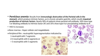
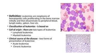
Hematologic disorders
Approach to patient with anemia
• Definition
• Functional – reduction in red cell mass with a corresponding decrease in 02
carrying capacity
• Laboratory - reduction in Hgb, red cell mass, or Hct
Hgb <12g/dl, & Hct <37% in females
Hgb <14g/dl, & Hct <40% in males)
• Prevalence : Ethiopia – 40.5%
• 3rd
leading cause of hematologic admission (Eth.)
• Fe deficiency anemia is the commonest globally
• Anemia is a manifestation of an underlying pathological condition.
• It results from:
1. Under production
2. Increased destruction/Hemolysis
3. Blood loss /bleeding
4. Multifactorial : a combination of these
• History: Accurate history provides information crucial to the diagnosis of the
underlying cause.
• Nutritional /Dietary history
• Underlying diseases
• Blood loss : GI or Genito uirinary blood loss
• Family history of anemia
• Exposure to drugs/toxins E.g. - Methyldopa
• Ggeographical location
• Pregnancy
Symptoms
• They are often non-specific
• Fatigue, dizziness, dyspnea, palpitation, syncope, exercise and cold
intolerance, angina, Tinnitus, vertigo, throbbing head ache, Anorexia,
indigestion, nausea, bowel irregularity
• Irritability, difficulty in concentration, worsened dementia
• Impotence or decreased libido
• Intermittent claudication
Physical Examination
• A comprehensive examination with emphasis to the following findings should be
made.
• Skin and mucous membrane: look for pallor, icterus, bleeding sites, leg ulcer
• Spooning of fingernails → in iron deficiency anemia
• Atrophy of the tongue, sore tongue (glossitis), angular stomatitis which may
be seen in iron deficiency anemia.
• Abdomen: splenomegaly, hepatomegaly, evidence of gallstone.
• Pelvic and rectal examination: to look for possible site of bleeding.
• Bone tenderness and Lymphadenopathy to rule our hematologic
malignancies
• Neurological: gait, reflexes, vibration and position sense which may
help to look for neurologic changes associated with Vit B-12
deficiency
• Fundoscopy: retinal hemorrhage
• Cardiovascular system; modest tachycardia, wide pulse pressure
hyper dynamic precordium, flow murmur. In severe form of Chronic
anemia patients may develop CHF with S3 gallops.
Laboratory study
1. Complete blood count Hgb, Hct, ESR, Platelet count, WBC with differential
2. Red blood cell indices
• Mean Corpuscular volume ( MCV): Hct/RBC : normal value is 80 – 100 fl
• Mean corpuscular Hemoglobin( MCH ) : Hgb/RBC : 27 – 32 pg
• Mean corpuscular Hemoglobin (MCHC): Hgb/Hct 32 – 36%3
3. Examination of the peripheral blood smear: examine a cellular morphology,
shape, size, color, abnormality of other cells.
• Red cell morphology
• Normochromic Normocytic RBCs are seen in normal individuals and in anemia
of chronic diseases
• Anisopoikilocytes: Variation in size and shape may be seen in iron deficiency
anemia
• Hypochromic microcytic anemia: is seen in iron defeciency anemia , anemia
of chronic diseases , thalassemia and sideroblastic anemia (SBA)
• Macrocytic RBCs : Macro-ovalocytes with hyper segmented neutrophils
indicate megaloblastic anemia and myelodysplasia
• Schistocytes (fragmented RBC/Helmentcells):- microangiopathies , DIC,
vasculitis, prosthetic heart valve
Classification of anemia
A. Pathophyisiologic Classifications (based on underlying disease)
1) Anemia associated with impaired RBC Production
a) Aplastic anemia
b) Iron deficiency
c) Myelodysplastic syndrome.
d) Megaloblastic anemia
e) Anemia of CRF
f) Anemia of chronic diseases
g) Drug related
2) Anemia associated with increased RBC loss or destruction
a) Bleeding
b) Hereditary hemolytic anemia
i) Hemoglobinopathies, sickle cell disease, Thalassemia
ii) Primary disorder of RBCs
iii) RBC erythropathies (G6 PDH, PK deficiency)
c) Acquired hemolytic anemia
i) Autoimmune
ii) Drug induced hemolytic anemia
iii) Microangiopathies (e.g. DIC)
iv) Traumatic
B. Morphological classification
1) Hypochromic microcytic anemia
a) Inherited: Thalassema, sideroblastic anemia
b) Acquired: IDA, Anemia of chronic diseases
2) Macrocytic anemia : (MCV > 100 fl)
a) With megaloblastic marrow: megaloblastic anemia
b) With normoblastic marrow: Hemolysis, acute bleeding,
3) Normchromic normocytic:-
a) Anemia of chronic disease
b) Early iron deficiency anemia
c) Aplastic anemia
d) Anemia of CRF
Microcytic Anemias
• Definition: - Iron deficiency anemia occurs when body iron stores become inadequate for the
needs of normal RBC production (erythropoiesis)
• It’s characterized by:
• Hypochromia and microcytosis of the circulating erythrocytes ( RBCs)
• Low plasma iron and ferretine concentration.
• A transferrin saturation of < 15% (Normally 35%)
∼
• It is the commonest cause of anemia world wide
Etiologies of Iron deficnecy Anemia
1. Chronic blood loss
• Uterine
• Gastrointestinal, e.g. esophageal varices; Hiatal hernia; peptic ulcer disease; aspirin ingestion;
Carcinoma of the stomach, ceacum, colon or rectum; hook worm infestation; colitis; piles;
Diverticulosis; etc
• Rarely hematuria, hemoglobulinuria, pulmonary hemosiderosis, self inflicted blood loss
• Disorders of hemeostasis, intravascular hemolysis
2. Increased demands
•Prematurity in newborns
• Rapid growth ( as in adolescent ) growth spurt
• Pregnancy
3. Mal absorption of iron
• Gastrectomy, Celiac disease
4. Poor diet
⇒ Contributory factor in many countries but rarely sole cause
• Clinical manifestation:
• Is insidious in onset and progressive in course
• Patients often present with nonspecific symptoms mentioned
Specific symptoms
• Pica: craving for unusual food substance (amylophagia, geophagia, pagophagia)
• Increased GI absorption of lead – lead poisoning
• Koilonychia – spooning of the finger nails
• Plummer – Vinson/Peterson - Kelly syndrome: characterized by IDA, koilonychia, and
dysphagia due to post cricoid esophageal web)
• Laboratory findings
A) Peripheral blood morphology and RBC indices
• ↓MCV, ↓MCH, ↓MCHC and hypochromic microcytic RBCs with occasional target cells and
pencil-shaped poikilocytes
• ↓ reticulocyte count
• Dimorphic peripheral films ( mixed Normal RBCs and microcytic hypochromic RBCs )
• Combined Iron and folate /B12 deficiency
• On those with recent iron therapy
• Those with recent blood transfusion
B) Serum iron and total iron – binding capacity (TIBC)
• ↓ serum iron and ↑ TIBC (hence IBC is < 10% saturated). This is in contrast to other forms (e.g.
ACD).
C) Serum ferritine( protien used to store fe) is decreased
D) Free erythrocyte protoporphyrine (FEP): non specific
• It increased during early phase of iron deficiency anemia, lead poisoning, sideroblastic anemia and
in erythropoietic porphyria.
E) Bone marrow iron
• Complete absence of iron from stores (macrophages) and absence of siderotic iron granules from
developing erythroblasts
F) Investigation to establish the underlying cause
• Stool for occult blood
• CXR to exclude pulmonary hemosiderosis
• U/A for hematuria or hemosiderinuria
• Pelvic – gynecologic studies
• Upper and lower GI radiologic and Endoscopic studies
• Bronchoscopy
• Differential diagnosis of hypochromic, microcytic anemia
A. With ↓ body iron stores – IDA
B. With normal body or ↑ body iron store
i) Impaired iron metabolism: Anemia of chronic diseases (ACD)
ii) Disorder of globin synthesis: Thalassemia
iii) Disorders of hem synthesis: sideroblastic anemia
iv) Both impaired iron metabolism and defective globin synthesis: lead
poisoning
• Treatment of Iron deficiency Anemia
1. Identify underlying cause and treat it
2. Correct anemia and replenish stores by oral iron, Ferous sulfate 325 mg (60 mg
elemental iron) 3x/day for 4-6 months
• Response to treatment
• An expected daily rise of hemoglobin by 0.1-0.2 gm /dl in
• Follow-up; reticulocytosis will start at 3-4 days and peak on the 10th day of
initiation of treatment.
• An increase in Hgb concentration of at least 2gm/dl after 3wks of therapy is
considered as a good response.
• Treatment should be continued for about 3 months after resolution of anemia to
replenish the iron store.
• Inadequate response may imply
• Continuing hemorrhage
• non compliance to therapy
• Wrong diagnosis
• Mixed deficiency – associated folate or vit.B12 deficiency
• Another cause for anemia e.g. malignancy, inflammation
• Malabsorption – rare cause
• Use of slow release preparations
Anemia of chronic diseases (ACD)
• It occurs in association with a variety of chronic inflammatory and malignant
diseases.
• Etiologies
1) Chronic inflammatory diseases
a) Infections e.g. pulmonary abscess, TB, Osteomyelitis, pneumonia, bacterial
endocarditis
b) Non infectious, e.g. Rheumatoid arthritis, SLE (systemic lupus erythematous)
and other
connective tissue diseases, sarcoidosis, Crohn’s disease
2) Malignant diseases
E.g. carcinoma, lymphoma, sarcoma
• Characteristic features are:
• NCNC or mildly hypochromic indices and red cell morphology
• Mild and non progressive anemia (Hgb rarely < 9.0g/dl) : Severity being related to severity of
underlying disease
• Both serum iron and TIBC are reduced
• Serum ferritine is normal or raised and
• Bone marrow storage iron is normal but erythroblast iron is reduced
• Treatment of Anemia of Chronic DiSeases:
1. Correction of underlying disease
2. Erythropoietin (40-80 % success rate)
3. Correction of reversible contributors (iron, folate, cobalamine supplements if necessary)
Macrocytic Anemia
• Pathogenesis: It’s a descriptive
morphologic term in which maturation of
the nucleus is delayed relative to that of
cytoplasm. The delay in maturation of the
nucleus is attributed to defective DNA
synthesis. This condition leads to
Megaloblastic marrow and ineffective
erythropoiesis.
• The immature erythroblasts are destroyed
within the bone marrow leading
(Intramedullary hemolysis) which results
Megaloblastic anemia.
• Causes of Vit. B12 deficiency
1) Nutritional: especially in vegans
2) Malabsorption
a) Gastric causes
i) Adult (addisonian) pernicious anemia
ii) Congenital lack or abnormality of intrinsic factor
iii) Total or partial gastrectomy
b) Intestinal causes
i) Intestinal stagnant loop syndrome, jejunal diverticulosis, blind loop, stricture etc.
ii) Chronic tropical sprue
iii) Ileal resection and Crohn’s disease
iv) Congenital selective mal absorption with proteinuria
v) Fish tapeworm : Diphlobotrium
• Pernicious anemia: Results from immunologic destruction of the Parietal cells in the
stomach, which produce intrinsic factors, as in chronic atrophic gastritis, which results impaired
production of intrinsic factors. Nearly 90% of patients show parietal cell antibody, 50% have type
I or blocking antibody to intrinsic factor (IF) and 35% show type II or precipitating antibody to IF.
• MCV is increases
• Bone marrow : Hyper cellular and megaloblasts
• Peripheral film : neutrophilic hypersegmentation indicated by
1 neutrophil with 7 segments
2-3 neutrophils with 6 segments or
> 5 neutrophils with 5 segments
• Treatment of megaloblastic anemia
• Vit B12 Deficiency: is treated with cyanocobalamine Dose: Initial dose:1mg IM dialey for 7
days then weekly for four weeks then monthly for 3 months
• Folate deficiency: is treated with Folic acid preparation which is given orally Dose: 5 mg Po
daily
• Additional measures:
• Correct underlying cause
• Antibiotics for bacterial over growth and treatment of fish tapeworm
• Response to therapy
• Feeling of general well being is restored in 48 hrs
• Reticulocytosis begins in 3-4 days and peaks in 7-10 days.
• If both folate and cobalamine deficiency present give cobalamine first
Leukemias
• Definition: Leukemias are neoplasms of
hematopoitic cells proliferating in the bone marrow
initially and then disseminate to peripheral blood,
lymph nodes, spleen, liver etc.
• Classification of leukemias - is based on
• Cell of origin : there are two types of leukemias
• Lymphoid leukemias
• Myeloid leukemias
• Clinical course of the disease - two forms of
presentation of leukemias
• Acute leukemias
• Chronic leukemias
• There are 4 main types of leukemias
A) Acute lymphoblastic leukemia (ALL)
B) Chronic lymphoid leukemia (CLL)
C) Acute myelogeneous leukemia (AML)
D) Chronic myeloid leukemia (CML)
Etiology
• Genetics
• increased incidence in identical twin.
• Down’s syndrome (trisomy 21).
• Environmental
• Ionizing radiation: Radiation exposure increases the risk of CML, AML and
ALL. No known relation with CLL.
• Chemicals like benzene, aromatic hydrocarbons, and treatment with
alkylating agents and other chemotherapeutic drugs are risks for AML.
• RNA based viral infections: like HTLV - I is related to human T-cell leukemia &
similarly Epstein Barr virus is related to ALL (T3 subtype) and aggressive
lymphoma.
Epidemiology
• Globally the incidence of all leukemias is 13/100,000/ year
• usually affecting men more than women.
• acute lymphoblastic leukemia (ALL) is common in children and young adults
• whereas AML, CLL and hairy cell leukemia are common in adults with peak
between the 6th & 7th
decade.
• A Study conducted in one of the teaching hospitals in Ethiopia has shown that:
leukemias were the commonest, accounting for about 35.5% of
hematological admissions
and acute leukemias were the leading cause of mortality, accounting for
78.1%of all hematological deaths.
• Acute Leukemias
• Acute leukemias are characterized by the presence of immature white blood cells
in the marrow and peripheral blood.
• There are two types of acute leukemias:
1. Acute lymphoblastic leukemia ( ALL )
2. Acute myeloblastic leukemia ( AML )
Acute lymphoblastic leukemia (ALL)
• 90 % of leukemia that occur in children is ALL.
• Lymphoblasts are abnormal white blood cells that are
found both in the bone marrow & the peripheral blood of
patients with ALL.
• They are characterized by
• Smaller size (10-15mm) than myeloblasts
• Thin rim of dark blue cytoplasm with no granules
• Nucleus is round or convoluted, and centrally located
and has 1 or 2 nucleoli which are less prominent
• Morphologically ALL is categorized into 3
subtypes (FAB classification)
1. L1 homogeneous population of small cells
(childhood), a small nucleolus
2. L2 - more heterogeneous population of
larger cells (more often seen in adults) with
≥1 prominent nucleoli
3. L3 - uncommon (constituting <5% of ALL),
more mature subtype, large vesicular
nucleus with basophilic often vacuolated
cytoplasm.
• Immuno phenotypic classification of ALL
1. Common ALL (L1, L2) account 75% of ALL, derived from precursors of B-cell.
2. T-ALL (L1, L2) account for 20%of ALL: common in adolescent males,
associated with high WBC count, anterior, mediastinal mass and central nervous
system involvement.
3. B-cell ALL (L3) accounts for 5% ALL. Extramedullary presentation with
metabolic abnormality
Acute myeloblastic leukemia(AML)
• Myeloblasts are abnormal cells that predominantly make up
AML. These cells are larger than lymphoblasts and have:-
Lower nuclear to cytoplasmic ratio
Prominent multiple nucleoli
 Auer rods (stick like structures in the cytoplasm) in 50% of
AML
 Granules in the cytoplasm
• The FAB group divided AML into 8 subtypes based on
1. Degree of differentiation
2. Maturation of predominant cells towards granulocytes,
monocytes erythrocytes or megakaryocytes
Pathophysiology
• Acute leukemias are characterized by clonal proliferation of immature
hematopoietic cells.
• The most important characteristic is a defect in maturation beyond the
myeloblast or promyelocyte level in AML, and beyond lymphoblast in ALL.
• These bone marrow changes will decrease the normal blood cells in the
circulation and cause
• Infection - due to decreased WBC
• Anemia - due to decreased RBC
• Bleeding - due to decreased platelets
Clinical Features
• Symptoms of anemia such as tiredness, weakness, shortness of breath on
exertion
• Recurrent infection of any part of the body is common due to
• Low neutrophil count in the circulation
• Functionally abnormal neutrophils
• Disruption of mucosal barriers
• Bruising and/or bleeding related to low platelet count
• Occasionally, lymph node enlargement and/or symptoms relating to
enlargement of the liver and spleen.
• Symptoms related to hypoperfusion of the lungs and brain due to occlusion of
microcirculation of these organs by blast cells.
• Physical findings:
• Pallor
• Bruises, petechial hemorrhages, purpura
• Signs of infection like fever
• Peripheral lymphadenopathy in ALL, not common in AML
• Enlarged liver and spleen in ALL and small percentage of AML
• Testicular involvement in ALL (T. variant)
• Bone pain and sternal tenderness which are due to expanding malignant cell
mass occur in more than half of the patients with acute leukemias.
• Symptoms related to involvement of the central nervous system (meningitis,
etc) and the kidneys.
• Laboratory Investigation
• The definitive diagnosis of acute leukemia is made on the basis of peripheral
blood film and bone marrow aspirate examination.
1. Complete blood count will show
• Hemoglobin -low
• WBC count -often very high, but occasionally decreased or normal
• Platelet count -low
2. Peripheral blood film shows characteristic leukemic cells, which are distinct
from each other morphologically,
3. Bone marrow aspiration is mostly done on the sternum using a special needle
and a smear prepared and stained with Wright stain , shows
• Increased cellularity with abnormal lymphoid or myeloid blast cell population.
• Replacement of the normal bone marrow elements
Management of acute leukemias
• Chemotherapeutic agents: Chemotherapy of acute leukemia’s is divided in to
four phases
a. Remission induction is characterized by intensive systemic chemotherapy
with the goal of reducing leukemic cell below the level of clinical detection
called complete remission.
b. Consolidation/ early intensification phase
After complete remission if there is no further treatment given, leukemia cells
will expand and lead to relapse. In this phase intensive chemotherapy is given.
c. Maintenance phase here lower dose chemotherapy is given over several
years.
d. Central nervous system prophylaxis in this phase local chemotherapy or
radiation is given to sites of frequent relapse like CNS. This prevents leukemic
meningitis.
• Supportive treatment is applicable for both ALL & AML
• For patients with severe anemia and thrombocytopenia (especially platelet
count below 20,000 /ML with the risk of bleeding) may be transfused with whole
blood and platelet concentrates.
• Infections are common in acute leukemias.
• Appropriate preventive measures should routinely be employed to prevent
infections in such immunocompromised patients.
• These include
• Isolation of staff and visitors by the use of face masks
• Practice careful hand washing before coming in contact with the patient
• Advise the patient to eat only cooked foods
• When infections occur, gram negative sepsis is the commonest
presentation, which requires prompt evaluation and empirical
antibiotic treatment until definitive diagnosis is made by blood
culture, after which the antibiotic(s) can be modified depending on
the organism identified & its drug sensitivity.
Chronic Leukemias
• There are two main types of chronic leukemias
1) Chronic lymphocytic leukemia
2) Chronic myelocytic leukemia
Chronic lymphocytic leukemia (CLL)
• Definition: It is an incurable disease of older people characterized by an
uncontrolled proliferation and accumulation of mature B-lymphocytes (98% of
CLL) and T-lymphocytes in rare instances.
• Although, the disease remains asymptomatic in a proportion of cases; symptoms
of anemia, infections and bleeding resulting from bone marrow failure are
common in the majority of patients at presentation.
• Classification:
• FAB suggested 3 types of CLL
1. Typical CLL: - where more than 95% of lymphocytes in the blood
are small lymphocytes.
2. Prolymphocytic CLL:- If ≥ 51% of lymphocytes in the blood are
prolymphocytes
3. Atypical CLL :- if < 10% of lymphocytes in the blood are small
lymphocytes
• Clinical Features
• In 25% the diagnosis is made incidentally in asymptomatic individuals when WBC count is done
for other reasons. However, the majority will have signs and symptoms resulting from:-
 tissue infiltration by leukemic cells
Bone marrow failure with peripheral blood cytopenias and immune suppression.
• Symptoms
• CLL is a disease of older adults and commonly present with
• Recurrent infections resulting from neutropenia & reduced immunoglobulin levels.
• Symptoms of anemia
• Painless lymph node enlargement
• Spleen and liver enlargement
• Signs
• Anemia
• Enlarged lymph nodes, liver and/or spleen
• Laboratory Investigation
1. CBC
• Hemoglobin is low or normal
• WBC count > 15,000/mm3 , of which 40% are lymphocytes, with a minimum of
5000/ mm3 mature lymphocytes in the circulation
• Platelets are low or normal
2. Bone marrow 30% of the cellular elements would be mature B cells
3. Serum immunoglobulins are low or normal
4. Coomb's test is positive in the presence of hemolysis.
• Staging of CLL
• Clinical course of the disease is variable, and prognosis is correlated directly with
the stage of the disease. There are two different staging classifications
1) RAl staging classification of CLL
Stage 0: Lymphocytosis only (in blood and bone marrow)
Stage I: Lymphocytosis with lymphadenopathy
Stage II: Lymphocytosis with liver and/or spleen enlargement
Stage III: Lymphocytosis with anemia (Hgb <11 gm/dl)
Stage IV: Lymphocytosis with thrombocytopenia (platelets < 100,000/ L)
Lymphocytosis is defined as WBC count > 15,000/ L, of which 40% are
lymphocytes
2) Binet staging classification of CLL
• Stage A = < 3 involved lymphoid areas Hgb>10g/dl
• Stage B= >3 involved lymphoid areas Platelets < 100,000/ L
• Stage C = Any number of involved Hgb < 10 g/dl and/or Platelets < 100,000/ L
lymphoid areas
•Prognosis
• Stage 0 and 1 CLL has good prognosis. If a patient has stage 0 disease without
other poor prognostic factors, the median survival would be more than 10 yrs
without treatment.
• Stage II has Intermediate prognosis - median survival is 5 yrs
• Stage III and IV has worse prognosis - Median survival is 3 years
• Treatment
• Patients with CLL should be referred to centers where they can be treated and followed.
• The disease may remain stable for several years without treatment. Therefore, no
advantage in starting drugs before there is any clinical indication for treatment:
• Indication for initiating treatment include:
1) Hemolytic anemia
2) Cytopenias (neutropenia, thrombocytopenia, etc) with recurrent infections, bleeding
3) Disfiguring lymphadenopathy
4) Symptomatic organomegally
5) Marked systemic symptoms
6) Advanced disease
• Drugs of choice:
• 1) Chlorambucil: is given as small daily doses or larger pulse (intermittent)
every 3-6 wks or
• 2) Cyclophosphamide is also used in the same way (daily or pulses)
• 3) Steroids (prednisolone) can be used in CLL if the patient develops
autoimmune hemolytic anemia, autoimmune thrombocytopenia etc
• Other modalities of treatment which are rarely employed include
• Combined chemotherapy (similar to lymphoma) is used for advanced disease
• Splenectomy/splenic irradiation
• IV immunoglobulin for life threatening infection
Chronic Myelocytic Leukemia (CML)
• Definition: It is disease of older adults and characterized by clonal expansion of
hematopoitic cells of myliod origin, possessing Philadelphia chromosome. It has a
progressive clinical course with three phases starting with chronic phase and
evolving to accelerated phase and then to blast transformation.
Epidemiology
• The incidence of CML is 1.3/100,000 population /year, with men
more affected than women,
• increasing slowly with age until middle forties when the incidence
rises rapidly.
• In the majority of cases the etiology is unknown. However, CML has
been one of the leukemias observed to have an increased prevalence
following atomic explosion at Hiroshima and Nagasaki.
• Clinical Features
• The clinical features of CML depend on the stages of the disease, and range from early
asymptomatic phase to severe manifestations of the accelerated phase and blast
transformation. Therefore, manifestation can be described as follows:-
• Chronic Phase:
• The onset is insidious and some patients can be diagnosed while asymptomatic
during health screening visits.
• Others may present with fatigue, anemia, night sweating, fever, weight loss and
symptoms related to enlarged spleen i.e. left upper abdominal dull pain and early
satiety.
• A few cases may show symptoms related to granulocytic or platelet dysfunction
• i.e. recurrent infections and thrombosis manifesting commonly with persistent
painful erection of the penis (priapism) and cerebrovascular accidents.
• Accelerated phase and blast transformation(or blast crisis)
• With progression of CML from chronic phase to accelerated phase and then to
blast transformation patients will present with severe symptoms such as
• Unexplained fever
• Progressive weight loss
• Bone and joint pain
• bleeding/ thrombosis
• Recurrent infections
• Increasing dose requirement of the drugs used for controlling the disease
• 10-15% of patients may present for the first time with either the accelerated
phase or blast transformation.
• Physical examination may show
• In the early stage 90% of or more of the cases may show
 Moderately pale conjunctivae
 Enlarged spleen and mild liver enlargement
• Late in the disease process patients may develop
 Lymph node enlargement
 Chloromas – leukemic deposits on the skin &
 Tender sternum
• Laboratory Findings
• 1) WBC count is increased of the granulocytic series with variable degrees of
maturity
• mature neutrophils and band forms are seen in the peripheral blood film.
• immature granulocytes such as promyelocytes, myelocytes and metamyelocytes
are seen in the peripheral film with increased number. Some myeloblasts are also
seen, and the percentage of blasts varies according to the stage of the disease is
• Chronic phase: blasts account for < 5% of granulocytes in the blood
• Accelerated phase: blasts account for ≥ 15% but < 30% of granulocytes in the
blood
• Blast crisis: blasts account for ≥ 30% granulocytes in the blood
2) Patelet count is elevated
3) RBC count and hemoglobin concentrations are low, presenting with
anemia
4) Bone marrow aspiration: shows increased cellularity primarily of the
myeloid and megakaryocytic lineage, but percentage of marrow blasts
remains normal or slightly increased
• Depending on the laboratory findings the three stages can be defined
as follows:-
• Chronic Phase
• <5% of circulating blasts
• < 10% blasts + promyelocytes in the blood
• Platelets are increased
• Mild anemia
• Increased cellularity of bone marrow with normal or increased
percentage of blasts.
• Accelerated Phase
• Worsening anemia
• Blasts 15-30% of granulocytes (in the blood or bone marrow)
• Blasts and promyelocytes ≥ 30% of granulocytes (in the blood or marrow)
• Basophils ≥ 20% of granulocytes
• Platelet counts < 100,000/L
• Blast Crisis
• Blasts ≥ 30% of granulocytes (in the blood or marrow)
• Hyposegmented neutrophils
• Blast cells may be
• Myeloid in 50% (AML)
• Lymphoid in 30% (ALL)
• Erythroid in 10% (erythroleukemia)
• Undifferentiated in 10%
• Treatment
• The goal of treatment is complete molecular remission and cure (i.e. achieving
prolonged, durable non-neoplastic and non clonal hematopoisis).
• The treatment includes the following (used singly or in combination)
1) Chemotherapy - hydroxyurea or busulphan - used in Ethiopia
2) Bone marrow transplantation - the only curative treatment but not available in this
country
Lymphomas
• Definition: Malignant transformation of cells residing predominantly
in lymphoid tissue. Lymphoma is broadly classified as
Hodgkin’s lymphomas
Non-Hodgkin’s lymphomas
• A. Hodgkin’s disease
• Epidemiology:
• It has bimodal age incidence (20-30 years and > 50 years).
• In developing countries young adults are commonly affected.
• Male to female ratio = 2:1
Clinical manifestations
• Most patients present with non-tender asymmetrical, firm, discrete and rubbery
enlargement of superficial nodes: Cervical (60-70%), axillary (10-15%) inguinal (6-
12%). Has waxing and waning feature.
• Mild spleenomegaly (50%), mediastinal Lymphadenopathy (6-11%) with or
without plural effusion and superior venacaval syndrome may be present.
• Constitutional symptoms like fever, weight loss and sweating are common in
widespread disease. Fever is found in 30% of patients, and it described as Pel
– ebsteins fever characterized by weeks of febrile period, interspersed by
several weeks of afebrile period.
• Pruritis is seen in 25%of patients
• Alcohol induced pain in the affected area
• Others symptoms include Weight loss, profuse sweating, weakness, fatigue,
anorexia,cachexia
• Hematological findings
• Normocytic normochromic anemia
• 1/3rd of patients may have leukocytosis
• Eosinophila
• Lymphopenia may be found in advanced disease
• Platelets count is variable (normal or increased in the early stage
and decreased in the late stage)
• ESR is elevated
• Bone marrow involvement – late in the course
• Immunologic findings
• Progressive loss of immunologicaly competent T-lymphocytes leads to
impaired cell mediated immunity which increases susceptibility to viral
infections ( herpes zoster, CMV) and fungal infections (e.g. Cryptococcus and
Candida) infections . Moreover patients will have increased risk of
reactivation of latent Tuberculosis infection.
• Biochemical findings: ↑Ca2+ , ↓ Po4-3, ↑Alkaline Phosphatase, ↑LDH indicates
poor prognosis Hyperuricemia,↑ Transaminases and ↑ bilirubin.
• Diagnosis
• Lymph node biopsy: histological examination of an excised lymph node.
• Feature: the distinctive multinucleated, polyploidy cell with a characteristic
Owel- eye appearing Reed-Stern berg (RS) cells on an appropriate
inflammatory background.
• Classification
• Histologically Hodgkin’s disease is classified in to four groups.
1. Lymphocytic predominant
2. Nodular sclerosis
3. Mixed cellularity
4. Lymphocytic depleted
• Clinical staging
•Stage 1: Only affecting one lymph node area
•Stage 2: 2 or more lymph node areas on the same side of the diaphragm involved
•Stage 3: Disease involving lymph nodes above and below diaphragm; splenic involvement
is included here.
•Stage 4: Extra-nodal site involvement (Liver, bone marrow and other extra nodal sites)
• Treatment
• Radiotherapy – mainstay for stage I and II diseases
• Chemotherapy
• Cyclical chemotherapy for stage III and IV diseases
• In stage I and II patient with bulky disease
• E.g. - Mediastinal widening by 1/3r, Lymph node > 10 cm in diameter
• The commnly used combinations of chemotherapy include
MOPP: mustine, vincristine (oncovine), procarbatine, predinisolone
ABVD : adramycin, bleomycine, vinblastin, decarbazine
This combination therapy is either used alone or MOPP – ABVD hybrid given
for 6 cycles (or 4 cycles after full remission).
Non Hodgkin’s Lymphoma (NHL)
• Predisposing diseases/ factors to NHL
• In HIV infected patients there is an increased incidence of lymphoma often at
unusual sites like CNS; usually B-cell origin with high or intermediate grading.
• Immunosuppression increases the risk of developing NHL
• Others : Celiac disease, dermatitis herpetiformis and angioimmunoblastic LAP
predispose to T-cell lymphomas
• Infections: e.g. Helicobacter infection is associated with ↑ frequency of
MALTOMAS
• Autoimmune diseases may cause NHL
Clinical features
• Superficial lymphadenopathy with a classic triangular distribution (the three
vertexes ling over the cervical and the two epitrochlear lymph nodes, while the
base lies over abdominal organs and lymph nodes.)
• Constitutional symptoms are encountered less commonly when compared with
HD and if present indicates dissemination.
• Oro-pharyngeal involvement is seen in 5-10% cases: have involvement of
Waldeyer’s ring including the tonsils adenoids and paratonsilar areas
• Anemia, neutropenia (infection) and thrombocytopenia may follow - bone
marrow involvement or may be autoimmune in origin
• Abdominal organs and lymphnode involvement is more common in NHL than HD
• Other organs: skin, brain, testis, GIT, or thyroid involvement is frequent.
• Laboratory findings:
• Hematological findings
• Normocytic normochromic anemia due to autoimmune hemolytic anemia
• Cytopenias or leukoerythroblastic features
• Lymphoma cells in may be seen in the peripheral blood sometimes
• Bone marrow biopsy may show focal or diffuse involvement
• Immunologic markers are used for classification
• Chromosomal findings: various forms of translocations identified
• Blood chemistry
•Uric acid level is elevated
• Liver enzymes are abnormally elevated
•LDH ↑ (in rapidly proliferating and extensive disease hence has prognostic
implication.)
• Prognostic features:
• Histology
• Staging
• Age > 60 years – unfavorable
• Poor performance status
• Multiple sites of extra nodal involvement
• ↑ LDH
• Bulky disease (mass > 5 cm in diameter)
• Prior history of low grade disease
• AIDS or other causes of imunosupression related
• Treatment
• Low – grade malignancy: No need of treatment if the patient is
asymptomatic
1) Local radiotherapy is indicated for stage I, Iε or stage II
2) Combination chemotherapy for advanced cases Chlorambucil or
cyclophosphamide or repeated courses of fludarabine
3) BMT (bone marrow transplantation)
4) INF – α
• Intermediate – grade malignancy
• Localized disease
•Initial combination therapy with CHOP (cyclophosphamide, Hydroxodunorubicin
(Adriamycin), Vincristine (oncovin) and prednisolone) followed by irradiation.
• Disseminated diseases: Combined Chemotherapy is the treatment of choice.
Approach to Bleeding Disorders
Evaluation of the patient
• History
• Physical Examination
• Laboratory Evaluation
• Are you a bleeder?
• surgical challenges
• accidents & injuries
• dental extractions
• menstrual history
• Type of Bleeding
• ecchymoses
• petechiae
• epistaxis
• deep soft tissue bleed
• hemarthroses
• GI bleeding
• Does it sound genetic?
• duration of bleeding history
• congenital v. acquired
• family history
• determine inheritance
• Medical History
• liver disease
• renal disease
• malignancies
• antibiotic therapy
• poor nutrition (Vit. K or C)
• Physical Examination
• current hemorrhage
• nature and extent
• intercurrent illnesses
• liver disease
• petechiae/ecchymoses
• Laboratory Assessment
• Guided by history
• Screening tests
• PT
• aPTT
• platelet count
• fibrinogen
• thrombin time
• Specific Laboratory Tests
• Mixing studies
• patient and PNP mixed 1:1
• incubated 2 hours at 37o
C
• perform clotting assay as usual
• Uncorrected - circulating
anticoagulant
• Corrected - factor deficiency
• Circulating Anticoagulant
• Lupus anticoagulant/APA syndrome
• rarely have associated bleeding
• tend to thrombose
• Acquired factor inhibitors
• Factor VIII most common
• tertiary care referral
• Factor deficiencies
• Hemophilia A or B
• Factor VIII or IX assays
• Probably mild unless bleeding
patient is an infant male
• Send to Hemophilia Treatment
Center
• von Willebrand’s disease
• most common genetic bleeding
disorder
• many different types
• von Willebrand’s Disease
• autosomal dominant except
Type III
• patients range from
asymptomatic to spontaneous
bleeding similar to a severe
hemophiliac
• characterized by
mucocutaneous bleeding
• von Willebrand’s Testing
• aPTT
• Factor VIII activity
• von Willebrand’s Factor
• Ristocetin Cofactor
• von Willebrand’s Factor
multimers
• von Willebrand’s Disease
• Type I
• normal molecule in abnormally
low quantities
• normal distribution of multimers
• Type II
• abnormal molecule
• abnormal distribution of
multimers with decrease in the
largest molecular weight forms
• Type III
• severe
von Willebrand’s Disease - Treatment
• DDAVP (Stimate)
• 0.3 micrograms/kg IV in 50cc NS over 30 minutes
• intranasally 2 puffs for adults, 1 puff for children
• Factor VIII product containing Vwf
• Humate P
• Koate HP
• Alphanate
• Cryoprecipitate ONLY IF VWF/VIII PRODUCT NOT AVAILABLE!
• 1 bag/10 kg q 12 to 24 hours depending upon the bleeding
• epsilon amino caproic acid (Amicar)
• Other Congenital Defects
• Other Factor deficiencies
• Platelet defects
• very rare
• platelet aggregation studies
• electron microscopy
• bleeding time
• What else could it be?
• Vitamin K deficiency
• drug-induced/malabsorption
• rarely nutritional in an outpatient
• Liver Disease
• long PT +/- aPTT
• poor clearance of coagulation
products
• DIC
• Liver Disease
• Decreased synthesis of factors
• Synthesis of abnormal factors
• Increased fibrinolysis
• Thrombocytopenia
• Fresh frozen plasma
• replete factors
• WILL NOT CORRECT THE PT
WILL NOT CORRECT THE PT
• Cryoprecipitate
• fibrinogen
• Platelets
• Disseminated Intravascular
Coagulation
• Treat the underlying cause
• Replete deficient factors
• FFP
• cryoprecipitate
• platelets
• Bone Marrow Diseases
• Acute leukemias
• Myelodysplasia
• Myeloproliferative disorders
• P. vera
• dysfunctional platelets
• Summary
• History & Physical Examination
• Laboratory tests
• screening tests
• specific diagnostic tests
• Diagnosis-specific therapy
• Factor replacement
• Drugs
Thank you

Hematologic Diseases had dwarf daddy card

  • 1.
  • 2.
    Approach to patientwith anemia • Definition • Functional – reduction in red cell mass with a corresponding decrease in 02 carrying capacity • Laboratory - reduction in Hgb, red cell mass, or Hct Hgb <12g/dl, & Hct <37% in females Hgb <14g/dl, & Hct <40% in males) • Prevalence : Ethiopia – 40.5% • 3rd leading cause of hematologic admission (Eth.) • Fe deficiency anemia is the commonest globally
  • 3.
    • Anemia isa manifestation of an underlying pathological condition. • It results from: 1. Under production 2. Increased destruction/Hemolysis 3. Blood loss /bleeding 4. Multifactorial : a combination of these
  • 4.
    • History: Accuratehistory provides information crucial to the diagnosis of the underlying cause. • Nutritional /Dietary history • Underlying diseases • Blood loss : GI or Genito uirinary blood loss • Family history of anemia • Exposure to drugs/toxins E.g. - Methyldopa • Ggeographical location • Pregnancy
  • 5.
    Symptoms • They areoften non-specific • Fatigue, dizziness, dyspnea, palpitation, syncope, exercise and cold intolerance, angina, Tinnitus, vertigo, throbbing head ache, Anorexia, indigestion, nausea, bowel irregularity • Irritability, difficulty in concentration, worsened dementia • Impotence or decreased libido • Intermittent claudication
  • 6.
    Physical Examination • Acomprehensive examination with emphasis to the following findings should be made. • Skin and mucous membrane: look for pallor, icterus, bleeding sites, leg ulcer • Spooning of fingernails → in iron deficiency anemia • Atrophy of the tongue, sore tongue (glossitis), angular stomatitis which may be seen in iron deficiency anemia. • Abdomen: splenomegaly, hepatomegaly, evidence of gallstone. • Pelvic and rectal examination: to look for possible site of bleeding.
  • 7.
    • Bone tendernessand Lymphadenopathy to rule our hematologic malignancies • Neurological: gait, reflexes, vibration and position sense which may help to look for neurologic changes associated with Vit B-12 deficiency • Fundoscopy: retinal hemorrhage • Cardiovascular system; modest tachycardia, wide pulse pressure hyper dynamic precordium, flow murmur. In severe form of Chronic anemia patients may develop CHF with S3 gallops.
  • 8.
    Laboratory study 1. Completeblood count Hgb, Hct, ESR, Platelet count, WBC with differential 2. Red blood cell indices • Mean Corpuscular volume ( MCV): Hct/RBC : normal value is 80 – 100 fl • Mean corpuscular Hemoglobin( MCH ) : Hgb/RBC : 27 – 32 pg • Mean corpuscular Hemoglobin (MCHC): Hgb/Hct 32 – 36%3 3. Examination of the peripheral blood smear: examine a cellular morphology, shape, size, color, abnormality of other cells.
  • 9.
    • Red cellmorphology • Normochromic Normocytic RBCs are seen in normal individuals and in anemia of chronic diseases • Anisopoikilocytes: Variation in size and shape may be seen in iron deficiency anemia • Hypochromic microcytic anemia: is seen in iron defeciency anemia , anemia of chronic diseases , thalassemia and sideroblastic anemia (SBA) • Macrocytic RBCs : Macro-ovalocytes with hyper segmented neutrophils indicate megaloblastic anemia and myelodysplasia • Schistocytes (fragmented RBC/Helmentcells):- microangiopathies , DIC, vasculitis, prosthetic heart valve
  • 10.
    Classification of anemia A.Pathophyisiologic Classifications (based on underlying disease) 1) Anemia associated with impaired RBC Production a) Aplastic anemia b) Iron deficiency c) Myelodysplastic syndrome. d) Megaloblastic anemia e) Anemia of CRF f) Anemia of chronic diseases g) Drug related
  • 11.
    2) Anemia associatedwith increased RBC loss or destruction a) Bleeding b) Hereditary hemolytic anemia i) Hemoglobinopathies, sickle cell disease, Thalassemia ii) Primary disorder of RBCs iii) RBC erythropathies (G6 PDH, PK deficiency) c) Acquired hemolytic anemia i) Autoimmune ii) Drug induced hemolytic anemia iii) Microangiopathies (e.g. DIC) iv) Traumatic
  • 12.
    B. Morphological classification 1)Hypochromic microcytic anemia a) Inherited: Thalassema, sideroblastic anemia b) Acquired: IDA, Anemia of chronic diseases 2) Macrocytic anemia : (MCV > 100 fl) a) With megaloblastic marrow: megaloblastic anemia b) With normoblastic marrow: Hemolysis, acute bleeding, 3) Normchromic normocytic:- a) Anemia of chronic disease b) Early iron deficiency anemia c) Aplastic anemia d) Anemia of CRF
  • 13.
    Microcytic Anemias • Definition:- Iron deficiency anemia occurs when body iron stores become inadequate for the needs of normal RBC production (erythropoiesis) • It’s characterized by: • Hypochromia and microcytosis of the circulating erythrocytes ( RBCs) • Low plasma iron and ferretine concentration. • A transferrin saturation of < 15% (Normally 35%) ∼ • It is the commonest cause of anemia world wide
  • 14.
    Etiologies of Irondeficnecy Anemia 1. Chronic blood loss • Uterine • Gastrointestinal, e.g. esophageal varices; Hiatal hernia; peptic ulcer disease; aspirin ingestion; Carcinoma of the stomach, ceacum, colon or rectum; hook worm infestation; colitis; piles; Diverticulosis; etc • Rarely hematuria, hemoglobulinuria, pulmonary hemosiderosis, self inflicted blood loss • Disorders of hemeostasis, intravascular hemolysis 2. Increased demands •Prematurity in newborns • Rapid growth ( as in adolescent ) growth spurt • Pregnancy 3. Mal absorption of iron • Gastrectomy, Celiac disease 4. Poor diet ⇒ Contributory factor in many countries but rarely sole cause
  • 15.
    • Clinical manifestation: •Is insidious in onset and progressive in course • Patients often present with nonspecific symptoms mentioned Specific symptoms • Pica: craving for unusual food substance (amylophagia, geophagia, pagophagia) • Increased GI absorption of lead – lead poisoning • Koilonychia – spooning of the finger nails • Plummer – Vinson/Peterson - Kelly syndrome: characterized by IDA, koilonychia, and dysphagia due to post cricoid esophageal web)
  • 16.
    • Laboratory findings A)Peripheral blood morphology and RBC indices • ↓MCV, ↓MCH, ↓MCHC and hypochromic microcytic RBCs with occasional target cells and pencil-shaped poikilocytes • ↓ reticulocyte count • Dimorphic peripheral films ( mixed Normal RBCs and microcytic hypochromic RBCs ) • Combined Iron and folate /B12 deficiency • On those with recent iron therapy • Those with recent blood transfusion B) Serum iron and total iron – binding capacity (TIBC) • ↓ serum iron and ↑ TIBC (hence IBC is < 10% saturated). This is in contrast to other forms (e.g. ACD).
  • 17.
    C) Serum ferritine(protien used to store fe) is decreased D) Free erythrocyte protoporphyrine (FEP): non specific • It increased during early phase of iron deficiency anemia, lead poisoning, sideroblastic anemia and in erythropoietic porphyria. E) Bone marrow iron • Complete absence of iron from stores (macrophages) and absence of siderotic iron granules from developing erythroblasts F) Investigation to establish the underlying cause • Stool for occult blood • CXR to exclude pulmonary hemosiderosis • U/A for hematuria or hemosiderinuria • Pelvic – gynecologic studies • Upper and lower GI radiologic and Endoscopic studies • Bronchoscopy
  • 18.
    • Differential diagnosisof hypochromic, microcytic anemia A. With ↓ body iron stores – IDA B. With normal body or ↑ body iron store i) Impaired iron metabolism: Anemia of chronic diseases (ACD) ii) Disorder of globin synthesis: Thalassemia iii) Disorders of hem synthesis: sideroblastic anemia iv) Both impaired iron metabolism and defective globin synthesis: lead poisoning • Treatment of Iron deficiency Anemia 1. Identify underlying cause and treat it 2. Correct anemia and replenish stores by oral iron, Ferous sulfate 325 mg (60 mg elemental iron) 3x/day for 4-6 months
  • 19.
    • Response totreatment • An expected daily rise of hemoglobin by 0.1-0.2 gm /dl in • Follow-up; reticulocytosis will start at 3-4 days and peak on the 10th day of initiation of treatment. • An increase in Hgb concentration of at least 2gm/dl after 3wks of therapy is considered as a good response. • Treatment should be continued for about 3 months after resolution of anemia to replenish the iron store.
  • 20.
    • Inadequate responsemay imply • Continuing hemorrhage • non compliance to therapy • Wrong diagnosis • Mixed deficiency – associated folate or vit.B12 deficiency • Another cause for anemia e.g. malignancy, inflammation • Malabsorption – rare cause • Use of slow release preparations
  • 21.
    Anemia of chronicdiseases (ACD) • It occurs in association with a variety of chronic inflammatory and malignant diseases. • Etiologies 1) Chronic inflammatory diseases a) Infections e.g. pulmonary abscess, TB, Osteomyelitis, pneumonia, bacterial endocarditis b) Non infectious, e.g. Rheumatoid arthritis, SLE (systemic lupus erythematous) and other connective tissue diseases, sarcoidosis, Crohn’s disease 2) Malignant diseases E.g. carcinoma, lymphoma, sarcoma
  • 22.
    • Characteristic featuresare: • NCNC or mildly hypochromic indices and red cell morphology • Mild and non progressive anemia (Hgb rarely < 9.0g/dl) : Severity being related to severity of underlying disease • Both serum iron and TIBC are reduced • Serum ferritine is normal or raised and • Bone marrow storage iron is normal but erythroblast iron is reduced • Treatment of Anemia of Chronic DiSeases: 1. Correction of underlying disease 2. Erythropoietin (40-80 % success rate) 3. Correction of reversible contributors (iron, folate, cobalamine supplements if necessary)
  • 23.
    Macrocytic Anemia • Pathogenesis:It’s a descriptive morphologic term in which maturation of the nucleus is delayed relative to that of cytoplasm. The delay in maturation of the nucleus is attributed to defective DNA synthesis. This condition leads to Megaloblastic marrow and ineffective erythropoiesis. • The immature erythroblasts are destroyed within the bone marrow leading (Intramedullary hemolysis) which results Megaloblastic anemia.
  • 24.
    • Causes ofVit. B12 deficiency 1) Nutritional: especially in vegans 2) Malabsorption a) Gastric causes i) Adult (addisonian) pernicious anemia ii) Congenital lack or abnormality of intrinsic factor iii) Total or partial gastrectomy b) Intestinal causes i) Intestinal stagnant loop syndrome, jejunal diverticulosis, blind loop, stricture etc. ii) Chronic tropical sprue iii) Ileal resection and Crohn’s disease iv) Congenital selective mal absorption with proteinuria v) Fish tapeworm : Diphlobotrium
  • 25.
    • Pernicious anemia:Results from immunologic destruction of the Parietal cells in the stomach, which produce intrinsic factors, as in chronic atrophic gastritis, which results impaired production of intrinsic factors. Nearly 90% of patients show parietal cell antibody, 50% have type I or blocking antibody to intrinsic factor (IF) and 35% show type II or precipitating antibody to IF. • MCV is increases • Bone marrow : Hyper cellular and megaloblasts • Peripheral film : neutrophilic hypersegmentation indicated by 1 neutrophil with 7 segments 2-3 neutrophils with 6 segments or > 5 neutrophils with 5 segments
  • 26.
    • Treatment ofmegaloblastic anemia • Vit B12 Deficiency: is treated with cyanocobalamine Dose: Initial dose:1mg IM dialey for 7 days then weekly for four weeks then monthly for 3 months • Folate deficiency: is treated with Folic acid preparation which is given orally Dose: 5 mg Po daily • Additional measures: • Correct underlying cause • Antibiotics for bacterial over growth and treatment of fish tapeworm • Response to therapy • Feeling of general well being is restored in 48 hrs • Reticulocytosis begins in 3-4 days and peaks in 7-10 days. • If both folate and cobalamine deficiency present give cobalamine first
  • 27.
  • 28.
    • Definition: Leukemiasare neoplasms of hematopoitic cells proliferating in the bone marrow initially and then disseminate to peripheral blood, lymph nodes, spleen, liver etc. • Classification of leukemias - is based on • Cell of origin : there are two types of leukemias • Lymphoid leukemias • Myeloid leukemias • Clinical course of the disease - two forms of presentation of leukemias • Acute leukemias • Chronic leukemias
  • 29.
    • There are4 main types of leukemias A) Acute lymphoblastic leukemia (ALL) B) Chronic lymphoid leukemia (CLL) C) Acute myelogeneous leukemia (AML) D) Chronic myeloid leukemia (CML)
  • 30.
    Etiology • Genetics • increasedincidence in identical twin. • Down’s syndrome (trisomy 21). • Environmental • Ionizing radiation: Radiation exposure increases the risk of CML, AML and ALL. No known relation with CLL. • Chemicals like benzene, aromatic hydrocarbons, and treatment with alkylating agents and other chemotherapeutic drugs are risks for AML. • RNA based viral infections: like HTLV - I is related to human T-cell leukemia & similarly Epstein Barr virus is related to ALL (T3 subtype) and aggressive lymphoma.
  • 31.
    Epidemiology • Globally theincidence of all leukemias is 13/100,000/ year • usually affecting men more than women. • acute lymphoblastic leukemia (ALL) is common in children and young adults • whereas AML, CLL and hairy cell leukemia are common in adults with peak between the 6th & 7th decade. • A Study conducted in one of the teaching hospitals in Ethiopia has shown that: leukemias were the commonest, accounting for about 35.5% of hematological admissions and acute leukemias were the leading cause of mortality, accounting for 78.1%of all hematological deaths.
  • 32.
    • Acute Leukemias •Acute leukemias are characterized by the presence of immature white blood cells in the marrow and peripheral blood. • There are two types of acute leukemias: 1. Acute lymphoblastic leukemia ( ALL ) 2. Acute myeloblastic leukemia ( AML )
  • 33.
    Acute lymphoblastic leukemia(ALL) • 90 % of leukemia that occur in children is ALL. • Lymphoblasts are abnormal white blood cells that are found both in the bone marrow & the peripheral blood of patients with ALL. • They are characterized by • Smaller size (10-15mm) than myeloblasts • Thin rim of dark blue cytoplasm with no granules • Nucleus is round or convoluted, and centrally located and has 1 or 2 nucleoli which are less prominent
  • 34.
    • Morphologically ALLis categorized into 3 subtypes (FAB classification) 1. L1 homogeneous population of small cells (childhood), a small nucleolus 2. L2 - more heterogeneous population of larger cells (more often seen in adults) with ≥1 prominent nucleoli 3. L3 - uncommon (constituting <5% of ALL), more mature subtype, large vesicular nucleus with basophilic often vacuolated cytoplasm.
  • 35.
    • Immuno phenotypicclassification of ALL 1. Common ALL (L1, L2) account 75% of ALL, derived from precursors of B-cell. 2. T-ALL (L1, L2) account for 20%of ALL: common in adolescent males, associated with high WBC count, anterior, mediastinal mass and central nervous system involvement. 3. B-cell ALL (L3) accounts for 5% ALL. Extramedullary presentation with metabolic abnormality
  • 36.
    Acute myeloblastic leukemia(AML) •Myeloblasts are abnormal cells that predominantly make up AML. These cells are larger than lymphoblasts and have:- Lower nuclear to cytoplasmic ratio Prominent multiple nucleoli  Auer rods (stick like structures in the cytoplasm) in 50% of AML  Granules in the cytoplasm • The FAB group divided AML into 8 subtypes based on 1. Degree of differentiation 2. Maturation of predominant cells towards granulocytes, monocytes erythrocytes or megakaryocytes
  • 37.
    Pathophysiology • Acute leukemiasare characterized by clonal proliferation of immature hematopoietic cells. • The most important characteristic is a defect in maturation beyond the myeloblast or promyelocyte level in AML, and beyond lymphoblast in ALL. • These bone marrow changes will decrease the normal blood cells in the circulation and cause • Infection - due to decreased WBC • Anemia - due to decreased RBC • Bleeding - due to decreased platelets
  • 38.
    Clinical Features • Symptomsof anemia such as tiredness, weakness, shortness of breath on exertion • Recurrent infection of any part of the body is common due to • Low neutrophil count in the circulation • Functionally abnormal neutrophils • Disruption of mucosal barriers • Bruising and/or bleeding related to low platelet count • Occasionally, lymph node enlargement and/or symptoms relating to enlargement of the liver and spleen. • Symptoms related to hypoperfusion of the lungs and brain due to occlusion of microcirculation of these organs by blast cells.
  • 39.
    • Physical findings: •Pallor • Bruises, petechial hemorrhages, purpura • Signs of infection like fever • Peripheral lymphadenopathy in ALL, not common in AML • Enlarged liver and spleen in ALL and small percentage of AML • Testicular involvement in ALL (T. variant) • Bone pain and sternal tenderness which are due to expanding malignant cell mass occur in more than half of the patients with acute leukemias. • Symptoms related to involvement of the central nervous system (meningitis, etc) and the kidneys.
  • 40.
    • Laboratory Investigation •The definitive diagnosis of acute leukemia is made on the basis of peripheral blood film and bone marrow aspirate examination. 1. Complete blood count will show • Hemoglobin -low • WBC count -often very high, but occasionally decreased or normal • Platelet count -low 2. Peripheral blood film shows characteristic leukemic cells, which are distinct from each other morphologically,
  • 41.
    3. Bone marrowaspiration is mostly done on the sternum using a special needle and a smear prepared and stained with Wright stain , shows • Increased cellularity with abnormal lymphoid or myeloid blast cell population. • Replacement of the normal bone marrow elements
  • 42.
    Management of acuteleukemias • Chemotherapeutic agents: Chemotherapy of acute leukemia’s is divided in to four phases a. Remission induction is characterized by intensive systemic chemotherapy with the goal of reducing leukemic cell below the level of clinical detection called complete remission. b. Consolidation/ early intensification phase After complete remission if there is no further treatment given, leukemia cells will expand and lead to relapse. In this phase intensive chemotherapy is given. c. Maintenance phase here lower dose chemotherapy is given over several years. d. Central nervous system prophylaxis in this phase local chemotherapy or radiation is given to sites of frequent relapse like CNS. This prevents leukemic meningitis.
  • 43.
    • Supportive treatmentis applicable for both ALL & AML • For patients with severe anemia and thrombocytopenia (especially platelet count below 20,000 /ML with the risk of bleeding) may be transfused with whole blood and platelet concentrates. • Infections are common in acute leukemias. • Appropriate preventive measures should routinely be employed to prevent infections in such immunocompromised patients. • These include • Isolation of staff and visitors by the use of face masks • Practice careful hand washing before coming in contact with the patient • Advise the patient to eat only cooked foods
  • 44.
    • When infectionsoccur, gram negative sepsis is the commonest presentation, which requires prompt evaluation and empirical antibiotic treatment until definitive diagnosis is made by blood culture, after which the antibiotic(s) can be modified depending on the organism identified & its drug sensitivity.
  • 45.
    Chronic Leukemias • Thereare two main types of chronic leukemias 1) Chronic lymphocytic leukemia 2) Chronic myelocytic leukemia
  • 46.
    Chronic lymphocytic leukemia(CLL) • Definition: It is an incurable disease of older people characterized by an uncontrolled proliferation and accumulation of mature B-lymphocytes (98% of CLL) and T-lymphocytes in rare instances. • Although, the disease remains asymptomatic in a proportion of cases; symptoms of anemia, infections and bleeding resulting from bone marrow failure are common in the majority of patients at presentation.
  • 47.
    • Classification: • FABsuggested 3 types of CLL 1. Typical CLL: - where more than 95% of lymphocytes in the blood are small lymphocytes. 2. Prolymphocytic CLL:- If ≥ 51% of lymphocytes in the blood are prolymphocytes 3. Atypical CLL :- if < 10% of lymphocytes in the blood are small lymphocytes
  • 48.
    • Clinical Features •In 25% the diagnosis is made incidentally in asymptomatic individuals when WBC count is done for other reasons. However, the majority will have signs and symptoms resulting from:-  tissue infiltration by leukemic cells Bone marrow failure with peripheral blood cytopenias and immune suppression. • Symptoms • CLL is a disease of older adults and commonly present with • Recurrent infections resulting from neutropenia & reduced immunoglobulin levels. • Symptoms of anemia • Painless lymph node enlargement • Spleen and liver enlargement • Signs • Anemia • Enlarged lymph nodes, liver and/or spleen
  • 49.
    • Laboratory Investigation 1.CBC • Hemoglobin is low or normal • WBC count > 15,000/mm3 , of which 40% are lymphocytes, with a minimum of 5000/ mm3 mature lymphocytes in the circulation • Platelets are low or normal 2. Bone marrow 30% of the cellular elements would be mature B cells 3. Serum immunoglobulins are low or normal 4. Coomb's test is positive in the presence of hemolysis.
  • 50.
    • Staging ofCLL • Clinical course of the disease is variable, and prognosis is correlated directly with the stage of the disease. There are two different staging classifications 1) RAl staging classification of CLL Stage 0: Lymphocytosis only (in blood and bone marrow) Stage I: Lymphocytosis with lymphadenopathy Stage II: Lymphocytosis with liver and/or spleen enlargement Stage III: Lymphocytosis with anemia (Hgb <11 gm/dl) Stage IV: Lymphocytosis with thrombocytopenia (platelets < 100,000/ L) Lymphocytosis is defined as WBC count > 15,000/ L, of which 40% are lymphocytes
  • 51.
    2) Binet stagingclassification of CLL • Stage A = < 3 involved lymphoid areas Hgb>10g/dl • Stage B= >3 involved lymphoid areas Platelets < 100,000/ L • Stage C = Any number of involved Hgb < 10 g/dl and/or Platelets < 100,000/ L lymphoid areas •Prognosis • Stage 0 and 1 CLL has good prognosis. If a patient has stage 0 disease without other poor prognostic factors, the median survival would be more than 10 yrs without treatment. • Stage II has Intermediate prognosis - median survival is 5 yrs • Stage III and IV has worse prognosis - Median survival is 3 years
  • 52.
    • Treatment • Patientswith CLL should be referred to centers where they can be treated and followed. • The disease may remain stable for several years without treatment. Therefore, no advantage in starting drugs before there is any clinical indication for treatment: • Indication for initiating treatment include: 1) Hemolytic anemia 2) Cytopenias (neutropenia, thrombocytopenia, etc) with recurrent infections, bleeding 3) Disfiguring lymphadenopathy 4) Symptomatic organomegally 5) Marked systemic symptoms 6) Advanced disease
  • 53.
    • Drugs ofchoice: • 1) Chlorambucil: is given as small daily doses or larger pulse (intermittent) every 3-6 wks or • 2) Cyclophosphamide is also used in the same way (daily or pulses) • 3) Steroids (prednisolone) can be used in CLL if the patient develops autoimmune hemolytic anemia, autoimmune thrombocytopenia etc • Other modalities of treatment which are rarely employed include • Combined chemotherapy (similar to lymphoma) is used for advanced disease • Splenectomy/splenic irradiation • IV immunoglobulin for life threatening infection
  • 54.
    Chronic Myelocytic Leukemia(CML) • Definition: It is disease of older adults and characterized by clonal expansion of hematopoitic cells of myliod origin, possessing Philadelphia chromosome. It has a progressive clinical course with three phases starting with chronic phase and evolving to accelerated phase and then to blast transformation.
  • 55.
    Epidemiology • The incidenceof CML is 1.3/100,000 population /year, with men more affected than women, • increasing slowly with age until middle forties when the incidence rises rapidly. • In the majority of cases the etiology is unknown. However, CML has been one of the leukemias observed to have an increased prevalence following atomic explosion at Hiroshima and Nagasaki.
  • 56.
    • Clinical Features •The clinical features of CML depend on the stages of the disease, and range from early asymptomatic phase to severe manifestations of the accelerated phase and blast transformation. Therefore, manifestation can be described as follows:- • Chronic Phase: • The onset is insidious and some patients can be diagnosed while asymptomatic during health screening visits. • Others may present with fatigue, anemia, night sweating, fever, weight loss and symptoms related to enlarged spleen i.e. left upper abdominal dull pain and early satiety. • A few cases may show symptoms related to granulocytic or platelet dysfunction • i.e. recurrent infections and thrombosis manifesting commonly with persistent painful erection of the penis (priapism) and cerebrovascular accidents.
  • 57.
    • Accelerated phaseand blast transformation(or blast crisis) • With progression of CML from chronic phase to accelerated phase and then to blast transformation patients will present with severe symptoms such as • Unexplained fever • Progressive weight loss • Bone and joint pain • bleeding/ thrombosis • Recurrent infections • Increasing dose requirement of the drugs used for controlling the disease • 10-15% of patients may present for the first time with either the accelerated phase or blast transformation.
  • 58.
    • Physical examinationmay show • In the early stage 90% of or more of the cases may show  Moderately pale conjunctivae  Enlarged spleen and mild liver enlargement • Late in the disease process patients may develop  Lymph node enlargement  Chloromas – leukemic deposits on the skin &  Tender sternum
  • 59.
    • Laboratory Findings •1) WBC count is increased of the granulocytic series with variable degrees of maturity • mature neutrophils and band forms are seen in the peripheral blood film. • immature granulocytes such as promyelocytes, myelocytes and metamyelocytes are seen in the peripheral film with increased number. Some myeloblasts are also seen, and the percentage of blasts varies according to the stage of the disease is • Chronic phase: blasts account for < 5% of granulocytes in the blood • Accelerated phase: blasts account for ≥ 15% but < 30% of granulocytes in the blood • Blast crisis: blasts account for ≥ 30% granulocytes in the blood
  • 60.
    2) Patelet countis elevated 3) RBC count and hemoglobin concentrations are low, presenting with anemia 4) Bone marrow aspiration: shows increased cellularity primarily of the myeloid and megakaryocytic lineage, but percentage of marrow blasts remains normal or slightly increased
  • 61.
    • Depending onthe laboratory findings the three stages can be defined as follows:- • Chronic Phase • <5% of circulating blasts • < 10% blasts + promyelocytes in the blood • Platelets are increased • Mild anemia • Increased cellularity of bone marrow with normal or increased percentage of blasts.
  • 62.
    • Accelerated Phase •Worsening anemia • Blasts 15-30% of granulocytes (in the blood or bone marrow) • Blasts and promyelocytes ≥ 30% of granulocytes (in the blood or marrow) • Basophils ≥ 20% of granulocytes • Platelet counts < 100,000/L • Blast Crisis • Blasts ≥ 30% of granulocytes (in the blood or marrow) • Hyposegmented neutrophils
  • 63.
    • Blast cellsmay be • Myeloid in 50% (AML) • Lymphoid in 30% (ALL) • Erythroid in 10% (erythroleukemia) • Undifferentiated in 10% • Treatment • The goal of treatment is complete molecular remission and cure (i.e. achieving prolonged, durable non-neoplastic and non clonal hematopoisis). • The treatment includes the following (used singly or in combination) 1) Chemotherapy - hydroxyurea or busulphan - used in Ethiopia 2) Bone marrow transplantation - the only curative treatment but not available in this country
  • 64.
    Lymphomas • Definition: Malignanttransformation of cells residing predominantly in lymphoid tissue. Lymphoma is broadly classified as Hodgkin’s lymphomas Non-Hodgkin’s lymphomas • A. Hodgkin’s disease • Epidemiology: • It has bimodal age incidence (20-30 years and > 50 years). • In developing countries young adults are commonly affected. • Male to female ratio = 2:1
  • 65.
    Clinical manifestations • Mostpatients present with non-tender asymmetrical, firm, discrete and rubbery enlargement of superficial nodes: Cervical (60-70%), axillary (10-15%) inguinal (6- 12%). Has waxing and waning feature. • Mild spleenomegaly (50%), mediastinal Lymphadenopathy (6-11%) with or without plural effusion and superior venacaval syndrome may be present. • Constitutional symptoms like fever, weight loss and sweating are common in widespread disease. Fever is found in 30% of patients, and it described as Pel – ebsteins fever characterized by weeks of febrile period, interspersed by several weeks of afebrile period. • Pruritis is seen in 25%of patients • Alcohol induced pain in the affected area • Others symptoms include Weight loss, profuse sweating, weakness, fatigue, anorexia,cachexia
  • 66.
    • Hematological findings •Normocytic normochromic anemia • 1/3rd of patients may have leukocytosis • Eosinophila • Lymphopenia may be found in advanced disease • Platelets count is variable (normal or increased in the early stage and decreased in the late stage) • ESR is elevated • Bone marrow involvement – late in the course
  • 67.
    • Immunologic findings •Progressive loss of immunologicaly competent T-lymphocytes leads to impaired cell mediated immunity which increases susceptibility to viral infections ( herpes zoster, CMV) and fungal infections (e.g. Cryptococcus and Candida) infections . Moreover patients will have increased risk of reactivation of latent Tuberculosis infection. • Biochemical findings: ↑Ca2+ , ↓ Po4-3, ↑Alkaline Phosphatase, ↑LDH indicates poor prognosis Hyperuricemia,↑ Transaminases and ↑ bilirubin. • Diagnosis • Lymph node biopsy: histological examination of an excised lymph node. • Feature: the distinctive multinucleated, polyploidy cell with a characteristic Owel- eye appearing Reed-Stern berg (RS) cells on an appropriate inflammatory background.
  • 68.
    • Classification • HistologicallyHodgkin’s disease is classified in to four groups. 1. Lymphocytic predominant 2. Nodular sclerosis 3. Mixed cellularity 4. Lymphocytic depleted • Clinical staging •Stage 1: Only affecting one lymph node area •Stage 2: 2 or more lymph node areas on the same side of the diaphragm involved •Stage 3: Disease involving lymph nodes above and below diaphragm; splenic involvement is included here. •Stage 4: Extra-nodal site involvement (Liver, bone marrow and other extra nodal sites)
  • 69.
    • Treatment • Radiotherapy– mainstay for stage I and II diseases • Chemotherapy • Cyclical chemotherapy for stage III and IV diseases • In stage I and II patient with bulky disease • E.g. - Mediastinal widening by 1/3r, Lymph node > 10 cm in diameter • The commnly used combinations of chemotherapy include MOPP: mustine, vincristine (oncovine), procarbatine, predinisolone ABVD : adramycin, bleomycine, vinblastin, decarbazine This combination therapy is either used alone or MOPP – ABVD hybrid given for 6 cycles (or 4 cycles after full remission).
  • 70.
    Non Hodgkin’s Lymphoma(NHL) • Predisposing diseases/ factors to NHL • In HIV infected patients there is an increased incidence of lymphoma often at unusual sites like CNS; usually B-cell origin with high or intermediate grading. • Immunosuppression increases the risk of developing NHL • Others : Celiac disease, dermatitis herpetiformis and angioimmunoblastic LAP predispose to T-cell lymphomas • Infections: e.g. Helicobacter infection is associated with ↑ frequency of MALTOMAS • Autoimmune diseases may cause NHL
  • 71.
    Clinical features • Superficiallymphadenopathy with a classic triangular distribution (the three vertexes ling over the cervical and the two epitrochlear lymph nodes, while the base lies over abdominal organs and lymph nodes.) • Constitutional symptoms are encountered less commonly when compared with HD and if present indicates dissemination. • Oro-pharyngeal involvement is seen in 5-10% cases: have involvement of Waldeyer’s ring including the tonsils adenoids and paratonsilar areas • Anemia, neutropenia (infection) and thrombocytopenia may follow - bone marrow involvement or may be autoimmune in origin • Abdominal organs and lymphnode involvement is more common in NHL than HD • Other organs: skin, brain, testis, GIT, or thyroid involvement is frequent.
  • 72.
    • Laboratory findings: •Hematological findings • Normocytic normochromic anemia due to autoimmune hemolytic anemia • Cytopenias or leukoerythroblastic features • Lymphoma cells in may be seen in the peripheral blood sometimes • Bone marrow biopsy may show focal or diffuse involvement • Immunologic markers are used for classification • Chromosomal findings: various forms of translocations identified • Blood chemistry •Uric acid level is elevated • Liver enzymes are abnormally elevated •LDH ↑ (in rapidly proliferating and extensive disease hence has prognostic implication.)
  • 73.
    • Prognostic features: •Histology • Staging • Age > 60 years – unfavorable • Poor performance status • Multiple sites of extra nodal involvement • ↑ LDH • Bulky disease (mass > 5 cm in diameter) • Prior history of low grade disease • AIDS or other causes of imunosupression related
  • 74.
    • Treatment • Low– grade malignancy: No need of treatment if the patient is asymptomatic 1) Local radiotherapy is indicated for stage I, Iε or stage II 2) Combination chemotherapy for advanced cases Chlorambucil or cyclophosphamide or repeated courses of fludarabine 3) BMT (bone marrow transplantation) 4) INF – α • Intermediate – grade malignancy • Localized disease •Initial combination therapy with CHOP (cyclophosphamide, Hydroxodunorubicin (Adriamycin), Vincristine (oncovin) and prednisolone) followed by irradiation. • Disseminated diseases: Combined Chemotherapy is the treatment of choice.
  • 75.
  • 76.
    Evaluation of thepatient • History • Physical Examination • Laboratory Evaluation • Are you a bleeder? • surgical challenges • accidents & injuries • dental extractions • menstrual history • Type of Bleeding • ecchymoses • petechiae • epistaxis • deep soft tissue bleed • hemarthroses • GI bleeding
  • 77.
    • Does itsound genetic? • duration of bleeding history • congenital v. acquired • family history • determine inheritance • Medical History • liver disease • renal disease • malignancies • antibiotic therapy • poor nutrition (Vit. K or C)
  • 78.
    • Physical Examination •current hemorrhage • nature and extent • intercurrent illnesses • liver disease • petechiae/ecchymoses • Laboratory Assessment • Guided by history • Screening tests • PT • aPTT • platelet count • fibrinogen • thrombin time
  • 79.
    • Specific LaboratoryTests • Mixing studies • patient and PNP mixed 1:1 • incubated 2 hours at 37o C • perform clotting assay as usual • Uncorrected - circulating anticoagulant • Corrected - factor deficiency • Circulating Anticoagulant • Lupus anticoagulant/APA syndrome • rarely have associated bleeding • tend to thrombose • Acquired factor inhibitors • Factor VIII most common • tertiary care referral
  • 80.
    • Factor deficiencies •Hemophilia A or B • Factor VIII or IX assays • Probably mild unless bleeding patient is an infant male • Send to Hemophilia Treatment Center • von Willebrand’s disease • most common genetic bleeding disorder • many different types • von Willebrand’s Disease • autosomal dominant except Type III • patients range from asymptomatic to spontaneous bleeding similar to a severe hemophiliac • characterized by mucocutaneous bleeding
  • 81.
    • von Willebrand’sTesting • aPTT • Factor VIII activity • von Willebrand’s Factor • Ristocetin Cofactor • von Willebrand’s Factor multimers • von Willebrand’s Disease • Type I • normal molecule in abnormally low quantities • normal distribution of multimers • Type II • abnormal molecule • abnormal distribution of multimers with decrease in the largest molecular weight forms • Type III • severe
  • 82.
    von Willebrand’s Disease- Treatment • DDAVP (Stimate) • 0.3 micrograms/kg IV in 50cc NS over 30 minutes • intranasally 2 puffs for adults, 1 puff for children • Factor VIII product containing Vwf • Humate P • Koate HP • Alphanate • Cryoprecipitate ONLY IF VWF/VIII PRODUCT NOT AVAILABLE! • 1 bag/10 kg q 12 to 24 hours depending upon the bleeding • epsilon amino caproic acid (Amicar)
  • 83.
    • Other CongenitalDefects • Other Factor deficiencies • Platelet defects • very rare • platelet aggregation studies • electron microscopy • bleeding time • What else could it be? • Vitamin K deficiency • drug-induced/malabsorption • rarely nutritional in an outpatient • Liver Disease • long PT +/- aPTT • poor clearance of coagulation products • DIC
  • 84.
    • Liver Disease •Decreased synthesis of factors • Synthesis of abnormal factors • Increased fibrinolysis • Thrombocytopenia • Fresh frozen plasma • replete factors • WILL NOT CORRECT THE PT WILL NOT CORRECT THE PT • Cryoprecipitate • fibrinogen • Platelets • Disseminated Intravascular Coagulation • Treat the underlying cause • Replete deficient factors • FFP • cryoprecipitate • platelets
  • 85.
    • Bone MarrowDiseases • Acute leukemias • Myelodysplasia • Myeloproliferative disorders • P. vera • dysfunctional platelets • Summary • History & Physical Examination • Laboratory tests • screening tests • specific diagnostic tests • Diagnosis-specific therapy • Factor replacement • Drugs
  • 86.