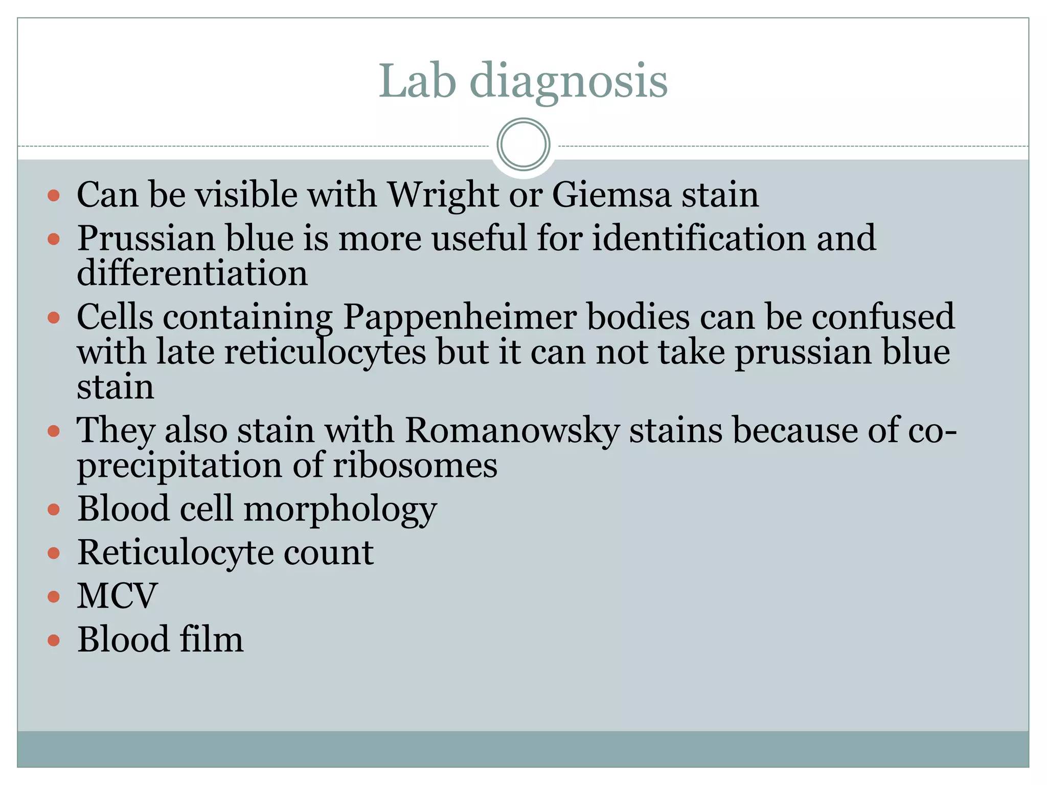
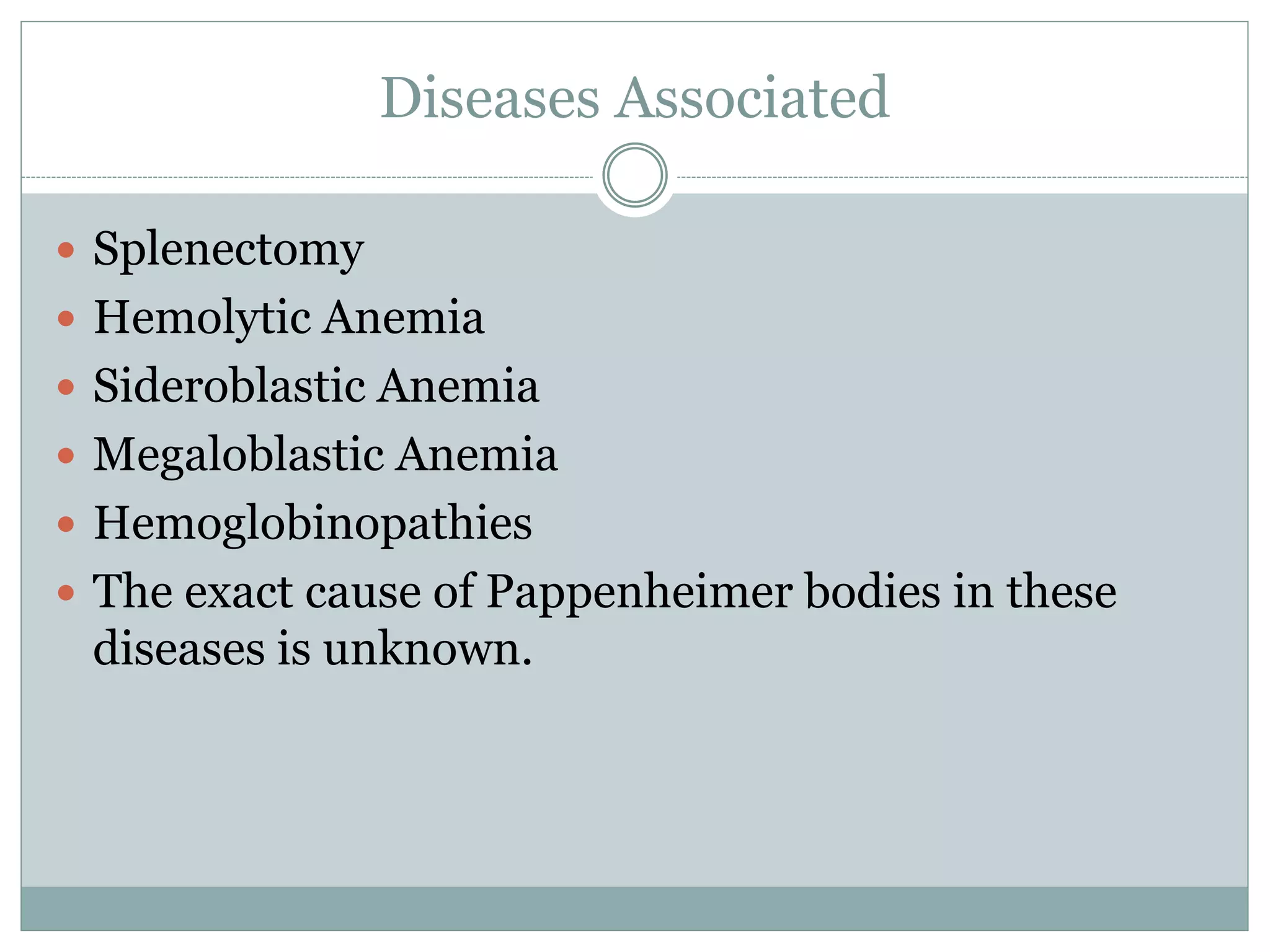
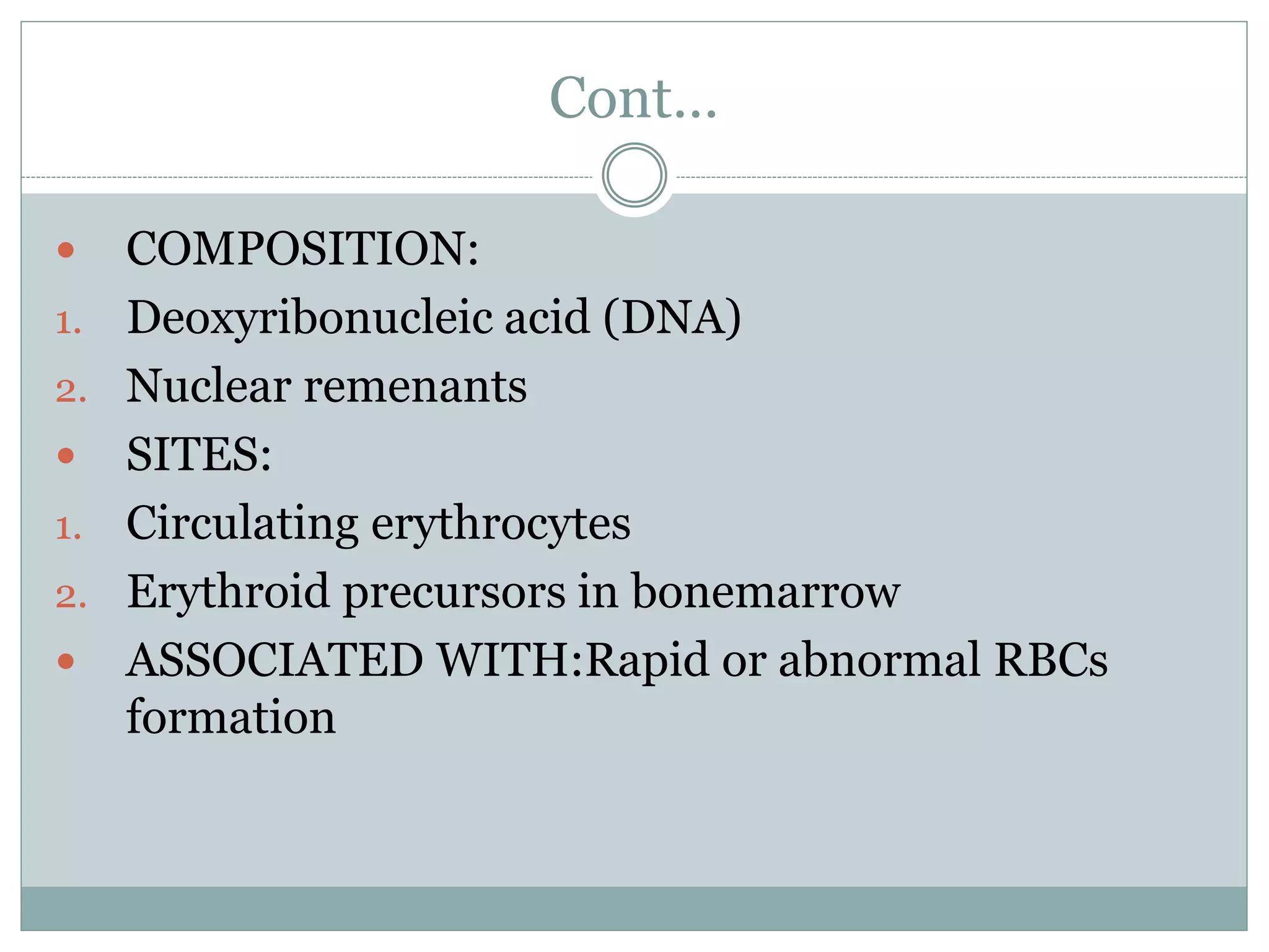
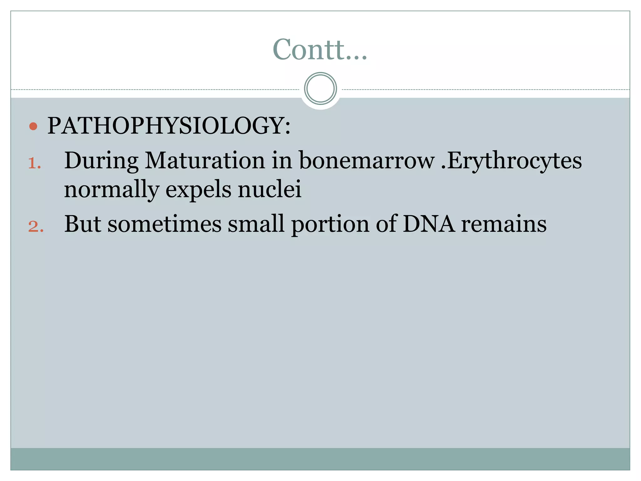
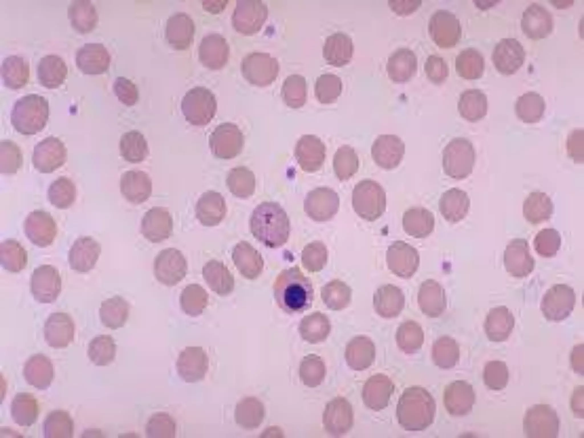
P R E S E N T E D B Y G R O U P 6
P R E S E N T E D T O M I S S M E H R E E N
Red blood cell inclusion
Introduction
 Inclusion bodies are those things that just appear in
cells and.. don’t really do anything. They’re just
included within the cell, for some purpose
 Typically, inclusion bodies are nuclear or
cytoplasmic aggregates of stainable substance,
usually proteins
 In RBC it is indication of some pathological
conditions
Introduction
 These bodies are never enclosed by a membrane and
serve as storage vessels.
 Glycogen, which is a polymer of glucose, is stored as
a reserve of carbohydrate and energy.
 it is useful to understand each inclusion body that
can occur within a red blood cell.
Contt…
 Following are the inclusion bodies with in RBC
 Howell-Jolly Body
 Heinz Body
 Pappenhemier Body
 Basophilic Stippling
 Cabot Ring
Pappenheimer Body
 Pappenheimer bodies are abnormal inclusions of
iron found inside RBC on a routine blood stain
 Formed by phagosomes that have engulfed excessive
amounts of iron
 Pappenheimer bodies are basophilic erythrocytic
inclusions usually Located at the periphery of the cell
 They appear as dense, blue-purple granules within
the red blood cell
 1 or 2 in number
Causes
 Pappenheimer bodies are most likely caused by
accelerated red cell division or impaired hemoglobin
synthesis
Lab diagnosis
 Can be visible with Wright or Giemsa stain
 Prussian blue is more useful for identification and
differentiation
 Cells containing Pappenheimer bodies can be confused
with late reticulocytes but it can not take prussian blue
stain
 They also stain with Romanowsky stains because of co-
precipitation of ribosomes
 Blood cell morphology
 Reticulocyte count
 MCV
 Blood film
Diseases Associated
 Splenectomy
 Hemolytic Anemia
 Sideroblastic Anemia
 Megaloblastic Anemia
 Hemoglobinopathies
 The exact cause of Pappenheimer bodies in these
diseases is unknown.
HOWELL-JOLLY BODIES
 DEFINITION:Histopathologic findings of basophilic
nuclear remenants in circulating erythrocytes
 TERMINOLOGY:Micronucleated Reticulocytes
 SIZE:1 micronmeter in diameter
 APPEARANCE:Small rounded,dense purple granules
Cont…
 COMPOSITION:
1. Deoxyribonucleic acid (DNA)
2. Nuclear remenants
 SITES:
1. Circulating erythrocytes
2. Erythroid precursors in bonemarrow
 ASSOCIATED WITH:Rapid or abnormal RBCs
formation
Contt…
 PATHOPHYSIOLOGY:
1. During Maturation in bonemarrow .Erythrocytes
normally expels nuclei
2. But sometimes small portion of DNA remains
contt…
 CONDITIONS:
1. Autospleenectomy due to sickle cell anemia
2. Hereditary spherocytosis
3. Megaloblastic anemia
4. Myelodysplastic syndrome .
5. Severe haemolytic anemia
6. Spleenectomy
 DIAGNOSIS:
1. Peripheral blood smear
2. Wright stain (methylene blue + eosine)
3. Remenants of nucleus (DNA) observes
Heinz Body
• H E I N Z B O D I E S A R E I N C L U S I O N S
W I T H I N R E D B L O O D C E L L S C O M P O S E D
O F D E N A T U R E D H E M O G L O B I N .
• H E I N Z B O D I E S A P P E A R A S S M A L L R O U N D
I N C L U S I O N S W I T H I N T H E R E D C E L L
B O D Y .
Contt…
 Heinz bodies are formed by damage and denaturing
to the hemoglobin component of red blood cells,
most commonly by oxidative stress, but also possibly
by genetic abnormalities in hemoglobin.
 Typically, during oxidative damage to hemoglobin,
an electron is transferred from the hemoglobin to
oxygen, resulting in the formation of a reactive
oxygen species (ROS). This ROS can lead to severe
damage within the cells, and can even cause lysis of
the entire cell.
Contt…
 The ROS denatures portions of the hemoglobin,
causing the to precipitate and produce Heinz Bodies,
which becomes an antigenic agent. Thus,
macrophages detect the antigen and remove the
damaged portions of the cell, its damaged membrane
and the denatured hemoglobin (now called the Heinz
Body).
Causes
 G6PD deficiency
 NADPH Deficiency
 Chronic Liver Disease
 Alpha Thalassemia
 Hyposplenism and Splenectomy
Cabot ring
 They are literally loop ,rings or figure 8 types
structure that are located with in the cytoplasm of
RBC.
 They are typically colored red purple under the
Wright stain
 They are quite rare
 They are simple microtubular remnants of mitotic
tubules that are involve in mitosis
DISEASES ASSOCIATED WITH CABOT RING
 MYELODYSPLASTIC SYNDROME
 MEGALOBLASTIC ANEMIA
 LEAD POISONING
 HEAVY METAL POISONING
 LIVER DISEASES
DIGNOSIS
 RARE CONDITION
 CHECK THE MORPHOLOGY OF RED BLOOD
CELL
Basophilic Stippling
 Basophilic stippling is also referred to
as punctuate *dots* basophilia
 Diseases Associated:
 can be divided into
 1 fine basophilic stippling
 polychromatophilia.
 2 coarse *rough* basophilic stippling.
 lead poisoning, and thalassemia
 impaired hemoglobin synthesis
 Stained by Wright stain
Rbc inclusion ()))))

Rbc inclusion ()))))

  • 2.
    P R ES E N T E D B Y G R O U P 6 P R E S E N T E D T O M I S S M E H R E E N Red blood cell inclusion
  • 3.
    Introduction  Inclusion bodiesare those things that just appear in cells and.. don’t really do anything. They’re just included within the cell, for some purpose  Typically, inclusion bodies are nuclear or cytoplasmic aggregates of stainable substance, usually proteins  In RBC it is indication of some pathological conditions
  • 4.
    Introduction  These bodiesare never enclosed by a membrane and serve as storage vessels.  Glycogen, which is a polymer of glucose, is stored as a reserve of carbohydrate and energy.  it is useful to understand each inclusion body that can occur within a red blood cell.
  • 5.
    Contt…  Following arethe inclusion bodies with in RBC  Howell-Jolly Body  Heinz Body  Pappenhemier Body  Basophilic Stippling  Cabot Ring
  • 6.
    Pappenheimer Body  Pappenheimerbodies are abnormal inclusions of iron found inside RBC on a routine blood stain  Formed by phagosomes that have engulfed excessive amounts of iron  Pappenheimer bodies are basophilic erythrocytic inclusions usually Located at the periphery of the cell  They appear as dense, blue-purple granules within the red blood cell  1 or 2 in number
  • 7.
    Causes  Pappenheimer bodiesare most likely caused by accelerated red cell division or impaired hemoglobin synthesis
  • 8.
    Lab diagnosis  Canbe visible with Wright or Giemsa stain  Prussian blue is more useful for identification and differentiation  Cells containing Pappenheimer bodies can be confused with late reticulocytes but it can not take prussian blue stain  They also stain with Romanowsky stains because of co- precipitation of ribosomes  Blood cell morphology  Reticulocyte count  MCV  Blood film
  • 9.
    Diseases Associated  Splenectomy Hemolytic Anemia  Sideroblastic Anemia  Megaloblastic Anemia  Hemoglobinopathies  The exact cause of Pappenheimer bodies in these diseases is unknown.
  • 11.
    HOWELL-JOLLY BODIES  DEFINITION:Histopathologicfindings of basophilic nuclear remenants in circulating erythrocytes  TERMINOLOGY:Micronucleated Reticulocytes  SIZE:1 micronmeter in diameter  APPEARANCE:Small rounded,dense purple granules
  • 12.
    Cont…  COMPOSITION: 1. Deoxyribonucleicacid (DNA) 2. Nuclear remenants  SITES: 1. Circulating erythrocytes 2. Erythroid precursors in bonemarrow  ASSOCIATED WITH:Rapid or abnormal RBCs formation
  • 13.
    Contt…  PATHOPHYSIOLOGY: 1. DuringMaturation in bonemarrow .Erythrocytes normally expels nuclei 2. But sometimes small portion of DNA remains
  • 15.
    contt…  CONDITIONS: 1. Autospleenectomydue to sickle cell anemia 2. Hereditary spherocytosis 3. Megaloblastic anemia 4. Myelodysplastic syndrome . 5. Severe haemolytic anemia 6. Spleenectomy  DIAGNOSIS: 1. Peripheral blood smear 2. Wright stain (methylene blue + eosine) 3. Remenants of nucleus (DNA) observes
  • 16.
    Heinz Body • HE I N Z B O D I E S A R E I N C L U S I O N S W I T H I N R E D B L O O D C E L L S C O M P O S E D O F D E N A T U R E D H E M O G L O B I N . • H E I N Z B O D I E S A P P E A R A S S M A L L R O U N D I N C L U S I O N S W I T H I N T H E R E D C E L L B O D Y .
  • 17.
    Contt…  Heinz bodiesare formed by damage and denaturing to the hemoglobin component of red blood cells, most commonly by oxidative stress, but also possibly by genetic abnormalities in hemoglobin.  Typically, during oxidative damage to hemoglobin, an electron is transferred from the hemoglobin to oxygen, resulting in the formation of a reactive oxygen species (ROS). This ROS can lead to severe damage within the cells, and can even cause lysis of the entire cell.
  • 18.
    Contt…  The ROSdenatures portions of the hemoglobin, causing the to precipitate and produce Heinz Bodies, which becomes an antigenic agent. Thus, macrophages detect the antigen and remove the damaged portions of the cell, its damaged membrane and the denatured hemoglobin (now called the Heinz Body).
  • 20.
    Causes  G6PD deficiency NADPH Deficiency  Chronic Liver Disease  Alpha Thalassemia  Hyposplenism and Splenectomy
  • 21.
    Cabot ring  Theyare literally loop ,rings or figure 8 types structure that are located with in the cytoplasm of RBC.  They are typically colored red purple under the Wright stain  They are quite rare  They are simple microtubular remnants of mitotic tubules that are involve in mitosis
  • 22.
    DISEASES ASSOCIATED WITHCABOT RING  MYELODYSPLASTIC SYNDROME  MEGALOBLASTIC ANEMIA  LEAD POISONING  HEAVY METAL POISONING  LIVER DISEASES
  • 23.
    DIGNOSIS  RARE CONDITION CHECK THE MORPHOLOGY OF RED BLOOD CELL
  • 25.
    Basophilic Stippling  Basophilicstippling is also referred to as punctuate *dots* basophilia  Diseases Associated:  can be divided into  1 fine basophilic stippling  polychromatophilia.  2 coarse *rough* basophilic stippling.  lead poisoning, and thalassemia  impaired hemoglobin synthesis  Stained by Wright stain

Editor's Notes

  • #9 Heinz bodies irregular shape and size (elevation of platlets on electronic counters) Increase low