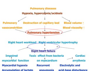
Cor  Pulmonale (Pulmonary Heart Disease) Guo Yubiao, M.D & Ph.D   Pulmonary & Critical Care Medicine  The first Affiliated Hospital of Sun-Yat Set University
 
Presentation Outline Definition Epidemiology/Classification Clinical manifestations Symptoms Signs   Diagnostic workup & Differential Diagnosis Diagnosis Differential Diagnosis Treatment & Prevention   Summary
Classification Acute  Cor Pulmonale Massive pulmonary embolism Chronic Cor Pulmonale
Epidemiology High prevalence(1992, 4.42 ‰) District difference Smoking  Acute exacerbation
Definition of Chronic Cor Pulmonale   Hypertrophy and dilatation of the right ventricle Secondary to the pulmonary hypertension  Caused by disease of the pulmonary parenchyma,  and/or chest wall, and/or pulmonary vascular system  With or without right heart failure Exclusion the causes of congenital heart disease and left heart disease
Etiology Pulmonary parenchyma disease COPD80%~90%,asthma,bronchiectasis,severe tuberculosis, idiopathic interstitial pneumonia,  sarcoidosis, eosinophilic granuloma,silicosis, etc . Disorders of the neuromuscular apparatus and chest wall Poliomyelitis ,Guillain-Barr é   syndrome, Kyphoscoliosis
Etiology Pulmonary vascular disorders chronic pulmonary thromboembolism, pulmonary arteriolitis, allergic granulomatosis, primary pulmonary hypertension  Others primary alveolar hypoventilation, sleep apnea syndromes
Pathophysiology The formation of pulmonary hypertension Functional factors of increase of vascular resistance Anatomical factors of increase of vascular resistance Increased blood volume and hyperviscosity Cardiac disorders and heart failure Damage to other vital organs
Functional factors of increase of vascular resistance Hypoxia is the most important factor for pulmonary hypertention. Hypoxic pulmonary vasoconstriction Determined mostly by the ratio of  vasoconstrictive substances to vasodilative substances Leukotriene, 5-HT, Ang II, PAF,EDCF/ NO, PGI 2   ,EDRF Direct effect of hypoxia on the increase of the smooth muscular cell membrane permeability to Ca 2+ Acidosis increases the sensitivity of vasoconstriction to hypoxia
Anatomical factors of increase of vascular resistance Vasculitis Emphysema, increased intra-alveolar pressure, compressed pulmonary capillaries  Reduction in pulmonary capillary bed Pulmonary vascular remodeling Multiple pulmonary micro-arteriole thrombosis
Increased blood volume and hyperviscosity Secondary polycythemia and hyperviscosity Water and sodium retention Aldosterone Renal arteriole constriction
Cardiac disorders and heart failure Right ventricular hypertrophy secondary to pulmonary hypertension. Sustained pulmonary hypertension exceeds the compensation of  right ventricle, and causes the increase of right ventricular end diastolic pressure,  and dilation and failure of right ventricle.  A few may develop left heart failure.
Damages to other vital organs Hypoxia and acidosis can also do damages to other vital organs, e.g. brain,liver,kidney,gastrointestine, endocrine system .
Pulmonary diseases Hypoxia, hypercapnia/acidosis Pulmonary  Destruction of capillary bed   Blood volume  ↑   vasoconstrition  Blood viscosity  ↑ Pulmonary hypertension Right heart workload ↑   Right ventricular hypertrophy   Right heart failure Impaired  Toxic  effect from bacteria   Cardiac myocardial  function   on myocardium  arrythmia Myocardial hypoxia  Recurrent  Electrolytic and  Accumulation of lactate  pneumonia  acid-base disturbance
Clinical manifestations Compensatory stage of the respiratory and cardiac function De-compensatory stage of the respiratory and cardiac function
Compensatory stage of the respiratory and cardiac function Symptoms:  cough, sputum, short of breath, dyspnea and palpitation on exertion, fatigue and  decrease of exercise tolerance more severe in acute exacerbation. A few with chest pain or hemoptysis .
Compensatory stage of the respiratory and  cardiac function Signs:  cyanosis, signs of emphysema, moist rales and/or rhonchi,  distal heart sound,   systolic murmur of tricuspid area ,  P2>A2  , subxiphoid visible/palpable cardiac   impulse, distended jugular venous pulsation
De-compensatory stage of the respiratory and cardiac function Respiratory failure Symtoms : severe dyspnea,especially at night, headache, insomnia,inappetence, somnolence, dizziness,confusion, even delirium. Signs : congunctiva  congestion and edema, retinal vasodilatation, optic papillary edema. weakness or disappear of deep reflexes, pathological reflexes, redness, sweaty
Right heart failure Symptoms:  predominant short of breath,palpitation, inappetence,nausea Signs:  cyanosis,tachycardia,arrhythmia,subxiphoid systolic murmur or even diastolic murmur. Tender hepatomegaly,Hepatojugular reflux, lower extremity edema, ascites
signs
Chest radiography Signs of pulmonary hypertension: Enlarged right descending pulmonary artery with diameter ≥15mm The ratio of the diameter of right descending pulmonary artery to trachea≥1.07 Bulge of pulmonary artery/with the height≥3mm dilation of the main pulmonary artery and its branches with concurrent underperfusion of the peripheral branches Signs of right ventricular enlargement Signs of underlying diseases and infection
 
Electrocardiography Right-axis deviation with a frontal plane axis greater than +90 ° Marked clockwise rotation of the electrical axis R v1 +S v5 ≥ 1.05mv P-pulmonale pattern Incomplete or rarely complete right bundle branch block  Low voltage QRS Occasional large Q wave or QS in V1,V2, even V3,suggesting healed myocardial infarction
Electrocardiography of Chronic Cor Pulmonale
Echocardiography The right ventricular outflow  ≥ 30 m m The   right ventricular internal dimension  ≥20mm Anterior RV wall thickness Ratio of left to right ventricular internal dimension <2 Increased right pulmonary artery or pulmonary artery dimension (Peak systolic pulmonary artery pressure) Increased right atrium dimension
Arterial blood gas analysis Hypoxemia and/or hypercapnia Respiratory failure: PaO2<60mmHg PaCO2>50mmHg
Serum assessment Increased RBC count and Hemoglobin Increased blood and plasma viscosity Increased WBC count and neutrophilic ratio when infection occurs Change of renal or hepatic function Electrolyte imbalance
Others Pulmonary function test For early stage or non-exacerbation stage patients Sputum culture For guidance of antibiotics selection in patients with acute exacerbation stage
Diagnosis History of COPD and pulmonary parenchymal or chest wall or pulmonary vascular disease, Symptoms and signs of pulmonary hypertension and right heart dilation or failure,  combined with the signs of right heart hypertrophy and dilation by ECG, X-ray,UCG.
Differential Diagnosis Coronary atherosclerotic heart   diseases History of left heart failure, hypertension, hyperlipoidemia  ; Symtoms of angina pectoris, myocardial infarction Left ventricular hypertrophy in P.E. and X-ray, myocardial ischemia in ECG Rheumatic heart diseases History of rheumatic arthritis and myocarditis Usu. involving other cardiac valves Special signs in X-ray, ECG and UCG Primary cardiomyopathy No history of chronic pulmonary diseases Enlargement of entire heart No signs of pulmonary hypertension in X-ray
Therapy  S tage of  Acute exacerbation Control of infection Oxygen therapy Control of heart failure Control of cardiac arrythmia Anticoagulatory therapy Patient care
Control of infection Antibiotics based on sputum culture  Before the culture, based on infection acquired location and sputum smear gram stain  CAP: G positive; HAP:G negative Penicillins, aminoglycerides,quinolones and cephalosporins Secondary fungal infection
Oxygen therapy Clearance of respiratory tract Correction of hypoxia and hypercapnia
Control of heart failure Different to heart failure caused by other cardiac disease May be improved after control of infection and normality of blood gas Diuretics Vasodilators Positive inotropic agents-digitalis
Diuretics Reduction of blood volume and right heart load,elimination of edema  Small dosage and short period Moderate degree:HCT .antisterone;Severe cases: furosemide  Adverse effect: alkalosis with low K + &CL - thickened sputum blood condensation
Vasodilators Reduction of cardiac pre- and after- load and oxygen consumption, improve the myocardial contractibility. Adverse effect: low blood pressure, tachycardia and hypoxemia and hypercapnia
Positive inotropic agents-digitalis Low tolerance to digitalis due to chronic hypoxia and infection: arrhythmia Correction of hypoxia and hypokalemia before use Low dosage, fast action and fast metabolite agents: lanatoside C(cedilanid),strophanthin K Indications: refractory edema after improvement of infection and respiratory function and no effect on diuretics right heart failure without obvious infection acute left heart failure
Control of arrhythmia:usually self-limited Anticoagulatory therapy:heparin or low molecular weight heparin Intensive  patient care:monitoring, airway secretion aspiration
Stage of non-exacerbation Combined with Chinese medicine Long-term home oxygen therapy Pulmonary Rehabilitation Regulation of immune system Nutrition
Complications Pulmonary encephalopathy Acid-base and electrolyte disturbance Cardiac arrhythmia:af,aF,at Shock Gastrointestional haemorrhage Disseminated intravascular coagulation
Prognosis Recurrent acute exacerbation, progressing with gradually impairment of pulmonary function  Motality of 10%-15% Prolong lifespan and improve quality of life
Prevention Smoking cessation Treatment of the predisposing factors respiratory tract infection, inhalation of noxious gases, occupational protection, etc  Education
Thank You!

7 cor pulmonale

  • 1.
    Cor Pulmonale(Pulmonary Heart Disease) Guo Yubiao, M.D & Ph.D Pulmonary & Critical Care Medicine The first Affiliated Hospital of Sun-Yat Set University
  • 2.
  • 3.
    Presentation Outline DefinitionEpidemiology/Classification Clinical manifestations Symptoms Signs Diagnostic workup & Differential Diagnosis Diagnosis Differential Diagnosis Treatment & Prevention Summary
  • 4.
    Classification Acute Cor Pulmonale Massive pulmonary embolism Chronic Cor Pulmonale
  • 5.
    Epidemiology High prevalence(1992,4.42 ‰) District difference Smoking Acute exacerbation
  • 6.
    Definition of ChronicCor Pulmonale Hypertrophy and dilatation of the right ventricle Secondary to the pulmonary hypertension Caused by disease of the pulmonary parenchyma, and/or chest wall, and/or pulmonary vascular system With or without right heart failure Exclusion the causes of congenital heart disease and left heart disease
  • 7.
    Etiology Pulmonary parenchymadisease COPD80%~90%,asthma,bronchiectasis,severe tuberculosis, idiopathic interstitial pneumonia, sarcoidosis, eosinophilic granuloma,silicosis, etc . Disorders of the neuromuscular apparatus and chest wall Poliomyelitis ,Guillain-Barr é syndrome, Kyphoscoliosis
  • 8.
    Etiology Pulmonary vasculardisorders chronic pulmonary thromboembolism, pulmonary arteriolitis, allergic granulomatosis, primary pulmonary hypertension Others primary alveolar hypoventilation, sleep apnea syndromes
  • 9.
    Pathophysiology The formationof pulmonary hypertension Functional factors of increase of vascular resistance Anatomical factors of increase of vascular resistance Increased blood volume and hyperviscosity Cardiac disorders and heart failure Damage to other vital organs
  • 10.
    Functional factors ofincrease of vascular resistance Hypoxia is the most important factor for pulmonary hypertention. Hypoxic pulmonary vasoconstriction Determined mostly by the ratio of vasoconstrictive substances to vasodilative substances Leukotriene, 5-HT, Ang II, PAF,EDCF/ NO, PGI 2 ,EDRF Direct effect of hypoxia on the increase of the smooth muscular cell membrane permeability to Ca 2+ Acidosis increases the sensitivity of vasoconstriction to hypoxia
  • 11.
    Anatomical factors ofincrease of vascular resistance Vasculitis Emphysema, increased intra-alveolar pressure, compressed pulmonary capillaries Reduction in pulmonary capillary bed Pulmonary vascular remodeling Multiple pulmonary micro-arteriole thrombosis
  • 12.
    Increased blood volumeand hyperviscosity Secondary polycythemia and hyperviscosity Water and sodium retention Aldosterone Renal arteriole constriction
  • 13.
    Cardiac disorders andheart failure Right ventricular hypertrophy secondary to pulmonary hypertension. Sustained pulmonary hypertension exceeds the compensation of right ventricle, and causes the increase of right ventricular end diastolic pressure, and dilation and failure of right ventricle. A few may develop left heart failure.
  • 14.
    Damages to othervital organs Hypoxia and acidosis can also do damages to other vital organs, e.g. brain,liver,kidney,gastrointestine, endocrine system .
  • 15.
    Pulmonary diseases Hypoxia,hypercapnia/acidosis Pulmonary Destruction of capillary bed Blood volume ↑ vasoconstrition Blood viscosity ↑ Pulmonary hypertension Right heart workload ↑ Right ventricular hypertrophy Right heart failure Impaired Toxic effect from bacteria Cardiac myocardial function on myocardium arrythmia Myocardial hypoxia Recurrent Electrolytic and Accumulation of lactate pneumonia acid-base disturbance
  • 16.
    Clinical manifestations Compensatorystage of the respiratory and cardiac function De-compensatory stage of the respiratory and cardiac function
  • 17.
    Compensatory stage ofthe respiratory and cardiac function Symptoms: cough, sputum, short of breath, dyspnea and palpitation on exertion, fatigue and decrease of exercise tolerance more severe in acute exacerbation. A few with chest pain or hemoptysis .
  • 18.
    Compensatory stage ofthe respiratory and cardiac function Signs: cyanosis, signs of emphysema, moist rales and/or rhonchi, distal heart sound, systolic murmur of tricuspid area , P2>A2 , subxiphoid visible/palpable cardiac impulse, distended jugular venous pulsation
  • 19.
    De-compensatory stage ofthe respiratory and cardiac function Respiratory failure Symtoms : severe dyspnea,especially at night, headache, insomnia,inappetence, somnolence, dizziness,confusion, even delirium. Signs : congunctiva congestion and edema, retinal vasodilatation, optic papillary edema. weakness or disappear of deep reflexes, pathological reflexes, redness, sweaty
  • 20.
    Right heart failureSymptoms: predominant short of breath,palpitation, inappetence,nausea Signs: cyanosis,tachycardia,arrhythmia,subxiphoid systolic murmur or even diastolic murmur. Tender hepatomegaly,Hepatojugular reflux, lower extremity edema, ascites
  • 21.
  • 22.
    Chest radiography Signsof pulmonary hypertension: Enlarged right descending pulmonary artery with diameter ≥15mm The ratio of the diameter of right descending pulmonary artery to trachea≥1.07 Bulge of pulmonary artery/with the height≥3mm dilation of the main pulmonary artery and its branches with concurrent underperfusion of the peripheral branches Signs of right ventricular enlargement Signs of underlying diseases and infection
  • 23.
  • 24.
    Electrocardiography Right-axis deviationwith a frontal plane axis greater than +90 ° Marked clockwise rotation of the electrical axis R v1 +S v5 ≥ 1.05mv P-pulmonale pattern Incomplete or rarely complete right bundle branch block Low voltage QRS Occasional large Q wave or QS in V1,V2, even V3,suggesting healed myocardial infarction
  • 25.
  • 26.
    Echocardiography The rightventricular outflow ≥ 30 m m The right ventricular internal dimension ≥20mm Anterior RV wall thickness Ratio of left to right ventricular internal dimension <2 Increased right pulmonary artery or pulmonary artery dimension (Peak systolic pulmonary artery pressure) Increased right atrium dimension
  • 27.
    Arterial blood gasanalysis Hypoxemia and/or hypercapnia Respiratory failure: PaO2<60mmHg PaCO2>50mmHg
  • 28.
    Serum assessment IncreasedRBC count and Hemoglobin Increased blood and plasma viscosity Increased WBC count and neutrophilic ratio when infection occurs Change of renal or hepatic function Electrolyte imbalance
  • 29.
    Others Pulmonary functiontest For early stage or non-exacerbation stage patients Sputum culture For guidance of antibiotics selection in patients with acute exacerbation stage
  • 30.
    Diagnosis History ofCOPD and pulmonary parenchymal or chest wall or pulmonary vascular disease, Symptoms and signs of pulmonary hypertension and right heart dilation or failure, combined with the signs of right heart hypertrophy and dilation by ECG, X-ray,UCG.
  • 31.
    Differential Diagnosis Coronaryatherosclerotic heart diseases History of left heart failure, hypertension, hyperlipoidemia ; Symtoms of angina pectoris, myocardial infarction Left ventricular hypertrophy in P.E. and X-ray, myocardial ischemia in ECG Rheumatic heart diseases History of rheumatic arthritis and myocarditis Usu. involving other cardiac valves Special signs in X-ray, ECG and UCG Primary cardiomyopathy No history of chronic pulmonary diseases Enlargement of entire heart No signs of pulmonary hypertension in X-ray
  • 32.
    Therapy Stage of Acute exacerbation Control of infection Oxygen therapy Control of heart failure Control of cardiac arrythmia Anticoagulatory therapy Patient care
  • 33.
    Control of infectionAntibiotics based on sputum culture Before the culture, based on infection acquired location and sputum smear gram stain CAP: G positive; HAP:G negative Penicillins, aminoglycerides,quinolones and cephalosporins Secondary fungal infection
  • 34.
    Oxygen therapy Clearanceof respiratory tract Correction of hypoxia and hypercapnia
  • 35.
    Control of heartfailure Different to heart failure caused by other cardiac disease May be improved after control of infection and normality of blood gas Diuretics Vasodilators Positive inotropic agents-digitalis
  • 36.
    Diuretics Reduction ofblood volume and right heart load,elimination of edema Small dosage and short period Moderate degree:HCT .antisterone;Severe cases: furosemide Adverse effect: alkalosis with low K + &CL - thickened sputum blood condensation
  • 37.
    Vasodilators Reduction ofcardiac pre- and after- load and oxygen consumption, improve the myocardial contractibility. Adverse effect: low blood pressure, tachycardia and hypoxemia and hypercapnia
  • 38.
    Positive inotropic agents-digitalisLow tolerance to digitalis due to chronic hypoxia and infection: arrhythmia Correction of hypoxia and hypokalemia before use Low dosage, fast action and fast metabolite agents: lanatoside C(cedilanid),strophanthin K Indications: refractory edema after improvement of infection and respiratory function and no effect on diuretics right heart failure without obvious infection acute left heart failure
  • 39.
    Control of arrhythmia:usuallyself-limited Anticoagulatory therapy:heparin or low molecular weight heparin Intensive patient care:monitoring, airway secretion aspiration
  • 40.
    Stage of non-exacerbationCombined with Chinese medicine Long-term home oxygen therapy Pulmonary Rehabilitation Regulation of immune system Nutrition
  • 41.
    Complications Pulmonary encephalopathyAcid-base and electrolyte disturbance Cardiac arrhythmia:af,aF,at Shock Gastrointestional haemorrhage Disseminated intravascular coagulation
  • 42.
    Prognosis Recurrent acuteexacerbation, progressing with gradually impairment of pulmonary function Motality of 10%-15% Prolong lifespan and improve quality of life
  • 43.
    Prevention Smoking cessationTreatment of the predisposing factors respiratory tract infection, inhalation of noxious gases, occupational protection, etc Education
  • 44.