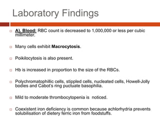
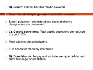
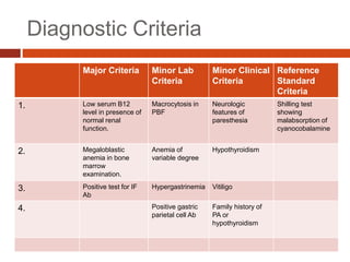
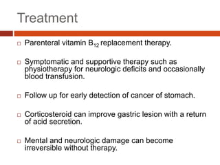
Vitamin B12 deficiency, also known as pernicious anemia, is an autoimmune disorder where antibodies block intrinsic factor and prevent vitamin B12 absorption from the stomach, leading to megaloblastic anemia. It presents with weakness, sore tongue, and neurological or gastrointestinal issues. Laboratory findings include macrocytic anemia, elevated MCV, hypersegmented neutrophils, and low serum B12 levels. The gold standard test is gastric biopsy showing parietal cell atrophy. Treatment is lifelong vitamin B12 supplementation by injection or oral administration.
Hemolytic anemia is characterized by accelerated red blood cell destruction and vigorous blood regeneration. It can be classified as intrinsic or extrinsic, congenital or acquired. The site of red blood cell destruction can be intravascular or extravascular. Common causes of hemolytic anemia include hereditary spherocytosis, thalassemias, sickle cell anemia, glucose-6-phosphate dehydrogenase deficiency, paroxysmal nocturnal hemoglobinuria, and immune-mediated hemolytic anemia. Evaluation of hemolytic anemia involves determining whether the anemia is hemolytic, the site of red blood cell destruction, the etiology, and severity through blood smears, reticulocyte counts, LDH and
Haemolytic anaemias are a group of anemias caused by the premature breakdown of red blood cells in the bloodstream or spleen. There are two main types - intrinsic defects that cause red blood cell damage from within, such as hereditary spherocytosis, and extrinsic defects that cause damage from outside factors like immune mediated hemolysis. Symptoms include anemia, jaundice, splenomegaly and gallstones. Laboratory tests show signs of increased red blood cell breakdown like elevated bilirubin and LDH, as well as signs of the bone marrow attempting to compensate with reticulocytosis and nucleated red blood cells. Intravascular hemolysis specifically causes hemoglobinemia,
This document defines anemia and provides a classification system. It begins by defining anemia as a low level of hemoglobin (Hb) and describes the functions of blood. It then outlines symptoms of anemia and discusses red blood cell indices used in evaluation. Three main classifications of anemia are described: morphological (based on cell appearance), pathological (based on cause), and etiological (based on specific cause). Various types of anemia are listed under each classification, such as iron deficiency under morphological and blood loss under acquired etiological anemia. Normal reference ranges for red blood cell parameters are also provided.
Megaloblastic Anaemia - Vit B12 deficiencyShahin Hameed
This document discusses megaloblastic anemia caused by vitamin B12 deficiency. It covers the normal metabolism and absorption of vitamin B12, the causes of deficiency including pernicious anemia, clinical features such as macrocytic anemia and neurological changes, diagnostic tests, and management with parenteral B12 injections. Deficiency results in defective DNA synthesis and affects all proliferating cells.
White Blood Cell Disorders can affect neutrophils, eosinophils, basophils and mast cells. Neutropenia is classified by severity based on absolute neutrophil count and risk of infection. Causes include acquired conditions like drugs/infections or congenital disorders. Hypereosinophilic syndrome is a broad condition caused by primary or secondary eosinophilia leading to tissue damage. Diagnosis involves ruling out secondary causes and identifying organ involvement. Treatment depends on etiology and includes steroids, hydroxyurea, interferon-alpha, imatinib or anti-IL-5 antibodies.
Megaloblastic anemias are caused by impaired DNA synthesis due to vitamin B12 or folate deficiency. The summary examines megaloblastic anemias, including causes such as vitamin B12 or folate metabolism defects, clinical features like pallor and neurological symptoms, investigation findings in peripheral blood and bone marrow showing megaloblasts and macroovalocytes, and treatment involving vitamin B12 or folate supplementation.
IDA is the most common form of anemia worldwide, affecting approximately 50% of anemia cases. It results from prolonged negative iron balance in the body due to factors like inadequate iron intake, decreased absorption, increased demand, or blood loss. Diagnosis involves a complete history, physical exam, and lab tests showing low indicators of iron stores like serum ferritin and iron, along with an elevated TIBC. Treatment aims to replenish iron stores and typically consists of oral iron supplementation of 200mg elemental iron per day for 3-6 months.
This document discusses megaloblastic anemia, its causes, symptoms, and treatments. It is characterized by abnormally large red blood cells due to a deficiency in vitamin B12 or folic acid, which is needed for DNA synthesis. The deficiencies can result from inadequate intake, malabsorption, or increased demand. Treatments include injections or supplements of vitamin B12, folic acid, or erythropoietin to stimulate red blood cell production. Adverse reactions are also discussed.
Hemolytic anemias are caused by increased red blood cell destruction. They are characterized by normochromic, normocytic anemia with reticulocytosis, increased indirect bilirubin and LDH, and absent haptoglobin. Causes include membrane defects, metabolic abnormalities, hemoglobinopathies, and immune or non-immune mechanisms. Specific conditions discussed include hereditary spherocytosis, glucose-6-phosphate dehydrogenase deficiency, paroxysmal nocturnal hemoglobinuria, drug-induced hemolysis, alloimmune hemolytic anemia, and warm or cold autoimmune hemolytic anemia. Management depends on the underlying cause and may involve transfusions, medications, or splenectomy.
Anemia is a decrease in red blood cells (RBCs), hemoglobin (Hgb), or hematocrit (HCT) levels compared to normal levels for age and sex. Anemias can be classified based on RBC size and hemoglobin content as normocytic normochromic, microcytic hypochromic, or macrocytic normocytic. Common causes of anemia include iron deficiency, anemia of chronic disease, thalassemia, vitamin B12 or folate deficiency, blood loss, and aplastic anemia.
This document provides an overview of blood disorders and summarizes key information about red blood cells and related disorders. It discusses the structure and function of red blood cells, variations in red blood cell count, and disorders involving too many or too few red blood cells such as polycythemia, anemia, sickle cell anemia, thalassemia, and erythroblastosis fetalis. Specific conditions are defined and their signs, symptoms, causes, and treatments are outlined.
This document discusses anemia, including its definition, classification, symptoms, diagnosis, and treatment. Anemia is characterized by low hemoglobin and red blood cell counts, resulting in reduced oxygen-carrying capacity of blood. It is classified based on cell morphology, etiology, and pathophysiology. Common symptoms include fatigue, weakness, and shortness of breath. Diagnosis involves laboratory tests of hemoglobin, hematocrit, red blood cell indices, iron, vitamin B12, and folate levels. Treatment depends on the underlying cause, and may involve oral or intravenous iron supplementation for iron-deficiency anemia.
Intracellular accumulations can occur through the buildup of various cellular constituents, including fat, proteins, carbohydrates, and pigments. Fatty change is the accumulation of triglycerides in cells, commonly seen in liver cells due to alcohol abuse, obesity, or other metabolic derangements. Proteins can accumulate intracellularly in conditions like myeloma or nephrotic syndrome. Glycogen storage diseases result in glycogen accumulation in cells. Pigment accumulations include lipofuscin, melanin, hemosiderin, and exogenous pigments like carbon. These accumulations can be transient or permanent and range from harmless to toxic.
This document provides an overview of hematopoiesis, erythropoiesis, and anemia. It discusses where blood cell formation occurs, the lifespan and production rate of red blood cells, and how hypoxia stimulates erythropoietin production. It defines anemia, lists global and country prevalence data, and compensatory mechanisms. It describes classifications of anemia including morphological and etiological, and covers causes such as blood loss, bone marrow disorders, nutritional deficiencies, and hemolytic anemias. Laboratory evaluation of anemia and peripheral blood smear findings are also summarized.
This document summarizes anemia, including its definition, classification, causes, signs and symptoms, diagnostic evaluation, and treatment. Anemia is defined as a deficiency in red blood cells, hemoglobin, or hematocrit. It is classified based on size, color, and cause of the red blood cells. Common causes include blood loss, decreased red blood cell production, and increased red blood cell breakdown. Signs and symptoms vary based on severity but can include fatigue, paleness, shortness of breath, and heart palpitations. Diagnostic testing includes complete blood count and iron studies. Treatment involves treating the underlying cause, oral or intravenous iron supplementation, blood transfusions, and medications.
This document provides an overview of anemia presented by Muhammad Abbas. It defines anemia as a decrease in hemoglobin below normal levels and discusses the pathophysiology and classification of anemia. Anemia is classified clinically based on severity, etiologically based on cause, and morphologically based on mean corpuscular volume. The main types are microcytic hypochromic anemia, macrocytic hyperchromic anemia, and normocytic normochromic anemia. Causes and diagnostic investigations for each type are also outlined.
1) Microcytic hypochromic anemia is characterized by small, pale red blood cells and can be caused by iron deficiency, thalassemia, sideroblastic anemia, or other conditions.
2) Iron deficiency anemia is the most common cause and results from inadequate iron intake or absorption. It disrupts hemoglobin synthesis and cellular proliferation.
3) Thalassemia is an inherited disorder of hemoglobin production that can range from mild to severe. Thalassemia major requires regular blood transfusions and causes severe anemia from ineffective erythropoiesis and hemolysis.
This document provides a classification and overview of hemolytic anemia. It discusses intracorpuscular defects like hereditary membrane defects (spherocytosis, elliptocytosis), enzyme defects (G6PD, pyruvate kinase), and hemoglobinopathies. Extracorpuscular defects include immune hemolytic anemia (autoimmune, alloimmune) and nonimmune causes. Evaluation of anemia involves hematological parameters. Thalassemias are classified based on affected globin chain (alpha, beta). Common hereditary spherocytosis causes premature RBC destruction and can be treated with splenectomy. G6PD deficiency results in drug-induced hemolysis.
The document discusses different types and causes of anemia. It classifies anemia into etiologic categories including impaired red blood cell production, excessive destruction of RBCs, and blood loss. It further describes morphologic classifications such as macrocytic, microcytic hypochromic, and normocytic normochromic anemia. Specific causes are provided for each category including deficiencies, diseases, and genetic disorders. Hemolytic anemia is discussed in more detail including hereditary and acquired causes. Laboratory findings associated with different types of anemia are also summarized.
Approach to microcytic hypochromic anemiaShinjan Patra
This document discusses the approach to evaluating and diagnosing microcytic hypochromic anemia. It begins by covering the basics of hemoglobin synthesis and iron metabolism. It then describes the morphological classification of anemias and discusses the main causes of microcytic anemia including iron deficiency anemia, anemia of chronic disease, thalassemia, sideroblastic anemia, and lead poisoning. For each condition, it outlines the pathogenesis, clinical features, diagnostic evaluation, and treatment approach. Throughout it emphasizes the importance of obtaining a thorough history and using iron studies, blood counts, and other tests to differentiate between the various microcytic anemia etiologies.
Iron is an essential mineral that is distributed throughout the body and is important for oxygen transport and cellular metabolism. Iron deficiency develops when requirements exceed supply and leads to iron deficient erythropoiesis and eventually iron deficiency anemia. It is one of the most common nutritional deficiencies worldwide, affecting toddlers, adolescent girls, pregnant women, and some minority groups. Treatment involves oral or parenteral iron supplementation depending on severity, with the goal of replenishing iron stores and repairing hemoglobin deficits.
Megaloblastic anemias are caused by deficiencies in vitamin B12 or folate which are needed for DNA synthesis. These deficiencies lead to large, immature red blood cells (megaloblasts) in the bone marrow. Symptoms include anemia, fatigue, weakness, and glossitis. The diagnosis involves blood tests to measure B12, folate, methylmalonate and homocysteine levels as well as examination of bone marrow cells which will show megaloblastic changes and chromosomal abnormalities. Treatment involves replacement of the deficient vitamin with either B12 or folate supplements.
This document summarizes aplastic and hypoplastic anemias. Aplastic anemia is a life-threatening condition where the bone marrow fails to produce red blood cells, white blood cells, and platelets, leading to pancytopenia. It can be acquired through exposure to radiation, chemicals, infections or idiopathically. Congenital causes include Fanconi's anemia and familial aplastic anemia. Clinical manifestations include fatigue, pallor, infections, and bleeding. Treatment involves removing causative agents if known, blood transfusions, and bone marrow transplant. Related disorders with pancytopenia include myelodysplastic syndromes and pure red cell aplasia.
This document discusses anemia and iron deficiency anemia. It defines anemia as a low level of hemoglobin and classifies it as mild, moderate, or severe based on hemoglobin levels. Iron deficiency anemia is described as the most common type worldwide, caused by too little iron in the body. Symptoms, causes like blood loss or poor diet, diagnostic tests, and iron metabolism are summarized. Iron is stored in the liver and spleen and transported by transferrin to support erythropoiesis when needed.
Megaloblastic anemia is caused by impaired DNA synthesis leading to ineffective red blood cell production. It results from vitamin B12 or folate deficiencies. Vitamin B12 is only produced by microorganisms and is obtained through animal products in the diet, while folate is found in many plant foods. Both are required as cofactors in important metabolic reactions. Deficiencies can be due to inadequate intake, impaired absorption, or genetic disorders affecting metabolism. Symptoms include megaloblastic changes in blood cells and pancytopenia.
This document provides an overview of several topics in hematology, including:
- Anemia, which is a reduction in red blood cells. Common types are discussed such as iron deficiency anemia and megaloblastic anemia.
- Leukemia, which are cancers of the white blood cells. The main types - acute and chronic leukemias - are defined. Acute leukemias like ALL and AML are discussed in more detail.
- Other blood disorders like lymphomas and lymphadenopathy are listed as learning objectives but not described further in this document.
This document discusses various types of anemia. It defines anemia as a condition with fewer than normal red blood cells or hemoglobin. The types discussed include iron deficiency anemia, thalassemia, anemia of chronic disease, sideroblastic anemia, and hemolytic anemia. For each type, the document outlines causes, pathophysiology, clinical presentation, laboratory findings, and management. Key points like ringed sideroblasts, ineffective erythropoiesis, and hereditary spherocytosis are explained. Treatment involves addressing the underlying cause, iron supplementation, blood transfusions, or splenectomy in some cases.
This document provides information on iron deficiency anemia, including its definition, causes, clinical features, diagnosis, and treatment. It defines iron deficiency anemia as a type of anemia that occurs when the supply of iron is inadequate to support optimal red blood cell production. The main causes listed are poor diet, increased demand, blood loss, and decreased iron absorption. Clinical features include general anemia symptoms as well as pale skin and abnormalities of the mouth, tongue, and nails. Diagnosis involves blood tests to check hemoglobin and iron levels. Treatment focuses on iron replacement through supplements or injections as well as treating the underlying cause.
This document discusses several hematological disorders including anemia, megaloblastic anemia, aplastic anemia, and provides details about their definitions, classifications, etiologies, pathophysiology, clinical features, diagnosis, and management. It describes how anemia can be classified based on morphology and etiology. Iron deficiency anemia is the most common type and can be caused by blood loss, insufficient iron intake or absorption. Megaloblastic anemia results from vitamin B12 or folate deficiencies, causing large immature red blood cells. Aplastic anemia is a condition where the bone marrow fails to produce sufficient new blood cells.
This document defines anemia and describes its causes and symptoms. Anemia is a reduction in red blood cells or hemoglobin below normal levels. It can be classified based on red blood cell morphology or etiology. Common causes include blood loss, red blood cell destruction, nutritional deficiencies, and bone marrow disorders. Symptoms result from reduced oxygen carrying capacity and include fatigue, paleness, weakness, and increased heart rate. Severe anemia places greater workload on the heart and can lead to cardiac failure during exercise due to extreme tissue hypoxia.
- Anemia is defined as a reduction in red blood cells (RBCs) or hemoglobin. It has many potential underlying causes and is a common complication in many disorders.
- A complete blood count provides important information about RBCs like hemoglobin levels, RBC count, MCV, and reticulocyte count. This helps classify anemias and determine if the bone marrow is responding appropriately.
- Taking a thorough history and physical exam can provide clues to the potential causes of anemia, like nutritional deficiencies, infections, hereditary disorders, medications, or other underlying illnesses. Certain physical findings may point to specific conditions like hepatosplenomegaly in hemolytic anem
This document discusses iron deficiency anemia. It begins by classifying anemias based on red blood cell morphology and etiology. Iron deficiency anemia is then explained in more detail. The symptoms, absorption of iron, and laboratory tests are summarized. The risks factors, treatment involving iron supplements, and prevention through an iron-rich diet are highlighted.
1. Erythropoiesis, the production of red blood cells, occurs in different sites throughout human development - in the yolk sac for the first 8 weeks, then the liver from 8 weeks to 6 months, and finally in the bone marrow from 6 months onwards in adults.
2. The bone marrow contains erythroid cells that develop into red blood cells, myeloid cells that develop into white blood cells, and megakaryocytic cells that produce platelets.
3. Anemia is defined as a decrease in hemoglobin concentration below the normal level for age and sex. It can be diagnosed through patient history, clinical examination, and laboratory investigations including complete blood count, blood indices, ret
Anemia is a condition where the hemoglobin level or red blood cell count is lower than normal. It can be caused by blood loss, decreased red blood cell production, or increased red blood cell destruction. Globally, anemia affects over 1.6 billion people. Common symptoms include fatigue, weakness, and pale skin. Diagnosis involves blood tests to measure hemoglobin, red blood cell count, and other indicators. Treatment depends on the underlying cause but may involve iron supplementation, vitamin B12/folate, blood transfusions, or treating the primary disease.
This document provides an overview of anemia, including its definition, cut-off levels used to diagnose it, common causes, classification approaches, and key details about specific types like iron deficiency anemia, megaloblastic anemias, sickle cell disease, and thalassemias. It covers diagnostic testing and clinical manifestations, emphasizing the importance of considering a patient's red blood cell morphology, erythropoiesis, and underlying pathophysiology when evaluating the cause of an anemia.
The document discusses the evaluation and diagnosis of pediatric blood disorders. It notes that the history, physical exam, and initial laboratory tests provide clues to diagnose blood diseases. Diagnosis requires knowledge of normal hematological values that vary by age. The initial workup involves tests like hemoglobin, red blood cell indices, blood smear exam. Further tests are used depending on the results to diagnose causes like anemia, hemolytic disorders, and hemoglobinopathies. Specific disorders discussed in more detail include iron deficiency anemia, thalassemias, sickle cell disease, and their typical laboratory findings and treatment approaches.
Anemia is the blood disorder, characterized by the reduction in:
1. Red blood cell (RBC) count
2. Hemoglobin content
3. Packed cell volume (PVC).
This occurs because of
Decreased production of RBC
Increased destruction of RBC
Excess loss of blood from the body
The document provides an overview of anemia and iron deficiency anemia. It discusses the typical signs and symptoms of iron deficiency anemia including fatigue, pale skin, spoon-shaped nails, and glossitis. Laboratory findings often show microcytic hypochromic anemia, low serum ferritin, and iron deficiency in bone marrow smears. Treatment involves oral or parenteral iron supplementation for 3-6 months to replenish iron stores.
Anemia overview document discusses red blood cell parameters, survival and production of RBCs, erythropoiesis process, classification of anemias, common causes, pathophysiology, bone marrow disorders, definitions of aplastic anemia, myelodysplastic syndromes, and acute leukemia. Diagnosis of anemia involves history, physical exam, labs including complete blood count and smears to evaluate RBC size and check for other abnormalities. Treatment focuses on supporting hematopoiesis, blood transfusions, and curing the underlying cause.
The approach to determining whether the cause of anemia is ineffective erythropoiesis versus increased red blood cell destruction involves examining the following:
- Reticulocyte count - Increased in hemolytic anemia, normal or low in ineffective erythropoiesis
- LDH and bilirubin levels - Increased in hemolytic anemia, normal in ineffective erythropoiesis
- Haptoglobin and hemoglobin levels - Decreased haptoglobin, increased free hemoglobin in plasma in hemolytic anemia.
Examining the red blood cell morphology on peripheral smear and markers of iron status can help determine if the cause is ineffective erythropoiesis related to nutritional deficiencies, bone marrow
Irion defitient and megaloblastic anemiasJasmine John
This document summarizes iron deficiency anemia and megaloblastic anemia. It discusses the causes, symptoms, laboratory findings, treatment, and prognosis of these conditions. Iron deficiency is the most common cause of anemia worldwide and results from inadequate iron intake or absorption. Megaloblastic anemia is caused by vitamin B12 or folate deficiencies and results in abnormal DNA synthesis and large, immature red blood cells. Treatment involves oral or intravenous iron supplementation for iron deficiency and vitamin B12/folate supplementation for megaloblastic anemia.
The document summarizes recent trends in endodontic devices presented by Dr. Pooja Chaturvedi. It discusses advances in diagnostic tools like ultrasound Doppler and CBCT for improved diagnosis. Newer loupes and microscopes provide enhanced visualization during access and treatment. Devices like SAF and laser-activated irrigation improve cleaning of the root canal system. Modern apex locators incorporate humidity sensing for more precise working length determination. Motorized gutta percha delivery systems and new obturation units allow for faster, easier filling of the root canal space. Overall the trend is towards smaller, more ergonomic devices that enhance treatment outcomes while reducing radiation exposure or hand fatigue.
Osteogenesis imperfecta and osteoporosis are heritable bone disorders characterized by fragile bones. Osteogenesis imperfecta arises from mutations affecting type 1 collagen production, resulting in bones that fracture easily. It is classified into 4 types based on severity, ranging from mild bone fragility to prenatal death. Osteoporosis is caused by failed bone resorption, leading to abnormally dense bones. It presents as infantile, intermediate, or adult-onset forms depending on age of symptoms onset. Treatment focuses on rehabilitation and surgery for osteogenesis imperfecta, while osteoporosis may be treated with medications to stimulate bone resorption.
The document discusses the muscles of mastication including the masseter, temporalis, lateral pterygoid, and medial pterygoid muscles. It describes the masticatory system and its components, the functions of mastication in breaking down food, and the chewing cycle. It also discusses the neurological control of mastication and additional muscles involved like the digastric. The importance of understanding the masticatory system for dentists is described for areas like prosthodontics and its relevance to jaw movements. Some clinical significance of lesions impacting structures involved in mastication are also noted.
This document discusses the development of occlusion from birth through adulthood. It describes the neonatal period where gum pads are present, the primary dentition period where baby teeth erupt, the mixed dentition period where permanent teeth begin to replace primary teeth, and the permanent dentition period. Key aspects of each developmental period are outlined such as the sequence of tooth eruption, characteristics of different malocclusions, and compensatory mechanisms involved in the transition between dentitions. The document also introduces Andrews' seven keys to normal occlusion.
This document discusses fibrous dysplasia and cemento-osseous dysplasia, two fibro-osseous lesions of bone. It covers the classification, etiology, clinical features, radiographic appearance, histopathology, and treatment of these conditions. Specifically, it describes how fibrous dysplasia is caused by a mutation leading to overproduction of cAMP, and can present as monostotic or polyostotic lesions. It also outlines the three types of cemento-osseous dysplasia - focal, periapical, and florid - based on clinical and radiographic characteristics.
Biomechanical considerations in removable partial denture designDr Pooja Chaturvedi
The document discusses biomechanical considerations in removable partial denture design. It notes that the primary goal is to control potentially damaging forces applied to the supporting structures, including bone and soft tissues. It describes the three classes of levers that a partial denture can act as when subjected to intraoral forces. Class I levers are most efficient while Class III are least efficient. Factors like extension base length can generate greater damaging loads. Removable partial dentures that are tooth-supported transmit forces axially like fixed bridges, while tissue-supported designs can result in non-axial loading and greater potential for damage over time. Control of forces is important through factors like tissue health and component positioning.
Bacterial infection is caused by disease-causing bacteria invading body tissues. This leads to bacteria multiplying and the body reacting to the microorganisms and toxins they produce. The document discusses many specific types of bacterial infections caused by different bacteria that affect various parts of the body, including their symptoms, pathogenesis, diagnosis and treatment.
This document provides information about teething and its management in infants and children. It discusses the typical timeline for teeth eruption, common signs and symptoms of teething, and potential complications. It describes conditions like eruption hematoma, eruption sequestrum, ectopic eruption, transposition, infraocclusion, and impacted teeth. Treatment options are outlined for managing teething problems and their associated conditions.
These lecture slides, by Dr Sidra Arshad, offer a quick overview of the physiological basis of a normal electrocardiogram.
Learning objectives:
1. Define an electrocardiogram (ECG) and electrocardiography
2. Describe how dipoles generated by the heart produce the waveforms of the ECG
3. Describe the components of a normal electrocardiogram of a typical bipolar lead (limb II)
4. Differentiate between intervals and segments
5. Enlist some common indications for obtaining an ECG
6. Describe the flow of current around the heart during the cardiac cycle
7. Discuss the placement and polarity of the leads of electrocardiograph
8. Describe the normal electrocardiograms recorded from the limb leads and explain the physiological basis of the different records that are obtained
9. Define mean electrical vector (axis) of the heart and give the normal range
10. Define the mean QRS vector
11. Describe the axes of leads (hexagonal reference system)
12. Comprehend the vectorial analysis of the normal ECG
13. Determine the mean electrical axis of the ventricular QRS and appreciate the mean axis deviation
14. Explain the concepts of current of injury, J point, and their significance
Study Resources:
1. Chapter 11, Guyton and Hall Textbook of Medical Physiology, 14th edition
2. Chapter 9, Human Physiology - From Cells to Systems, Lauralee Sherwood, 9th edition
3. Chapter 29, Ganong’s Review of Medical Physiology, 26th edition
4. Electrocardiogram, StatPearls - https://www.ncbi.nlm.nih.gov/books/NBK549803/
5. ECG in Medical Practice by ABM Abdullah, 4th edition
6. Chapter 3, Cardiology Explained, https://www.ncbi.nlm.nih.gov/books/NBK2214/
7. ECG Basics, http://www.nataliescasebook.com/tag/e-c-g-basics
Muktapishti is a traditional Ayurvedic preparation made from Shoditha Mukta (Purified Pearl), is believed to help regulate thyroid function and reduce symptoms of hyperthyroidism due to its cooling and balancing properties. Clinical evidence on its efficacy remains limited, necessitating further research to validate its therapeutic benefits.
Recomendações da OMS sobre cuidados maternos e neonatais para uma experiência pós-natal positiva.
Em consonância com os ODS – Objetivos do Desenvolvimento Sustentável e a Estratégia Global para a Saúde das Mulheres, Crianças e Adolescentes, e aplicando uma abordagem baseada nos direitos humanos, os esforços de cuidados pós-natais devem expandir-se para além da cobertura e da simples sobrevivência, de modo a incluir cuidados de qualidade.
Estas diretrizes visam melhorar a qualidade dos cuidados pós-natais essenciais e de rotina prestados às mulheres e aos recém-nascidos, com o objetivo final de melhorar a saúde e o bem-estar materno e neonatal.
Uma “experiência pós-natal positiva” é um resultado importante para todas as mulheres que dão à luz e para os seus recém-nascidos, estabelecendo as bases para a melhoria da saúde e do bem-estar a curto e longo prazo. Uma experiência pós-natal positiva é definida como aquela em que as mulheres, pessoas que gestam, os recém-nascidos, os casais, os pais, os cuidadores e as famílias recebem informação consistente, garantia e apoio de profissionais de saúde motivados; e onde um sistema de saúde flexível e com recursos reconheça as necessidades das mulheres e dos bebês e respeite o seu contexto cultural.
Estas diretrizes consolidadas apresentam algumas recomendações novas e já bem fundamentadas sobre cuidados pós-natais de rotina para mulheres e neonatos que recebem cuidados no pós-parto em unidades de saúde ou na comunidade, independentemente dos recursos disponíveis.
É fornecido um conjunto abrangente de recomendações para cuidados durante o período puerperal, com ênfase nos cuidados essenciais que todas as mulheres e recém-nascidos devem receber, e com a devida atenção à qualidade dos cuidados; isto é, a entrega e a experiência do cuidado recebido. Estas diretrizes atualizam e ampliam as recomendações da OMS de 2014 sobre cuidados pós-natais da mãe e do recém-nascido e complementam as atuais diretrizes da OMS sobre a gestão de complicações pós-natais.
O estabelecimento da amamentação e o manejo das principais intercorrências é contemplada.
Recomendamos muito.
Vamos discutir essas recomendações no nosso curso de pós-graduação em Aleitamento no Instituto Ciclos.
Esta publicação só está disponível em inglês até o momento.
Prof. Marcus Renato de Carvalho
www.agostodourado.com
Histololgy of Female Reproductive System.pptxAyeshaZaid1
Dive into an in-depth exploration of the histological structure of female reproductive system with this comprehensive lecture. Presented by Dr. Ayesha Irfan, Assistant Professor of Anatomy, this presentation covers the Gross anatomy and functional histology of the female reproductive organs. Ideal for students, educators, and anyone interested in medical science, this lecture provides clear explanations, detailed diagrams, and valuable insights into female reproductive system. Enhance your knowledge and understanding of this essential aspect of human biology.
Promoting Wellbeing - Applied Social Psychology - Psychology SuperNotesPsychoTech Services
A proprietary approach developed by bringing together the best of learning theories from Psychology, design principles from the world of visualization, and pedagogical methods from over a decade of training experience, that enables you to: Learn better, faster!
Cell Therapy Expansion and Challenges in Autoimmune DiseaseHealth Advances
There is increasing confidence that cell therapies will soon play a role in the treatment of autoimmune disorders, but the extent of this impact remains to be seen. Early readouts on autologous CAR-Ts in lupus are encouraging, but manufacturing and cost limitations are likely to restrict access to highly refractory patients. Allogeneic CAR-Ts have the potential to broaden access to earlier lines of treatment due to their inherent cost benefits, however they will need to demonstrate comparable or improved efficacy to established modalities.
In addition to infrastructure and capacity constraints, CAR-Ts face a very different risk-benefit dynamic in autoimmune compared to oncology, highlighting the need for tolerable therapies with low adverse event risk. CAR-NK and Treg-based therapies are also being developed in certain autoimmune disorders and may demonstrate favorable safety profiles. Several novel non-cell therapies such as bispecific antibodies, nanobodies, and RNAi drugs, may also offer future alternative competitive solutions with variable value propositions.
Widespread adoption of cell therapies will not only require strong efficacy and safety data, but also adapted pricing and access strategies. At oncology-based price points, CAR-Ts are unlikely to achieve broad market access in autoimmune disorders, with eligible patient populations that are potentially orders of magnitude greater than the number of currently addressable cancer patients. Developers have made strides towards reducing cell therapy COGS while improving manufacturing efficiency, but payors will inevitably restrict access until more sustainable pricing is achieved.
Despite these headwinds, industry leaders and investors remain confident that cell therapies are poised to address significant unmet need in patients suffering from autoimmune disorders. However, the extent of this impact on the treatment landscape remains to be seen, as the industry rapidly approaches an inflection point.
Here is the updated list of Top Best Ayurvedic medicine for Gas and Indigestion and those are Gas-O-Go Syp for Dyspepsia | Lavizyme Syrup for Acidity | Yumzyme Hepatoprotective Capsules etc
4. Pathophysiology
Subnormal level of Hb causes lowered oxygen carrying
capacity of the blood. This in turn, initiates
compensatory physiologic adaptations such as:
Increased release of oxygen from Hb
Increased blood flow volume
Maintenance of the blood volume
Redistribution of blood flow to maintain the cerebral
blood supply.
5. Clinical Features
The speed of onset of anemia: rapidly progressive anemia causes
more symptoms than anemia of slow-onset as there is less time for
physiologic adaptation.
The severity of anemia: mild anemia produces no symptoms or
signs but a rapidly developing severe anemia (Hb<6.0 g/dl) may
produce significant clinical features.
The age of the patient: the young patient due to good
cardiovascular compensation tolerate anemia quite well as
compared to the elderly.
The heamoglobin dissociation curve: in anemia, the affinity of
heamoglobin for oxygen is depressed as 2,3-BPG in the red cells
increase.
6. Symptoms
The presenting features are:
Tiredness
Easy fatiguability
Generalized muscular weakness
Lethargy
Headache
In older patients, there may be symptoms of
cardiac failure, angina pectoris, intermittent
claudication, confusion and visual disturbances.
8. Investigations
A). Haemoglobin Estimation:
Cynmethaemoglobin (HCN) method employing Drabkin’s solution and a
spectrophotometer.
If pregnancy, there is haemodilution and, therefore, the lower limit in normal pregnant
women is less (10.5 g/dl) than in the non pregnant state.
B).Peripheral Blood Film Examination:
Stain with Romanowsky dyes (Leishman’s stain, May-Grunwal’s stain, Jenner-Giemsa
stain, Wright’s stain)
C). Red Cell Indices:
In Fe deficiency and thalassaemia, MCV, MCH and MCHC are reduced.
In anemia due to acute blood loss and haemolytic anemia, MCV, MCH and MCHC
are all within normal limits.
In megaloblastic anemia, MCV is raised above the normal range.
D). Leucocyte Count and Platelet Count
E). Reticulocyte Count
F). Erythrocyte Sedimentation Rate
G). Bone Marrow Examination
9.
10. Classification
A). Pathophysiology
I. Anemias due to increased
blood loss
a). Acute post-haemorrhagic blood
loss
b). Chronic blood loss
II. Anemias due to impaired red
cell production
a).Cytoplasmic maturation defects
i. Fe deficiency anaemia
ii.Thallassaemia syndromes
b).Nuclear Maturation Syndromes
i. Vitamin B12/ Folic acid
deficiency
ii. Megaloblastic anemia
c).Defect In Stem cell proliferation
i. Aplastic Anemia
pure red cell aplasia
d).Anaemia of chronic disorders
e).Bone Marrow Infiltration
f). Congenital anaemias
III. Anaemias due to increased
red cell destruction(Haemolytic
anaemias)
a). Extrinsic red cell
abnormalities
b). Intrinsic red cell abnormalities
11. B).Morphologic
I. Microcytic, hypochromic:
MCV, MCH, MCHC are all reduced e.g. after Fe
deficiency anaemia and in certain non-iron deficient
anaemias
II. Normocytic, normochromic:
MCV, MCH, MCHC are all normal e.g after acute blood
loss, haemolytic anaemias, bone marrow failure,
anaemia of chronic disorders
III. Macrocytic, normochromic:
MCV is raised e.g in megaloblastic anaemia due to
deficiency of vitamin B12 or folic acid.
12. ANAEMIA OF BLOOD
LOSS
Depending upon the rate of blood loss due to
haemorrhage, the effects of post-
haemmorhagic anaemia appear.
13. When the loss of blood
occurs suddenly,
i. Immediate threat to life due
to hypovolemia which may
result in shock and death.
ii. If patient survives, shifting
of interstitial fluid to
intravascular compartment
with consequent
haemodilution with low
haematocrit.
iii. Hypoxia stimulates
production of
erythropoeitin resulting in
increased marrow
erythropoeisis.
When the loss of blood is slow
and insidious, the effects of
anaemia will become apparent
only when the rate of loss is
more than the rate of
production and the iron stores
are depleted.
This results in Fe deficiency
anaemia as seen in other
clinical conditions.
Acute Blood Loss Chronic Blood Loss
14. Hypochromic Anaemia
Hypochromic anaemia due to Fe deficiency is the
commonest cause of anaemia the world over.
Fe deficiency is the most important cause of microcytic
hypochromic anemia which all the three red cell indices
(MCV, MCH, MCHC) are reduced and occurs due to
defective Hb synthesis.
It is of two types:
a).Hypochromic anaemia due to Fe deficiency
b).hypochromic anaemia other than Fe deficiency
15. The commonest nutritional deficiency disorder present
throughout the world is iron deficiency but its prevalence is
higher in developing countries.
The factors responsible for iron deficiency in different
populations are variable and are best understood in the
context of normal iron metabolism.
Iron Deficiency Anemia
17. Pathophysiology
Iron deficiency anemia develops when the supply of iron
is inadequate for the requirement of Hb synthesis.
Initially, negative iron balance is covered by mobilization
from the tissue stores so as to maintain Hb synthesis.
It is only after the tissue stores of iron are exhausted
that the supply of iron to the marrow becomes
insufficient for Hb formation and thus a state of iron
deficiency develops.
19. Etiology
A).Females in reproductive period of life:
Highest incidence in women.
i. Blood Loss: Most important cause of anemia in women
during child bearing age group.
due to persistant and heavy menstrual blood loss.
young girls at the start of menstruation may develop mild
anemia due to blood loss.
ii. Inadequate Intake: prevalent in women of lower economic
status.
besides diet deficient in iron, other factors such as
anorexia, impaired absorption and diminished bioavailability
may act as contributory factors.
iii. Increased Requirements: During adolescence and
pregnancy the body’s iron demands are increased.
during a normal pregnancy, 750 mg of iron may be
siphoned off from the mother - 400 mg to foetus + 150 mg to
placenta + 200 mg lost at parturition and lactation
21. B).Post Menopausal Females:
i. Post menopausal uterine bleeding: due to
carcinoma of uterus.
i. Bleeding from alimentary tract: due to
carcinoma of stomach and large bowel and
hiatus hernia.
22. C).Adult Males:
i. Gastrointestinal Tract: due to peptic ulcer,
haemarrhoids, hook worm infestation,
carcinoma of stomach and large bowel,
oesophageal varices, hiatus hernia, chronic
aspirin ingestion and ulcerative colitis.
ii. Urinary Tract: due to haematuria and
haemoglobinurea.
iii. Nose: repeated epistaxis
iv. Lungs: haemoptysis from various causes.
23. D).Infants and Children:
Peak incidence at 1-2 years of age.
Principal cause is increased demand of iron
which is not met by inadequate intake of iron in
the diet.
Normal full term infants have sufficient iron
stores, while premature infants have
inadequate reserves because iron stores from
the mother are mainly laid down during the last
trimester of pregnancy.
24. Clinical Features
Anemia: onset of iron deficiency anemia is
generally slow.
Usual symptoms are weakness, fatigue,
dyspnoea on exertion, palpitations and pallor of
the skin, mucous membranes and sclerae.
Older patients may develop angina and
congestive cardiac failure.
Patients may have dietary cravings such as
pica.
Menorrhagia is a common symptom in iron
deficient women.
25. Epithelial Tissue Changes:
Long standing iron deficiency anemia causes
epithelial tissue changes in some patients.
Koilonychia (spoon shaped nails)
Atrophic glossitis
Angular stomatitis
Dysphagia (a feature of Plummer Vinson
Syndrome)
27. Laboratory Findings
Stages:
Stage 1: Storage iron depletion
Stage 2: Iron deficient erythropoeisis
Stage 3: Frank iron deficiency anemia where
the RBCs become microcytic and
hypochromic.
28. Blood Picture and Red Cell Indices
i. Haemoglobin
ii. Red Cells: Hypochromic and Microcytic and there is Anisocytosis and
poikilocytosis.
Target cells, elliptical forms and polychromatic cells are often present.
Normoblasts are uncommon.
RBC count is below normal level but is proportionate to the fall in Hb
values.
iii. Reticulocyte Count: Normal or reduced but may be slightly raised (2-
5%) in cases after haemorrhage.
iv. Absolute Values: MCV, MCH, MCHC are reduced.
v. Leucocytes: TLC, DLC are usually normal.
vi. Platelets: usually normal but may be slightly to moderately raised in
patients who have had recent bleeding.
29.
30. Bone Marrow Findings
i. Marrow Cellularity: Increased due to erythroid hyperplasia
(M:E decreased)
ii. Erythropoeisis: Normoblastic erythropoeisis with
predominance of small polychromatic normoblasts
(micronormoblasts). These normoblasts have a thin rim of
cytoplasm around the nucleus and ragged and irregular cell
border.
iii. Other Cells: Myeloid, Lymphoid and Megakaryocytic cells
are normal in number and morphology.
iv. Marrow Iron: Iron staining (Prussian blue reaction) shows
deficient reticuloendothelial iron stores and absence of
siderotic iron granules from developing normoblasts.
31. Treatment
1. Correction of the disorder: Appropriate surgical,
medical and preventive measures are instituted to
correct the cause of blood loss.
2. Correction of iron deficiency:
a. Oral therapy: Administration of iron salts such as
ferrous sulfate, ferrous fumerate, ferrous gluconate
and polysaccharide iron.
These preparations having varying amounts of
elemental iron ranging from 39 to 105 mg.
Optimal absorption is obtained by giving iron fasting,
but if side effects occur (nausea, abdominal
discomfort, diarrhoea) iron can be given along with
food or with a lower iron preparation.
32. 3. Parenteral Therapy:
Indicated in cases who are:-
i. Intolerant to oral iron therapy
ii. In GIT disorders (Malabsorption)
iii. Women with severe anemia a few weeks before expected
date of delivery
A common preparation is Iron Dextran which may be given
as a single IM or as IV infusion after dilution with dextrose or
saline.
Adverse effects of dextran are anaphylactoid reaction,
haemolysis, hypotension, circulatory collapse, vomiting and
muscle pain.
Newer iron complexes such as sodium ferric gluconate and
iron sucrose can be administered as repeated IV injections
with much lower side effects.
35. Was first described by Addison in 1885 as a
chronic disorder of middle-aged and elderly
indivisual of either sex in which IF secretion
ceases owing to atrophy of gastric mucosa.
The average age at presentation is 60 years but
rarely can be seen in children under 10 years
(Juvenile Pernicious Anemia)
Vitamin B12 is an ‘Erythrocyte Maturing Factor’ or
‘Haemopoeitic Factor’ and present in foods(liver,
beef, milk and dairy products).
36. Pathogenesis
Probably an autoimmune disorder with a genetic predisposition and
the disease is associated with HLA types A2, A3 and B7 and blood
group A.
Similarly binding and blocking antibodies (Anti Parietal Ab) to IF are
found in most patients.
Also associated with other autoimmune disorders like thyroid
disorders, type I diabetes mellitus, ulcerative colitis, Addisons
disease and acquired agammaglobulinemia.
Relatives of patients have increased incidence.
Corticosteroids have been reported to be beneficial in curing this
disease.
38. Morphologic Features
Most characteristic pathologic finding is gastric
atrophy affecting the acid-pepsin secreting
portion of stomach, sparing the antrum.
Gastric epithelium may show cellular atypia.
Complications include: peripheral neuropathy,
spinal cord damage.
39. Clinical Features
Onset is insidous and progresses slowly.
Characterized by a triad of symptoms: generalized
weakness, a sore, painful tongue.
Anemia, glossitis, neurological(subacute degeneration
of spinal cord, retrobulbar neuritis), gastrointestinal
manifestations(diarrhoea, anorexia, weight loss,
dyspepsia), hepatosplenomegaly, CHF and
haemorrhagic manifestations.
40. Oral Manifestations
Glossitis
Pain and burning sensation in lingual areas
‘Beefy red’ tongue (entirely or in patches)
Shallow ulcers resembling apthous ulcers.
Glossodynia, glossopyrosis
Gradual atrophy of pappilae of tongue referred
to as ‘Bald Tongue’; ‘Hunter’s glossitis’;
’Moeller’s tongue’.
Dysguesia
Patients become intolerant to dentures.
41.
42. Laboratory Findings
A). Blood: RBC count is decreased to 1,000,000 or less per cubic
millimeter.
Many cells exhibit Macrocytosis.
Poikilocytosis is also present.
Hb is increased in proportion to the size of the RBCs.
Polychromatophillic cells, stippled cells, nucleated cells, Howell-Jolly
bodies and Cabot’s ring puctuate basophilia.
Mild to moderate thrombocytopenia is noticed.
Coexistent iron deficiency is common because achlorhydria prevents
solubilisation of dietery ferric iron from foodstuffs.
43. B). Serum: Indirect bilirubin maybe elevated.
Serum lactate dehydrogenase is markedly increased.
Serum pottasium, cholesterol and skeletal alkaline
phosphatase are decreased.
C). Gastric secretions: Total gastric secretions are reduced
to about 10%.
Most patients are achlorhydric.
IF is absent or markedly decreased.
D). Bone Marrow: biopsy and aspirate are hypercellular and
show trilineage differentiation.
44. Diagnostic Criteria
Major Criteria Minor Lab
Criteria
Minor Clinical
Criteria
Reference
Standard
Criteria
1. Low serum B12
level in presence of
normal renal
function.
Macrocytosis in
PBF
Neurologic
features of
paresthesia
Shilling test
showing
malabsorption of
cyanocobalamine
2. Megaloblastic
anemia in bone
marrow
examination.
Anemia of
variable degree
Hypothyroidism
3. Positive test for IF
Ab
Hypergastrinemia Vitiligo
4. Positive gastric
parietal cell Ab
Family history of
PA or
hypothyroidism
45. Treatment
Parenteral vitamin B12 replacement therapy.
Symptomatic and supportive therapy such as
physiotherapy for neurologic deficits and occasionally
blood transfusion.
Follow up for early detection of cancer of stomach.
Corticosteroid can improve gastric lesion with a return
of acid secretion.
Mental and neurologic damage can become
irreversible without therapy.
46. 1. Shafer’s textbook of oral pathology
2. Navel’s textbook of oral pathology
3. Textbook of pathology-Harsh Mohan
4. Burket’s oral medicine
References