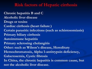
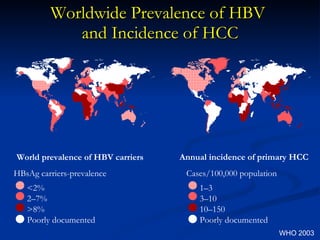
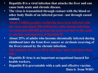
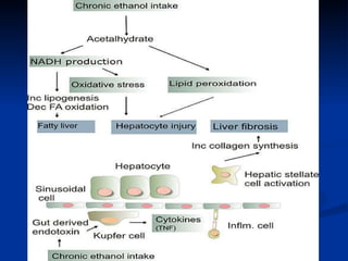
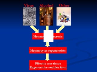
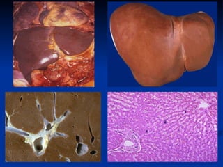
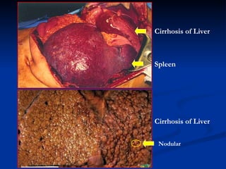
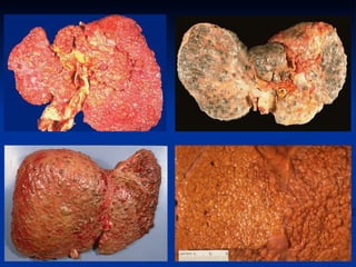
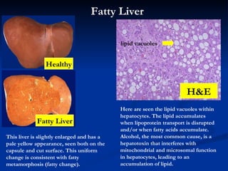
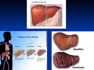
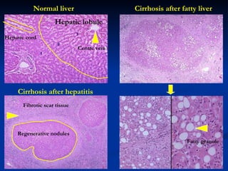
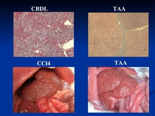
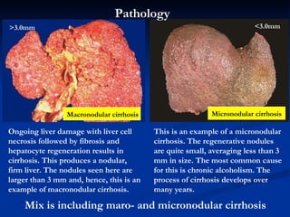
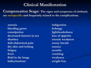
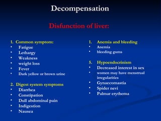
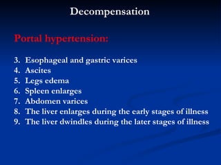
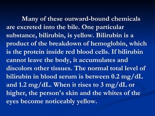
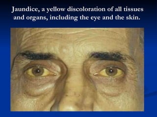
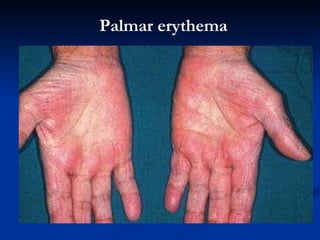
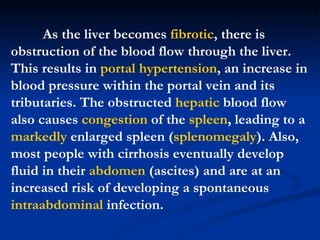
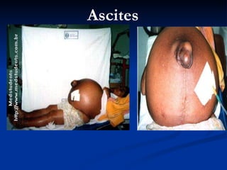
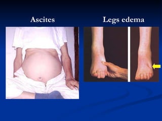
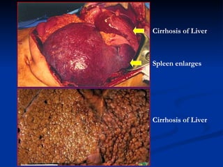
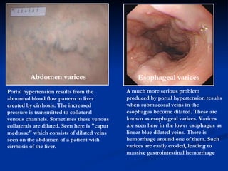
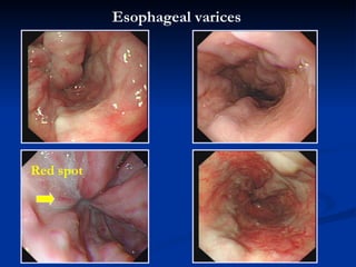
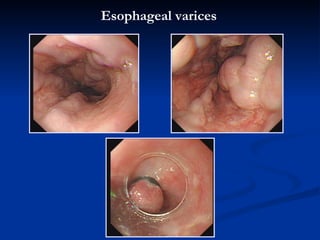
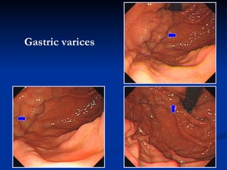
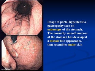
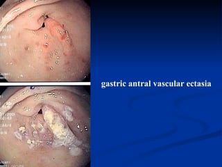
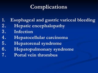
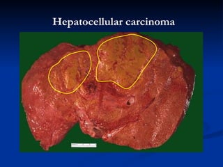
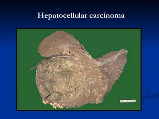
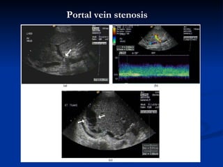
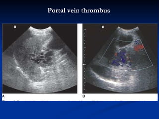
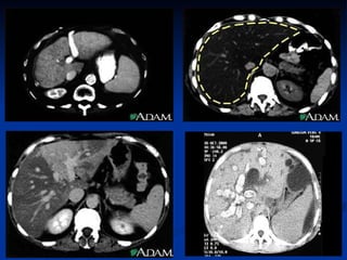
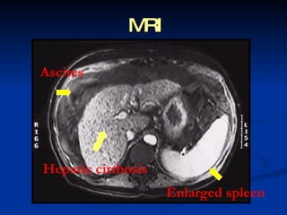
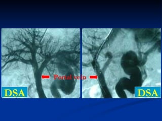
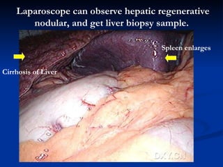
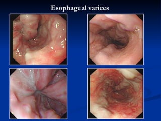
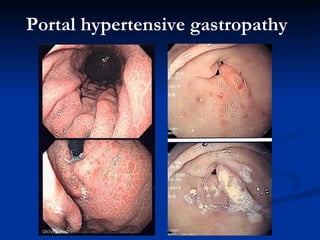
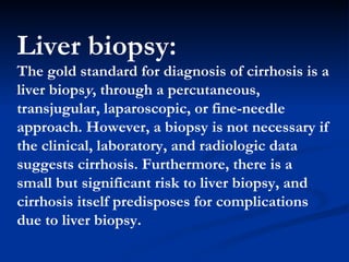
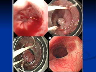
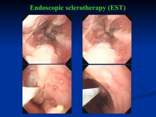
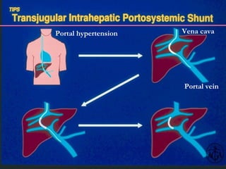
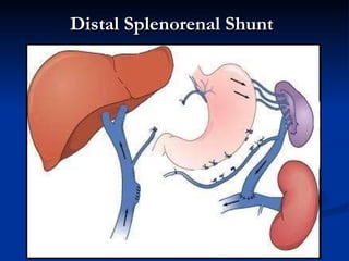
Hepatic Cirrhosis Bin Wu, MD & PhD Professor and Head Department of Gastroenterology  The Third Affiliated Hospital Sun Yat-Sen University
Aim:   Master:   Clinical Manifestation and  Diagnosis of Hepatic cirrhosis   Acquaint:   Distinguish Diagnosis and  Complication as well as Rule of  Treatment Know:   Causes and Risk Factors ,   Mechanism. Times:  2 hours
Definition  Hepatic c irrhosis  is a chronic, degenerative disease in which normal liver cells are damaged and are then replaced by scar tissue.
Description  Hepatic  Cirrhosis  changes the structure of the liver and the blood vessels that nourish it. The disease reduces the liver's ability to manufacture proteins and process hormones, nutrients, medications, and poisons.
Cirrhosis gets worse over time and can become potentially life  injury .  This disease can cause:   E xcessive bleeding (hemorrhage) I mpotence  L iver cancer  C oma due to accumulated ammonia   and body wastes (liver failure)  5.  D eath
Long-term alcoholism is the primary cause of cirrhosis in the United States. Men and women respond differently to alcohol. Although most men can safely consume two to five drinks a day, one to two drinks a day can cause liver damage in women. Individual tolerance to alcohol varies, but people who drink more and drink more often have a higher risk of developing cirrhosis. In some people, one drink a day can cause liver scarring.
Chronic liver infections, such as hepatitis B and particularly hepatitis C, are commonly linked to cirrhosis. People at high risk of contracting hepatitis B include those exposed to the virus through contact with blood and body fluids. This includes healthcare workers and intravenous (IV) drug users. In the past, people have contracted hepatitis C through blood transfusions. As of 2003, cirrhosis resulting from chronic hepatitis has emerged as a leading cause of death among HIV-positive patients; in Europe, about 30% of HIV-positive patients are coinfected with a hepatitis virus.
Liver injury, reactions to prescription medications, certain autoimmune disorders, exposure to toxic substances, and repeated episodes of heart failure with liver congestion can cause cirrhosis. A family history of diseases can genetically predispose a person to develop cirrhosis.
Cirrhosis is the seventh leading cause of disease   related death in the  United States . It is twice as common in men as in women. The disease occurs in more than half of all chronic alcoholics and kills about 25,000 peoples a year. It is the third most common cause of death in adults between the ages of 45 and 65.
Hepatitis  is the  first  leading cause of disease   related death in the  China .  So far, HBsAg +  peoples are about 1200 million (1, 200, 000, 000),  and peoples who are infected HCV are about 38  million   (38, 000, 000).
Types of cirrhosis  Portal or nutritional cirrhosis is the form of the disease most common in the United States. About 30–50% of all cases of cirrhosis are this type. Nine out of every 10 people who have nutritional cirrhosis have a history of  alcoholism .  Biliary cirrhosis  is caused by intrahepatic bile-duct diseases that impede bile flow. Bile is formed in the liver and is carried by ducts to the intestines. Bile then helps digest fats in the intestines. Biliary cirrhosis can scar or block these ducts. It represents 15–20% of all cirrhosis. Various types of chronic hepatitis, especially  hepatitis B  and  hepatitis C , can cause post-necrotic cirrhosis. This form of the disease affects up to 40% of all patients who have cirrhosis.
Chronic hepatitis  B and C Alcoholic liver disease Drugs or toxins Cardiac cirrhosis (heart failure   ) Certain parasitic infections (such as schistosomiasis) Primary biliary cirrhosis Autoimmune hepatitis Primary sclerosing cholangitis Other: such as  Wilson's disease ,  Hereditary Hemochromatosis ,  Alpha 1-antitrypsin deficiency ,  Galactosemia ,  Cystic fibrosis In China, the chronic hepatitis is common cause, but not the alcoholic liver disease. Risk factors of Hepatic cirrhosis
Worldwide Prevalence of HBV  and Incidence of HCC World prevalence of HBV carriers HBsAg carriers-prevalence <2% 2–7% >8% Poorly documented Annual incidence of primary HCC Cases/100,000 population 1–3 3–10 10–150 Poorly documented WHO 2003
Hepatitis B Related Death in China * >85% related to HBV infection He et al.  NEJM  2006 Per 100,000 person/yr 10 1.3 16.0 113 Women 8 1.7 26.7 193 Men 8 1.5 21.3 306 Total Chronic liver diseases* Rank order Percentage of total death Mortality No. of Deaths Cause of death
Hepatitis B is a viral infection that attacks the liver and can cause both acute and chronic disease.  The virus is transmitted through contact with the blood or other body fluids of an infected person - not through casual contact.  About 2 billion peoples worldwide have been infected with the virus and about 350 million live with chronic infection. An estimated 600 000 persons die each year due to the acute or chronic consequences of hepatitis B.   About 25% of adults who become chronically infected during childhood later die from liver cancer or cirrhosis (scarring of the liver) caused by the chronic infection.  The hepatitis B virus is 50 to 100 times more infectious than HIV.  Hepatitis B virus is an important occupational hazard for health workers.  Hepatitis B is preventable with a safe and effective vaccine.  Data is  from WHO
 
Hepatocytes necrosis Hepatocytes  regenerati on F ibrotic scar tissue R egenerative nodules  form Virus Alcohol Other
 
Cirrhosis of Liver Spleen Cirrhosis of Liver N odular
 
This liver is slightly enlarged and has a pale yellow appearance, seen both on the capsule and cut surface. This uniform change is consistent with fatty metamorphosis (fatty change). Here are seen the lipid vacuoles within hepatocytes. The lipid accumulates when lipoprotein transport is disrupted and/or when fatty acids accumulate. Alcohol, the most common cause, is a hepatotoxin that interferes with mitochondrial and microsomal function in hepatocytes, leading to an accumulation of lipid. Fatty Liver H&E Fatty Liver lipid vacuoles Healthy
 
Normal liver Cirrhosis after hepatitis Cirrhosis after fatty liver R egenerative nodules F ibrotic scar tissue Hepatic  lobule   Centre vein Fatty  granule H epatic cord
CBDL TAA CCl4 TAA
Pathology  Type of hepatic cirrhosis: M acronodular cirrhosis ,  ф >3.0 mm M icronodular cirrhosis ,  ф <3.0 mm Mix: Macro- +Micro-nodular cirrhosis
Ongoing liver damage with liver cell necrosis followed by fibrosis and hepatocyte regeneration results in cirrhosis. This produces a nodular, firm liver. The nodules seen here are larger than 3 mm and, hence, this is an example of macronodular cirrhosis.  This is an example of a micronodular cirrhosis. The regenerative nodules are quite small, averaging less than 3 mm in size. The most common cause for this is chronic alcoholism. The process of cirrhosis develops over many years.  M acronodular cirrhosis M icronodular cirrhosis >3.0mm <3.0mm Mix is including maro- and micronodular cirrhosis  Pathology
Clinical Manifestation   Compensation Stage:  The signs and symptoms of cirrhosis are  nonspecific  and frequently related to the complications.   anemia bleeding gums  constipation  decreased interest in sex  diarrhea  dull abdominal pain  dry skin and itching  fatigue  fever  fluid in the lungs  hallucinations  indigestion lethargy  lightheadedness  loss of appetite  muscle weakness  musty breath  nausea neuritis  vomiting  weakness  weight loss
Decompensation 1.  Common s ymptom :   Fatigue  Lethargy W eakness weight loss F ever D ark yellow or brown urine 2.  Digest system s ymptoms D iarrhea Constipation D ull abdominal pain I ndigestion N ausea A nemia  and bleeding Anemia bleeding gums   Hypoendocrinism D ecreased interest in sex women may have menstrual irregularities   Gynaecomastia   Spider nevi Palmar erythema Disfunction of liver:
P ortal hypertension :   Esophageal  and gastric  varices  A scites  L egs edema S pleen enlarges  A bdomen varices The liver enlarges during the early stages of illness  The liver  dwindles  during the  later  stages of illness  Decompensation
Jaundice  is a condition in which a person's skin and the whites of the eyes are discolored yellow due to an increased level of bile pigments in the blood resulting from liver disease. Jaundice is sometimes called icterus, from a Greek word for the condition.
Many of these outward-bound chemicals are excreted into the bile. One particular substance, bilirubin, is yellow. Bilirubin is a product of the breakdown of hemoglobin, which is the protein inside red blood cells. If bilirubin cannot leave the body, it accumulates and discolors other tissues. The normal total level of bilirubin in blood serum is between 0.2 mg/dL and 1.2 mg/dL. When it rises to 3 mg/dL or higher, the person's skin and the whites of the eyes become noticeably yellow.
J aundice ,  a yellow discoloration of all tissues and organs, including  the eye and  the skin.
After liver is damaged, the hepatocytes are not able to treat  estrogen , resulting in Spider nevi and Palmar erythema.
Spider nevi
Palmar erythema
As the liver becomes  fibrotic , there is obstruction of the blood flow through the liver. This results in  portal hypertension , an increase in blood pressure within the portal vein and its tributaries. The obstructed  hepatic  blood flow also causes  congestion  of the  spleen , leading to a  markedly  enlarged spleen ( splenomegaly ). Also, most people with cirrhosis eventually develop fluid in their  abdomen  (ascites) and are at an increased risk of developing a spontaneous  intraabdominal  infection.
A scites
A scites L egs edema
Cirrhosis of Liver Spleen  enlarges Cirrhosis of Liver
Portal hypertension results from the abnormal blood flow pattern in liver created by cirrhosis. The increased pressure is transmitted to collateral venous channels. Sometimes these venous collaterals are dilated. Seen here is &quot;caput medusae&quot; which consists of dilated veins seen on the abdomen of a patient with cirrhosis of the liver.  A bdomen varices Esophageal   varices A much more serious problem produced by portal hypertension results when submucosal veins in the esophagus become dilated. These are known as esophageal varices. Varices are seen here in the lower esophagus as linear blue dilated veins. There is hemorrhage around one of them. Such varices are easily eroded, leading to massive gastrointestinal hemorrhage
Esophageal   varices Red spot
Esophageal   varices
Gastric  varices
Image of portal hypertensive gastropathy seen on  endoscopy  of the stomach. The normally smooth mucosa of the stomach has developed a  mosaic  like appearance, that resembles  snake -skin
gastric antral vascular ectasia
Complications Esophageal  and gastric  varice al bleeding   Hepatic encephalopathy   I nfection   Hepatocellular carcinoma   Hepatorenal syndrome   Hepatopulmonary syndrome   P ortal vein  thrombus
Esophageal   varice al bleeding
Hepatocellular carcinoma
P ortal vein   stenosis
P ortal vein  thrombus
Lab findings  Aminotransferases  -  AST and ALT  are moderately elevated, with AST > ALT. However, normal aminotransferases do not preclude cirrhosis.  Alkaline  phosphatase  – ( AKP )usually slightly elevated.  GGT  -- correlates with AP levels. Typically much higher in chronic liver disease from alcohol.  Bilirubin   - may elevate as cirrhosis progresses.  Albumin  - levels fall as the synthetic function of the liver declines with worsening cirrhosis since albumin is exclusively synthesized in the liver  Prothrombin  time  - increases since the liver synthesizes clotting factors.  Globulins  - increased due to shunting of bacterial antigens away from the liver to lymphoid tissue.  Serum  sodium  -  hyponatremia  due to inability to excrete free water resulting from high levels of  ADH  and  aldosterone .  Thrombocytopenia  - due to both congestive splenomegaly as well as decreased  thrombopoietin  from the liver. However this rarely results in platelet count < 50,000/mL.  Leukopenia  and neutropenia  - due to splenomegaly with splenic margination.  Coagulation defects  - the liver produces most of the coagulation factors and thus coagulopathy correlates with worsening liver disease.
Other laboratory studies performed in newly diagnosed cirrhosis may include: Serology for  hepatitis  viruses,  autoantibodies  ( ANA , anti-smooth muscle,  anti-mitochondria , anti-LKM)  Ferritin  and  transferrin  saturation  (markers of iron overload),  copper  and  ceruloplasmin  (markers of copper overload)  Immunoglobulin levels (IgG, IgM, IgA) - these are non-specific but may assist in distinguishing various causes  Cholesterol  and  glucose   Alpha 1-antitrypsin
Imaging  Ultrasound   ,  CT and MRI are  routinely used in the evaluation of cirrhosis, where it may show a small and nodular liver in advanced cirrhosis along with increased echogenicity with irregular appearing areas. Ultrasound may also screen for hepatocellular carcinoma, portal hypertension and Budd-Chiari syndrome (by assessing flow in the hepatic vein).
 
MRI A scites Hepatic cirrhosis Enlarged spleen
DSA DSA Portal vein
Laparoscope can observe hepatic  regenerative nodular , and get liver biopsy sample.   Cirrhosis of Liver Spleen  enlarges
Endoscopy Gastroscopy  (endoscopic examination of the esophagus, stomach and duodenum) is performed in patients with established cirrhosis to exclude the possibility of esophageal varices. If these are found, prophylactic local therapy may be applied (sclerotherapy or banding) and beta blocker treatment may be commenced.
Esophageal   varices
Portal hypertensive gastropathy
Liver biopsy:  The gold standard for diagnosis of cirrhosis is a liver biops y , through a percutaneous, transjugular, laparoscopic, or fine-needle approach.   However, a biopsy is not necessary if the clinical, laboratory, and radiologic data suggests cirrhosis. Furthermore, there is a small but significant risk to liver biopsy, and cirrhosis itself predisposes for complications due to liver biopsy .
A patient's medical history can reveal illnesses or lifestyles likely to lead to cirrhosis. Liver changes can be seen during a physical examination. A doctor who suspects cirrhosis may order blood and urine tests to measure liver function. Because only a small number of healthy cells are needed to carry out essential liver functions, test results may be normal even when cirrhosis is present.  Diagnosis
In about 10 out of every 100 patients, the cause of cirrhosis cannot be determined. Many people who have cirrhosis do not have any symptoms (often called compensated cirrhosis). Their disease is detected during a routine physical or when tests for an unrelated medical problem are performed. This type of cirrhosis can also be detected when complications occur (decompensated cirrhosis).
Computed tomography scans (CT), ultrasound, and other imaging techniques can be used during diagnosis. They can help determine the size of the liver, indicate healthy and scarred areas of the organ, and detect gallstones. Cirrhosis is sometimes diagnosed during surgery or by examining the liver with a laparoscope. This viewing device is inserted into the patient's body through a tiny incision in the abdomen.
Liver biopsy is usually needed to confirm a diagnosis of cirrhosis. In this procedure, a tissue sample is removed from the liver and examined under a microscope in order to learn more about the organ's condition and to properly diagnose it.
A newer and less invasive test involves the measurement of  hyaluronic acid (HA)  in the patient's blood serum. As of 2003, however, the serum  HA  test is most useful in monitoring the progress of liver disease; it is unlikely to completely replace liver biopsy in the diagnosis of cirrhosis.
Prevention Eliminating alcohol abuse could prevent 75–80% of all cases of cirrhosis. Other preventive measures include: M aintaining a healthy diet that includes whole foods and grains, vegetable, and fruits  O btaining counseling or other treatment for alcoholism  T aking precautions ( practicing safe sex, avoiding dirty needles ) to prevent hepatitis  G etting immunizations against hepatitis if a person is in a high-risk group  receiving appropriate medical treatment quickly when diagnosed with hepatitis B or hepatitis C  H aving blood drawn at regular intervals to rid the body of excess iron from hemochromatosis  U sing medicines (chelating agents) to rid the body of excess copper from Wilson's disease  W earing protective clothing and following product directions when using toxic chemicals at work, at home, or in the garden
General treatment 1.  E tiological treatment  No drink alcoholic Treatment of HBV and HCV and so on P rotecting liver  treatment Treatment of ascites Treatment of jaundice
Common treatment: Rest, n utritional  and supportable t herapy . Treating underlying causes :  Alcoholic cirrhosis caused by alcohol abuse is treated by abstaining from alcohol. Treatment for hepatitis-related cirrhosis involves medications used to treat the different types of hepatitis, such as interferon for viral hepatitis and corticosteroids for autoimmune hepatitis. Cirrhosis caused by Wilson's disease, in which copper builds up in organs, is treated with chelation therapy (e.g. penicillamine) to remove the copper . Preventing further liver damage : Drug application of ameliorate liver function. Treatment of  complications : Please see other part. Surgery  treatment: Please see surgery Transplantation :  If complications cannot be controlled or when the liver ceases functioning, liver transplantation is necessary.
The therapy of cirrhosis is aimed primarily at preventing or reducing the complications. Bleeding esophageal  varices  (collateral  venous  channels) are a frequent serious complication of cirrhosis. Various techniques are used to control the bleeding. In some individuals with severe portal  hypertension , vascular shunts are made to reduce the pressure in the portal vein by bypassing the liver. Most frequently the portal vein is surgically connected to the  inferior vena cava  so that some of the blood in the portal vein does not pass through the liver.  Treatment
E ndoscope band ligation  ( EBL ) can prevent and treat  Esophageal  and gastric  varices  or with bleeding
 
Ref:  De Franchis R. Digestive and liver disease 2004;36(S1):S93 Endoscopic sclerotherapy (EST) is an established method for controlling and preventing bleeding from oesophageal varices
Endoscopic sclerotherapy (EST)
AT Injection
P ortal hypertension Portal vein V ena cava
Distal Splenorenal Shunt
Harvard University
Harvard University
Harvard University
Harvard Medical School
The Third Affiliated Hospital Sun Yat-Sen University
Position Open Postdoctoral Fellow Ph.D. Program  Master’s Program
Thank you

19 hepatic cirrhosis

  • 1.
    Hepatic Cirrhosis BinWu, MD & PhD Professor and Head Department of Gastroenterology The Third Affiliated Hospital Sun Yat-Sen University
  • 2.
    Aim: Master: Clinical Manifestation and Diagnosis of Hepatic cirrhosis Acquaint: Distinguish Diagnosis and Complication as well as Rule of Treatment Know: Causes and Risk Factors , Mechanism. Times: 2 hours
  • 3.
    Definition Hepaticc irrhosis is a chronic, degenerative disease in which normal liver cells are damaged and are then replaced by scar tissue.
  • 4.
    Description Hepatic Cirrhosis changes the structure of the liver and the blood vessels that nourish it. The disease reduces the liver's ability to manufacture proteins and process hormones, nutrients, medications, and poisons.
  • 5.
    Cirrhosis gets worseover time and can become potentially life injury . This disease can cause: E xcessive bleeding (hemorrhage) I mpotence L iver cancer C oma due to accumulated ammonia and body wastes (liver failure) 5. D eath
  • 6.
    Long-term alcoholism isthe primary cause of cirrhosis in the United States. Men and women respond differently to alcohol. Although most men can safely consume two to five drinks a day, one to two drinks a day can cause liver damage in women. Individual tolerance to alcohol varies, but people who drink more and drink more often have a higher risk of developing cirrhosis. In some people, one drink a day can cause liver scarring.
  • 7.
    Chronic liver infections,such as hepatitis B and particularly hepatitis C, are commonly linked to cirrhosis. People at high risk of contracting hepatitis B include those exposed to the virus through contact with blood and body fluids. This includes healthcare workers and intravenous (IV) drug users. In the past, people have contracted hepatitis C through blood transfusions. As of 2003, cirrhosis resulting from chronic hepatitis has emerged as a leading cause of death among HIV-positive patients; in Europe, about 30% of HIV-positive patients are coinfected with a hepatitis virus.
  • 8.
    Liver injury, reactionsto prescription medications, certain autoimmune disorders, exposure to toxic substances, and repeated episodes of heart failure with liver congestion can cause cirrhosis. A family history of diseases can genetically predispose a person to develop cirrhosis.
  • 9.
    Cirrhosis is theseventh leading cause of disease related death in the United States . It is twice as common in men as in women. The disease occurs in more than half of all chronic alcoholics and kills about 25,000 peoples a year. It is the third most common cause of death in adults between the ages of 45 and 65.
  • 10.
    Hepatitis isthe first leading cause of disease related death in the China . So far, HBsAg + peoples are about 1200 million (1, 200, 000, 000), and peoples who are infected HCV are about 38 million (38, 000, 000).
  • 11.
    Types of cirrhosis Portal or nutritional cirrhosis is the form of the disease most common in the United States. About 30–50% of all cases of cirrhosis are this type. Nine out of every 10 people who have nutritional cirrhosis have a history of alcoholism . Biliary cirrhosis is caused by intrahepatic bile-duct diseases that impede bile flow. Bile is formed in the liver and is carried by ducts to the intestines. Bile then helps digest fats in the intestines. Biliary cirrhosis can scar or block these ducts. It represents 15–20% of all cirrhosis. Various types of chronic hepatitis, especially hepatitis B and hepatitis C , can cause post-necrotic cirrhosis. This form of the disease affects up to 40% of all patients who have cirrhosis.
  • 12.
    Chronic hepatitis B and C Alcoholic liver disease Drugs or toxins Cardiac cirrhosis (heart failure ) Certain parasitic infections (such as schistosomiasis) Primary biliary cirrhosis Autoimmune hepatitis Primary sclerosing cholangitis Other: such as Wilson's disease , Hereditary Hemochromatosis , Alpha 1-antitrypsin deficiency , Galactosemia , Cystic fibrosis In China, the chronic hepatitis is common cause, but not the alcoholic liver disease. Risk factors of Hepatic cirrhosis
  • 13.
    Worldwide Prevalence ofHBV and Incidence of HCC World prevalence of HBV carriers HBsAg carriers-prevalence <2% 2–7% >8% Poorly documented Annual incidence of primary HCC Cases/100,000 population 1–3 3–10 10–150 Poorly documented WHO 2003
  • 14.
    Hepatitis B RelatedDeath in China * >85% related to HBV infection He et al. NEJM 2006 Per 100,000 person/yr 10 1.3 16.0 113 Women 8 1.7 26.7 193 Men 8 1.5 21.3 306 Total Chronic liver diseases* Rank order Percentage of total death Mortality No. of Deaths Cause of death
  • 15.
    Hepatitis B isa viral infection that attacks the liver and can cause both acute and chronic disease. The virus is transmitted through contact with the blood or other body fluids of an infected person - not through casual contact. About 2 billion peoples worldwide have been infected with the virus and about 350 million live with chronic infection. An estimated 600 000 persons die each year due to the acute or chronic consequences of hepatitis B. About 25% of adults who become chronically infected during childhood later die from liver cancer or cirrhosis (scarring of the liver) caused by the chronic infection. The hepatitis B virus is 50 to 100 times more infectious than HIV. Hepatitis B virus is an important occupational hazard for health workers. Hepatitis B is preventable with a safe and effective vaccine. Data is from WHO
  • 16.
  • 17.
    Hepatocytes necrosis Hepatocytes regenerati on F ibrotic scar tissue R egenerative nodules form Virus Alcohol Other
  • 18.
  • 19.
    Cirrhosis of LiverSpleen Cirrhosis of Liver N odular
  • 20.
  • 21.
    This liver isslightly enlarged and has a pale yellow appearance, seen both on the capsule and cut surface. This uniform change is consistent with fatty metamorphosis (fatty change). Here are seen the lipid vacuoles within hepatocytes. The lipid accumulates when lipoprotein transport is disrupted and/or when fatty acids accumulate. Alcohol, the most common cause, is a hepatotoxin that interferes with mitochondrial and microsomal function in hepatocytes, leading to an accumulation of lipid. Fatty Liver H&E Fatty Liver lipid vacuoles Healthy
  • 22.
  • 23.
    Normal liver Cirrhosisafter hepatitis Cirrhosis after fatty liver R egenerative nodules F ibrotic scar tissue Hepatic lobule Centre vein Fatty granule H epatic cord
  • 24.
  • 25.
    Pathology Typeof hepatic cirrhosis: M acronodular cirrhosis , ф >3.0 mm M icronodular cirrhosis , ф <3.0 mm Mix: Macro- +Micro-nodular cirrhosis
  • 26.
    Ongoing liver damagewith liver cell necrosis followed by fibrosis and hepatocyte regeneration results in cirrhosis. This produces a nodular, firm liver. The nodules seen here are larger than 3 mm and, hence, this is an example of macronodular cirrhosis. This is an example of a micronodular cirrhosis. The regenerative nodules are quite small, averaging less than 3 mm in size. The most common cause for this is chronic alcoholism. The process of cirrhosis develops over many years. M acronodular cirrhosis M icronodular cirrhosis >3.0mm <3.0mm Mix is including maro- and micronodular cirrhosis Pathology
  • 27.
    Clinical Manifestation Compensation Stage: The signs and symptoms of cirrhosis are nonspecific and frequently related to the complications. anemia bleeding gums constipation decreased interest in sex diarrhea dull abdominal pain dry skin and itching fatigue fever fluid in the lungs hallucinations indigestion lethargy lightheadedness loss of appetite muscle weakness musty breath nausea neuritis vomiting weakness weight loss
  • 28.
    Decompensation 1. Common s ymptom : Fatigue Lethargy W eakness weight loss F ever D ark yellow or brown urine 2. Digest system s ymptoms D iarrhea Constipation D ull abdominal pain I ndigestion N ausea A nemia and bleeding Anemia bleeding gums Hypoendocrinism D ecreased interest in sex women may have menstrual irregularities Gynaecomastia Spider nevi Palmar erythema Disfunction of liver:
  • 29.
    P ortal hypertension: Esophageal and gastric varices A scites L egs edema S pleen enlarges A bdomen varices The liver enlarges during the early stages of illness The liver dwindles during the later stages of illness Decompensation
  • 30.
    Jaundice isa condition in which a person's skin and the whites of the eyes are discolored yellow due to an increased level of bile pigments in the blood resulting from liver disease. Jaundice is sometimes called icterus, from a Greek word for the condition.
  • 31.
    Many of theseoutward-bound chemicals are excreted into the bile. One particular substance, bilirubin, is yellow. Bilirubin is a product of the breakdown of hemoglobin, which is the protein inside red blood cells. If bilirubin cannot leave the body, it accumulates and discolors other tissues. The normal total level of bilirubin in blood serum is between 0.2 mg/dL and 1.2 mg/dL. When it rises to 3 mg/dL or higher, the person's skin and the whites of the eyes become noticeably yellow.
  • 32.
    J aundice , a yellow discoloration of all tissues and organs, including the eye and the skin.
  • 33.
    After liver isdamaged, the hepatocytes are not able to treat estrogen , resulting in Spider nevi and Palmar erythema.
  • 34.
  • 35.
  • 36.
    As the liverbecomes fibrotic , there is obstruction of the blood flow through the liver. This results in portal hypertension , an increase in blood pressure within the portal vein and its tributaries. The obstructed hepatic blood flow also causes congestion of the spleen , leading to a markedly enlarged spleen ( splenomegaly ). Also, most people with cirrhosis eventually develop fluid in their abdomen (ascites) and are at an increased risk of developing a spontaneous intraabdominal infection.
  • 37.
  • 38.
    A scites Legs edema
  • 39.
    Cirrhosis of LiverSpleen enlarges Cirrhosis of Liver
  • 40.
    Portal hypertension resultsfrom the abnormal blood flow pattern in liver created by cirrhosis. The increased pressure is transmitted to collateral venous channels. Sometimes these venous collaterals are dilated. Seen here is &quot;caput medusae&quot; which consists of dilated veins seen on the abdomen of a patient with cirrhosis of the liver. A bdomen varices Esophageal varices A much more serious problem produced by portal hypertension results when submucosal veins in the esophagus become dilated. These are known as esophageal varices. Varices are seen here in the lower esophagus as linear blue dilated veins. There is hemorrhage around one of them. Such varices are easily eroded, leading to massive gastrointestinal hemorrhage
  • 41.
    Esophageal varices Red spot
  • 42.
    Esophageal varices
  • 43.
  • 44.
    Image of portalhypertensive gastropathy seen on endoscopy of the stomach. The normally smooth mucosa of the stomach has developed a mosaic like appearance, that resembles snake -skin
  • 45.
  • 46.
    Complications Esophageal and gastric varice al bleeding   Hepatic encephalopathy   I nfection   Hepatocellular carcinoma   Hepatorenal syndrome   Hepatopulmonary syndrome   P ortal vein thrombus
  • 47.
    Esophageal varice al bleeding
  • 48.
  • 49.
  • 50.
    P ortal vein stenosis
  • 51.
    P ortal vein thrombus
  • 52.
    Lab findings Aminotransferases - AST and ALT are moderately elevated, with AST > ALT. However, normal aminotransferases do not preclude cirrhosis. Alkaline phosphatase – ( AKP )usually slightly elevated. GGT -- correlates with AP levels. Typically much higher in chronic liver disease from alcohol. Bilirubin - may elevate as cirrhosis progresses. Albumin - levels fall as the synthetic function of the liver declines with worsening cirrhosis since albumin is exclusively synthesized in the liver Prothrombin time - increases since the liver synthesizes clotting factors. Globulins - increased due to shunting of bacterial antigens away from the liver to lymphoid tissue. Serum sodium - hyponatremia due to inability to excrete free water resulting from high levels of ADH and aldosterone . Thrombocytopenia - due to both congestive splenomegaly as well as decreased thrombopoietin from the liver. However this rarely results in platelet count < 50,000/mL. Leukopenia and neutropenia - due to splenomegaly with splenic margination. Coagulation defects - the liver produces most of the coagulation factors and thus coagulopathy correlates with worsening liver disease.
  • 53.
    Other laboratory studiesperformed in newly diagnosed cirrhosis may include: Serology for hepatitis viruses, autoantibodies ( ANA , anti-smooth muscle, anti-mitochondria , anti-LKM) Ferritin and transferrin saturation (markers of iron overload), copper and ceruloplasmin (markers of copper overload) Immunoglobulin levels (IgG, IgM, IgA) - these are non-specific but may assist in distinguishing various causes Cholesterol and glucose Alpha 1-antitrypsin
  • 54.
    Imaging Ultrasound , CT and MRI are routinely used in the evaluation of cirrhosis, where it may show a small and nodular liver in advanced cirrhosis along with increased echogenicity with irregular appearing areas. Ultrasound may also screen for hepatocellular carcinoma, portal hypertension and Budd-Chiari syndrome (by assessing flow in the hepatic vein).
  • 55.
  • 56.
    MRI A scitesHepatic cirrhosis Enlarged spleen
  • 57.
  • 58.
    Laparoscope can observehepatic regenerative nodular , and get liver biopsy sample. Cirrhosis of Liver Spleen enlarges
  • 59.
    Endoscopy Gastroscopy (endoscopic examination of the esophagus, stomach and duodenum) is performed in patients with established cirrhosis to exclude the possibility of esophageal varices. If these are found, prophylactic local therapy may be applied (sclerotherapy or banding) and beta blocker treatment may be commenced.
  • 60.
    Esophageal varices
  • 61.
  • 62.
    Liver biopsy: The gold standard for diagnosis of cirrhosis is a liver biops y , through a percutaneous, transjugular, laparoscopic, or fine-needle approach. However, a biopsy is not necessary if the clinical, laboratory, and radiologic data suggests cirrhosis. Furthermore, there is a small but significant risk to liver biopsy, and cirrhosis itself predisposes for complications due to liver biopsy .
  • 63.
    A patient's medicalhistory can reveal illnesses or lifestyles likely to lead to cirrhosis. Liver changes can be seen during a physical examination. A doctor who suspects cirrhosis may order blood and urine tests to measure liver function. Because only a small number of healthy cells are needed to carry out essential liver functions, test results may be normal even when cirrhosis is present. Diagnosis
  • 64.
    In about 10out of every 100 patients, the cause of cirrhosis cannot be determined. Many people who have cirrhosis do not have any symptoms (often called compensated cirrhosis). Their disease is detected during a routine physical or when tests for an unrelated medical problem are performed. This type of cirrhosis can also be detected when complications occur (decompensated cirrhosis).
  • 65.
    Computed tomography scans(CT), ultrasound, and other imaging techniques can be used during diagnosis. They can help determine the size of the liver, indicate healthy and scarred areas of the organ, and detect gallstones. Cirrhosis is sometimes diagnosed during surgery or by examining the liver with a laparoscope. This viewing device is inserted into the patient's body through a tiny incision in the abdomen.
  • 66.
    Liver biopsy isusually needed to confirm a diagnosis of cirrhosis. In this procedure, a tissue sample is removed from the liver and examined under a microscope in order to learn more about the organ's condition and to properly diagnose it.
  • 67.
    A newer andless invasive test involves the measurement of hyaluronic acid (HA) in the patient's blood serum. As of 2003, however, the serum HA test is most useful in monitoring the progress of liver disease; it is unlikely to completely replace liver biopsy in the diagnosis of cirrhosis.
  • 68.
    Prevention Eliminating alcoholabuse could prevent 75–80% of all cases of cirrhosis. Other preventive measures include: M aintaining a healthy diet that includes whole foods and grains, vegetable, and fruits O btaining counseling or other treatment for alcoholism T aking precautions ( practicing safe sex, avoiding dirty needles ) to prevent hepatitis G etting immunizations against hepatitis if a person is in a high-risk group receiving appropriate medical treatment quickly when diagnosed with hepatitis B or hepatitis C H aving blood drawn at regular intervals to rid the body of excess iron from hemochromatosis U sing medicines (chelating agents) to rid the body of excess copper from Wilson's disease W earing protective clothing and following product directions when using toxic chemicals at work, at home, or in the garden
  • 69.
    General treatment 1. E tiological treatment No drink alcoholic Treatment of HBV and HCV and so on P rotecting liver treatment Treatment of ascites Treatment of jaundice
  • 70.
    Common treatment: Rest,n utritional and supportable t herapy . Treating underlying causes : Alcoholic cirrhosis caused by alcohol abuse is treated by abstaining from alcohol. Treatment for hepatitis-related cirrhosis involves medications used to treat the different types of hepatitis, such as interferon for viral hepatitis and corticosteroids for autoimmune hepatitis. Cirrhosis caused by Wilson's disease, in which copper builds up in organs, is treated with chelation therapy (e.g. penicillamine) to remove the copper . Preventing further liver damage : Drug application of ameliorate liver function. Treatment of complications : Please see other part. Surgery treatment: Please see surgery Transplantation : If complications cannot be controlled or when the liver ceases functioning, liver transplantation is necessary.
  • 71.
    The therapy ofcirrhosis is aimed primarily at preventing or reducing the complications. Bleeding esophageal varices (collateral venous channels) are a frequent serious complication of cirrhosis. Various techniques are used to control the bleeding. In some individuals with severe portal hypertension , vascular shunts are made to reduce the pressure in the portal vein by bypassing the liver. Most frequently the portal vein is surgically connected to the inferior vena cava so that some of the blood in the portal vein does not pass through the liver. Treatment
  • 72.
    E ndoscope bandligation ( EBL ) can prevent and treat Esophageal and gastric varices or with bleeding
  • 73.
  • 74.
    Ref: DeFranchis R. Digestive and liver disease 2004;36(S1):S93 Endoscopic sclerotherapy (EST) is an established method for controlling and preventing bleeding from oesophageal varices
  • 75.
  • 76.
  • 77.
    P ortal hypertensionPortal vein V ena cava
  • 78.
  • 79.
  • 80.
  • 81.
  • 82.
  • 83.
    The Third AffiliatedHospital Sun Yat-Sen University
  • 84.
    Position Open PostdoctoralFellow Ph.D. Program Master’s Program
  • 85.