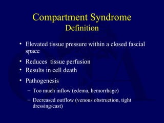
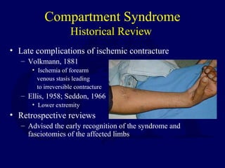
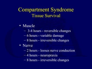
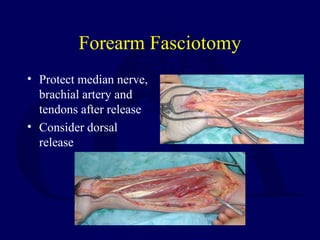
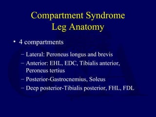
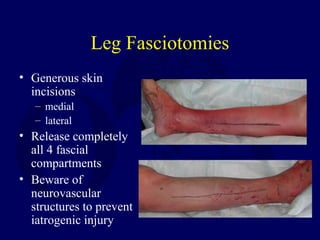
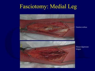
Compartment
Syndromes
Robert M. Harris, MD
Compartment Syndrome
Definition
• Elevated tissue pressure within a closed fascial
space
• Reduces tissue perfusion
• Results in cell death
• Pathogenesis
– Too much inflow (edema, hemorrhage)
– Decreased outflow (venous obstruction, tight
dressing/cast)
Compartment Syndrome
Historical Review
• Late complications of ischemic contracture
– Volkmann, 1881
• Ischemia of forearm
venous stasis leading
to irreversible contracture
– Ellis, 1958; Seddon, 1966
• Lower extremity
• Retrospective reviews
– Advised the early recognition of the syndrome and
fasciotomies of the affected limbs
Compartment Syndrome
Pathophysiology
• Normal tissue pressure
– 0-4 mm Hg
– 8-10 with exertion
• Absolute pressure theory
– 30 mm Hg - Mubarak
– 45 mm Hg - Matsen
• Pressure gradient theory
– < 20 mm Hg of diastolic pressure – Whitesides
– McQueen, et al
Compartment Syndrome
Tissue Survival
• Muscle
– 3-4 hours - reversible changes
– 6 hours - variable damage
– 8 hours - irreversible changes
• Nerve
– 2 hours - looses nerve conduction
– 4 hours - neuropraxia
– 8 hours - irreversible changes
Compartment Syndrome
Etiology
• Fractures-closed and open
• Blunt trauma
• Temp vascular
occlusion
• Cast/dressing
• Closure of fascial
defects
• Burns/electrical
• Exertional states
• GSW
• IV/A-lines
• Hemophiliac/coag
• Intraosseous IV(infant)
• Snake bite
• Arterial injury
Compartment Syndrome
Diagnosis
• Pain out of proportion
• Palpably tense compartment
• Pain with passive stretch
• Paresthesia/hypoesthesia
• Paralysis
• Pulselessness/pallor
Compartment Syndrome
Differential diagnosis
• Arterial occlusion
• Peripheral nerve injury
• Muscle rupture
Compartment Syndrome
Pressure Measurements
• Suspected compartment syndrome
• Equivocal or unreliable exam
• Clinical adjunct
• Contraindication
– Clinically evident compartment
syndrome
Compartment Syndrome
Pressure Measurements
• Infusion
– manometer
– saline
– 3-way stopcock
(Whitesides, CORR
1975)
• Catheter
– wick
– slit wick
• Arterial line
– 16 - 18 ga. Needle
(5-19 mm Hg higher)
– transducer
– monitor
• Stryker device
– Side port needle
Compartment Syndrome
Pressure Measurements
• Arterial line
– Zero at the
level of the
affected limb
Compartment Syndrome
Pressure Measurements
• Needle
– 18 gauge
– Side ported
• Catheter
– wick
– slit wick
• Performed within 5
cm of the injury if
possible-
Whitesides,
Heckman Side port
Compartment Syndrome
Emergent Treatment
• Remove cast or dressing
• Place at level of heart
(DO NOT ELEVATE to optimize
perfusion)
• Alert OR and Anesthesia
• Bedside procedure
• Medical treatment
Compartment Syndrome
Surgical Treatment
• Fasciotomy - prophylactic release
of pressure before permanent
damage occurs. Will not reverse
injury from trauma.
• Fracture care – rigid
stabilization
– Ex-fix
– IM Nail
Compartment Syndrome
Indications for Fasciotomy
• Unequivocal clinical findings
• Pressure within 15-20 mm hg of DBP
• Rising tissue pressure
• Significant tissue injury or high risk pt
• > 6 hours of total limb ischemia
• Injury at high risk of compartment
syndrome
• CONTRAINDICATION - Missed CS (>24-
48 hrs)
Fasciotomy Principles
• Make early diagnosis
• Long extensile incisions
• Release all fascial compartments
• Preserve neurovascular structures
• Debride necrotic tissues
• Coverage within 7-10 days
Compartment Syndrome
Forearm Anatomy
• Anatomy-3 compartments
– Mobile wad-BR,ECRL,ECRB
– Volar-Superficial and deep flexors, Pronator
teres, Supinator
– Dorsal-Extensors
Forearm Fasciotomy
• Volar-Henry approach
– Include a carpal tunnel
release
• Release lacertus
fibrosus and fascia
Forearm Fasciotomy
• Protect median nerve,
brachial artery and
tendons after release
• Consider dorsal
release
Compartment Syndrome
Leg Anatomy
• 4 compartments
– Lateral: Peroneus longus and brevis
– Anterior: EHL, EDC, Tibialis anterior,
Peroneus tertius
– Posterior-Gastrocnemius, Soleus
– Deep posterior-Tibialis posterior, FHL, FDL
Leg Fasciotomies
• Generous skin
incisions
– medial
– lateral
• Release completely
all 4 fascial
compartments
• Beware of
neurovascular
structures to prevent
iatrogenic injury
Fasciotomy: Medial Leg
Flexor digitorum
longus
Gastroc-soleus
Fasciotomy: Lateral Leg
Superficial peroneal nerve
Intermuscular septum
Compartment Syndrome
Thigh
• Lateral to release
anterior and
posterior
compartments
• May require medial
incision for
adductor
compartment
Lateral septum
Vastus lateralis
Compartment Syndrome
Foot
• Four major compartments
• Multiple layers
• Careful exam with any swelling
• Clinical suspicion with certain mechanisms
of injury
– Lisfranc fracture dislocation
– Calcaneus fracture
Compartment Syndrome
Foot Fasciotomies
• Dorsal incision-to
release the
interosseous, central
and lateral
compartments
• Medial incision-to
release the medial
compartment
Compartment Syndrome
Other Areas
• Can occur anywhere in the body
• Hand-dorsal incisions, thenar, hypothenar
• Arm-lateral incision
• Buttock-posterior (Kocher) approach
• Abdominal- with the Trauma surgeons
Interim Coverage Techniques
• Simple absorbent dressing
• Semipermeable skin-like
membrane
• Vessel loop “bootlace”
• Sutures progressively
tightened in ensuing days
• “VAC” (Vacuum Assisted
Closure)
Aftercare
• Wound is not closed at initial surgery
• Second look debridement with consideration for
coverage after 48-72 hrs
– Limb should not be at risk for further swelling
– Pt should be adequately stabilized
– Usually requires skin graft
– DPC possible if residual swelling is minimal
• Goal is to obtain definitive coverage within 7-10
days
– DPC/STSG/flap if nerves, vessels, bone exposed
Compartment Syndrome
Medical-Legal
• Most frequent cause of litigation
• 1992-average award $225K-Hennepin
County, Minn.
• Medical health care providers
• Lawyers
• Mass media
Questions
Return to
General Index

G04 compartment syndrome

  • 1.
  • 2.
    Compartment Syndrome Definition • Elevatedtissue pressure within a closed fascial space • Reduces tissue perfusion • Results in cell death • Pathogenesis – Too much inflow (edema, hemorrhage) – Decreased outflow (venous obstruction, tight dressing/cast)
  • 3.
    Compartment Syndrome Historical Review •Late complications of ischemic contracture – Volkmann, 1881 • Ischemia of forearm venous stasis leading to irreversible contracture – Ellis, 1958; Seddon, 1966 • Lower extremity • Retrospective reviews – Advised the early recognition of the syndrome and fasciotomies of the affected limbs
  • 4.
    Compartment Syndrome Pathophysiology • Normaltissue pressure – 0-4 mm Hg – 8-10 with exertion • Absolute pressure theory – 30 mm Hg - Mubarak – 45 mm Hg - Matsen • Pressure gradient theory – < 20 mm Hg of diastolic pressure – Whitesides – McQueen, et al
  • 5.
    Compartment Syndrome Tissue Survival •Muscle – 3-4 hours - reversible changes – 6 hours - variable damage – 8 hours - irreversible changes • Nerve – 2 hours - looses nerve conduction – 4 hours - neuropraxia – 8 hours - irreversible changes
  • 6.
    Compartment Syndrome Etiology • Fractures-closedand open • Blunt trauma • Temp vascular occlusion • Cast/dressing • Closure of fascial defects • Burns/electrical • Exertional states • GSW • IV/A-lines • Hemophiliac/coag • Intraosseous IV(infant) • Snake bite • Arterial injury
  • 7.
    Compartment Syndrome Diagnosis • Painout of proportion • Palpably tense compartment • Pain with passive stretch • Paresthesia/hypoesthesia • Paralysis • Pulselessness/pallor
  • 8.
    Compartment Syndrome Differential diagnosis •Arterial occlusion • Peripheral nerve injury • Muscle rupture
  • 9.
    Compartment Syndrome Pressure Measurements •Suspected compartment syndrome • Equivocal or unreliable exam • Clinical adjunct • Contraindication – Clinically evident compartment syndrome
  • 10.
    Compartment Syndrome Pressure Measurements •Infusion – manometer – saline – 3-way stopcock (Whitesides, CORR 1975) • Catheter – wick – slit wick • Arterial line – 16 - 18 ga. Needle (5-19 mm Hg higher) – transducer – monitor • Stryker device – Side port needle
  • 11.
    Compartment Syndrome Pressure Measurements •Arterial line – Zero at the level of the affected limb
  • 12.
    Compartment Syndrome Pressure Measurements •Needle – 18 gauge – Side ported • Catheter – wick – slit wick • Performed within 5 cm of the injury if possible- Whitesides, Heckman Side port
  • 13.
    Compartment Syndrome Emergent Treatment •Remove cast or dressing • Place at level of heart (DO NOT ELEVATE to optimize perfusion) • Alert OR and Anesthesia • Bedside procedure • Medical treatment
  • 14.
    Compartment Syndrome Surgical Treatment •Fasciotomy - prophylactic release of pressure before permanent damage occurs. Will not reverse injury from trauma. • Fracture care – rigid stabilization – Ex-fix – IM Nail
  • 15.
    Compartment Syndrome Indications forFasciotomy • Unequivocal clinical findings • Pressure within 15-20 mm hg of DBP • Rising tissue pressure • Significant tissue injury or high risk pt • > 6 hours of total limb ischemia • Injury at high risk of compartment syndrome • CONTRAINDICATION - Missed CS (>24- 48 hrs)
  • 16.
    Fasciotomy Principles • Makeearly diagnosis • Long extensile incisions • Release all fascial compartments • Preserve neurovascular structures • Debride necrotic tissues • Coverage within 7-10 days
  • 17.
    Compartment Syndrome Forearm Anatomy •Anatomy-3 compartments – Mobile wad-BR,ECRL,ECRB – Volar-Superficial and deep flexors, Pronator teres, Supinator – Dorsal-Extensors
  • 18.
    Forearm Fasciotomy • Volar-Henryapproach – Include a carpal tunnel release • Release lacertus fibrosus and fascia
  • 19.
    Forearm Fasciotomy • Protectmedian nerve, brachial artery and tendons after release • Consider dorsal release
  • 20.
    Compartment Syndrome Leg Anatomy •4 compartments – Lateral: Peroneus longus and brevis – Anterior: EHL, EDC, Tibialis anterior, Peroneus tertius – Posterior-Gastrocnemius, Soleus – Deep posterior-Tibialis posterior, FHL, FDL
  • 21.
    Leg Fasciotomies • Generousskin incisions – medial – lateral • Release completely all 4 fascial compartments • Beware of neurovascular structures to prevent iatrogenic injury
  • 22.
    Fasciotomy: Medial Leg Flexordigitorum longus Gastroc-soleus
  • 23.
    Fasciotomy: Lateral Leg Superficialperoneal nerve Intermuscular septum
  • 24.
    Compartment Syndrome Thigh • Lateralto release anterior and posterior compartments • May require medial incision for adductor compartment Lateral septum Vastus lateralis
  • 25.
    Compartment Syndrome Foot • Fourmajor compartments • Multiple layers • Careful exam with any swelling • Clinical suspicion with certain mechanisms of injury – Lisfranc fracture dislocation – Calcaneus fracture
  • 26.
    Compartment Syndrome Foot Fasciotomies •Dorsal incision-to release the interosseous, central and lateral compartments • Medial incision-to release the medial compartment
  • 27.
    Compartment Syndrome Other Areas •Can occur anywhere in the body • Hand-dorsal incisions, thenar, hypothenar • Arm-lateral incision • Buttock-posterior (Kocher) approach • Abdominal- with the Trauma surgeons
  • 28.
    Interim Coverage Techniques •Simple absorbent dressing • Semipermeable skin-like membrane • Vessel loop “bootlace” • Sutures progressively tightened in ensuing days • “VAC” (Vacuum Assisted Closure)
  • 29.
    Aftercare • Wound isnot closed at initial surgery • Second look debridement with consideration for coverage after 48-72 hrs – Limb should not be at risk for further swelling – Pt should be adequately stabilized – Usually requires skin graft – DPC possible if residual swelling is minimal • Goal is to obtain definitive coverage within 7-10 days – DPC/STSG/flap if nerves, vessels, bone exposed
  • 30.
    Compartment Syndrome Medical-Legal • Mostfrequent cause of litigation • 1992-average award $225K-Hennepin County, Minn. • Medical health care providers • Lawyers • Mass media
  • 31.

Editor's Notes

  • #4 The classic descriptions of late complications of ischemic contractures of the extremities due to compartment syndrome are described in the retrospective reviews of Seddon, Owen and Tsimboukis. They recognized the need for early recognition, surgical decompression and found the classic signs of physical exam were unreliable in compartment syndrome. Volkmann is credited with the description of ischemic contracture of the upper extremity associated with compartment syndrome without treatment
  • #5 Normal resting muscle tissue pressure is up to 4 mm Hg and 8-10 with exertion. Exercise induced CS may have a resting based line of 10-15 mmHg. Many studies utilizing clinical evaluations and animal models by Whitesides, Mubarak, Matsen, Heckman, Heppenstall and Matava have help to establish a better understanding of the pathophysiology and thresholds of ischemia. Two schools of though prevail: 1. The Absolute Pressure Theory of Murbarak and Matsen who suggest surgical decompression in CS with pressures that reach or exceed these thresholds. 2. The Perfusion Theory of Whitesides who demonstrated in animal models and human subjects the relationship of tissue perfusion and diastolic blood pressure. His group recommends surgical decompression when the tissue pressure is within 20mm Hg of the DBP. McQueen suggested a differential &amp;lt;30mmHg of the diastolic pressure and the intramuscular pressure as a threshold for release as being more reliable. Mean arterial pressure can also serve as a benchmark with a release suggested when intramuscular pressure is within 45 mm Hg. Caution must be exercised in traumatized tissue and especially in hypotensive patients
  • #6 Studies have shown that nerve tissue is the most sensitive to ischemic changes. Nerve conduction is lost in 1-2 hours of total ischemia and survive up to 4 hrs with only neuropraxia changes, while axonotmesis and irreversible changes occur after 8 hrs. Muscle may survive up to 4 hours with reversible changes, variable damage occurs by 6 hrs, and irreversible changes after 8 hrs under conditions of warm ischemia.
  • #7 Causes range from minor trauma to major injuries and interosseous infusion of IV fluids. Open fractures can have a 9% incidence of Compartment Syndrome-(Brumback et al). The incidence of CS in electrical injuries is proportional to the amount of voltage the patient was exposed to: minimal risk with low voltage (normal household current) and can be as high as 40% in higher voltage. Most burn literature uses the loss of pulses to decide when to perform escharotomies, however, tissue perfusion may still be compromised. Temporary vascular occlusion can occur in obtunded states(drug abuse), operative positioning (hemi and full lithotomy), and prolonged tourniquet use. If compartment syndrome is suspected, tissue pressures measurement is warranted and fasciotomies performed as indicated and supported in the literature.
  • #8 These physical findings have been described as the clinical hallmarks of CS. They are not very sensitive and if seen in the later stages it may be too late to change the underlying pathology. CS may be present with good pulses and no pallor and loss of pulses rarely occur unless arterial damage is present. Pain out of proportion and pain with passive stretch of a muscle in the compartment in question may be the most sensitive clinical finding before the onset of ischemic dysfunction of the nerves and muscles. These findings are useful only in a conscious cooperative patient and once paresthesia begin the pain may decrease. One important point to make is of CS is a possibility then regional anesthesia, continuous epidurals and PCA intravenous opiate analgesia should be avoided since they may mask the symptoms of compartment syndrome. Otherwise monitoring of the tissue pressure is warranted. There exist reports of missed compartment syndrome in tibia fracture and other surgical patients at risk managed postoperatively with these techniques and therefore they are generally avoided.
  • #9 These are in the differential but CS must be ruled out first
  • #10 CS can many times be made by PE without tissue-pressure measurements. Pressure measurements can help the treating surgeon in his clinical decision making process in these situations, but in itself does not make the diagnosis of CS. If a CS is clinically evident do not waste valuable time trying to locate the equipment or set up for the pressure measurement, perform the indicated surgical decompression ASAP
  • #11 Whitesides described the use of a 3-way stop cock connected to a mercury manometer(now against JCAH rules-biohazard) An arterial line using a large bore needle hooked up to a transducer and monitor in any ICU, OR or the recovery room will work. Remember that a standard needle will give higher results than a side port (Srtyker) or wick catheter. (Moed and Thorderson, JBJS(A), 1993) The stryker device is one of the more commonly used portable hand-held devices used for the tissue pressure measurements and since the redesign of the side port needle is very accurate. All devices must have the transducer at the level of the needle to be zeroed for an accurate reading.
  • #13 Whitesides et al have demonstrated that the pressure measurements should be done within 5 cm of the fracture (tibia) to obtain a true pressure reading within the suspected compartment.
  • #14 Initial steps in treating an extremity with elevated pressures or evolving CS. Because tissue viability depends on arterial inflow, elevating the extremity will decrease the inflow and time to prevent the secondary effects of CS. Although ideally performed in the OR, fasciotomy may have to be performed at the bedside after appropriate surgical prep. Animal studies have show some efficacy of extending muscle ischemia tolerance with the use of anticoagulants, steroids and hypothermia. Clinically most pharmacological agents are ineffective unless perfusion to the muscle tissue has been reestablished. Hypothermia may be useful to extend the time period until reperfusion or fasciotomies can be performed
  • #15 Surgical decompression does not reverse the damage present but can prevent secondary sequella of the CS. Fasciotomies destabilize any long bone or extremity fracture. Studies have shown ex-fix and URN in tibias may provide temporary or permanent fixation for treatment of the fracture.
  • #16 These are indications for surgical decompression. A missed CS &amp;gt; 24-48 hours should not be opened. (see Rockwood and Green 5th edition and Campbell’s 10 th edition) The damage cannot be reversed and there is a significant infection rate when the dead tissue is exposed to the hospital environment. The surgeon must deal with the residual contractures of the ischemic muscle and not risk the chance of infection. Some have suggested that the scarred ant tib muscle can serve as a check rein and limit foot drop sequelae.
  • #19 Volar approach use Henry Approach to release superficial and deep flexors flexors followed by the pronators and supinator. The dorsal approach is centered over the proximal forearm and can be used to release the Mobile Wad (BR, ECRL,ECRB). It is important to remember when treating and electrical injury with a compartment syndrome, the most damage is deep along the bone (i.e. the Pro and Sup muscles) and may require debridement at the initial surgery…Do not miss this!
  • #20 Volar approach use Henry Approach to release superficial and deep flexors flexors followed by the pronators and supinator. The dorsal approach is centered over the proximal forearm and can be used to release the Mobile Wad (BR, ECRL,ECRB). It is important to remember when treating and electrical injury with a compartment syndrome, the most damage is deep along the bone (i.e. the Pro and Sup muscles) and may require debridement at the initial surgery…Do not miss this!
  • #21 Four compartments of the leg contain these names muscles and corresponding arteries and nerves. Complete release of all four compartments is mandatory. Physical exam based on sensory loss may be useful in exercise induced CS. The nerves are the most sensitive to ischemic changes.
  • #22 Four compartments of the lower leg contain these named muscles and corresponding arteries and nerves. Complete release of all four compartments is mandatory. For major trauma cases the intramuscular septum may be difficult to ID on the lateral incision, after the skin incision a transverse incision centered over the muscle mass is made to find the septum, then the anterior and lateral compartments are reliably released under direct visualization in a longitudinal fashion. The fascial release looks like an “H” on its side.  
  • #26 Compartment syndromes do occur in the foot and must not be overlooked in the polytrauma patient, neurologically impaired, or assumed to be swelling and edema. Authors disagree about the number of actual compartments in the multiple layers of the foot. Clinical suspicion should be heightened with crush injuries, LisFranc injuries and looked for in the polytrauma or unconscious patient.
  • #27 Dorsal incisions placed over 1st and 3rd web space, can be used to decompress and reduce and fix fractures. Medial incision releases medial compartment and affords access to the base of the hallux DeCoster, T. Miller, R. Management of Traumatic Foot Wounds. J of AAOS 12; 4 226-230 Jul/Aug 1994.
  • #28 CS can occur anywhere in the body where muscle tissue is contained within fascia. These are examples of the locations and the type of incisions used to perform the surgical decompression. Abdominal CS is now becoming well recognized in the Gen Surg and Trauma literature, the Orthopaedic surgeon should know how to measure bladder pressures with the foley.
  • #31 One of the most common causes of medical legal litigation for a missed diagnosis or incomplete treatment of an established compartment syndrome i.e. incomplete fasciotomies. The diagnosis of compartment syndrome has to be taught to all levels of health care providers and the treating physician must be notified if suspected and treat appropriately. Many malpractice lawyers and the media are extremely educated in this diagnosis, treatments, and sequelae necessitating even higher awareness and expeditious treatment on the part of the medical team.