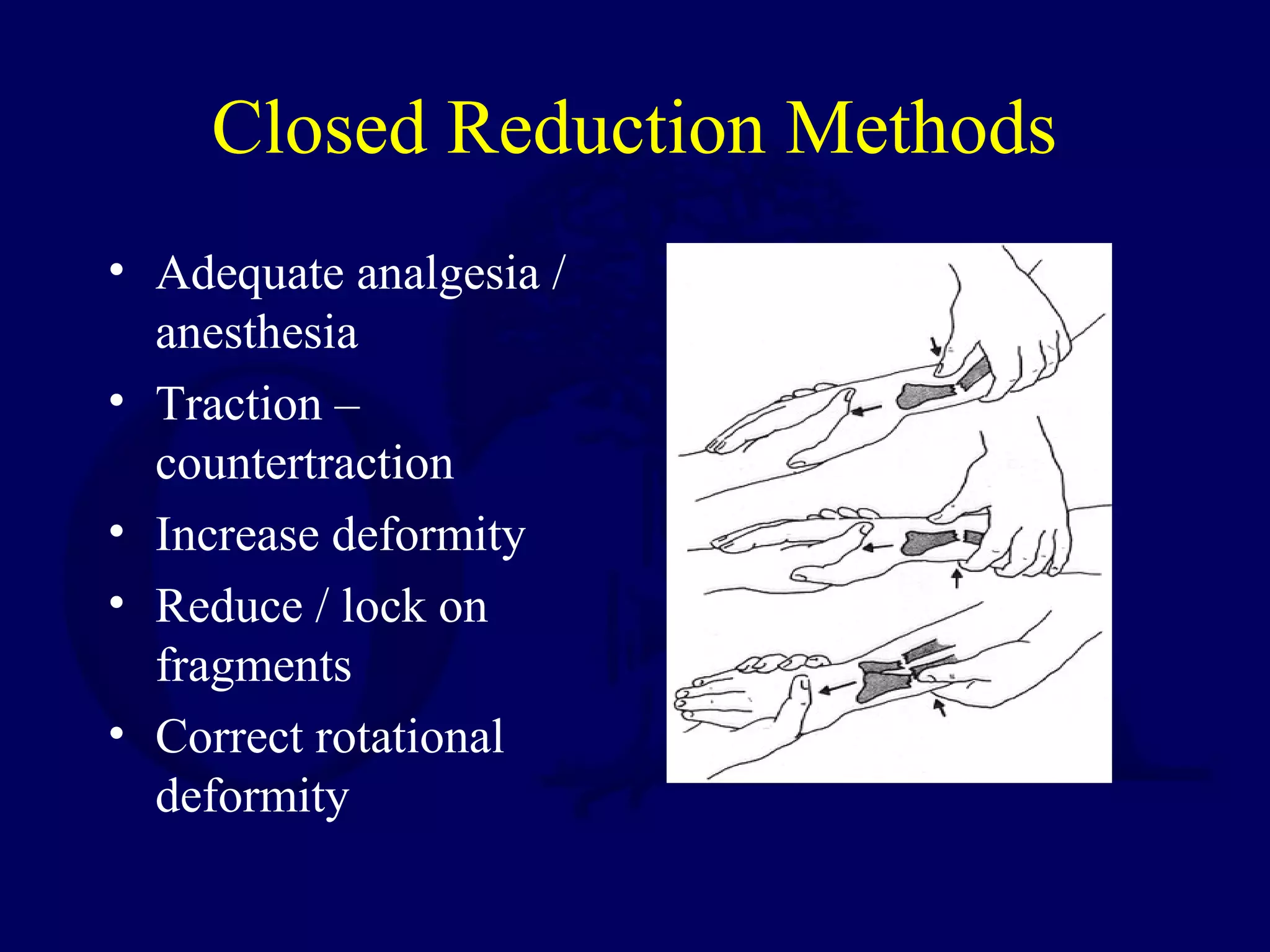
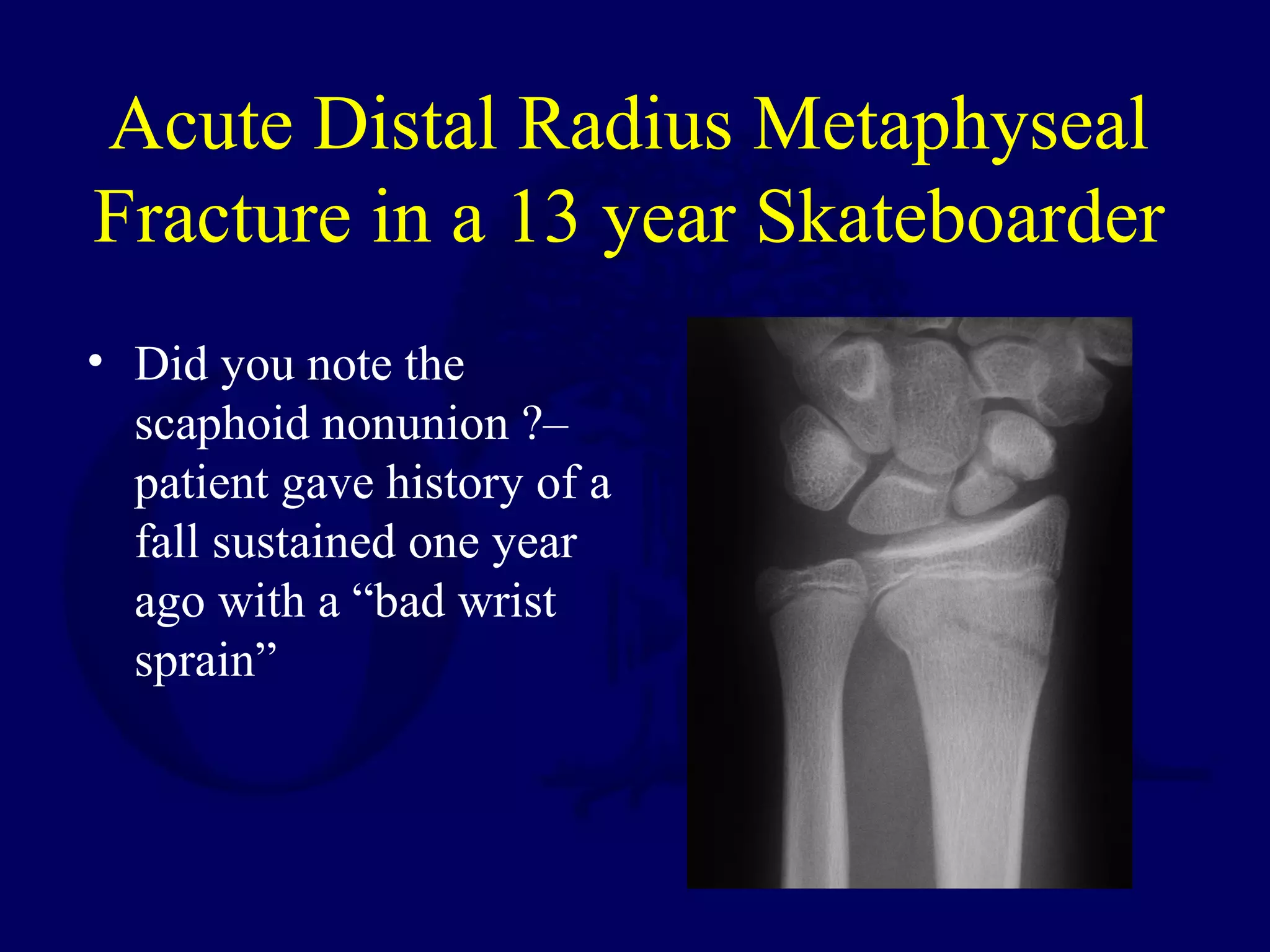
SlideShare a Scribd company logo
Pediatric Fractures of the
Forearm, Wrist and Hand
Amanda Marshall, MD
Pediatric Forearm Fractures-
Radial and Ulnar Shafts
• Approximately 4% of children’s fractures
• Middle and proximal radius more protected
by musculature than distal
• Ulna subcutaneous and susceptible to
trauma when raised for self protection
• Most fractures are from fall on an
outstretched arm
Forearm Developmental
Anatomy
• Primary ossification centers at 8 weeks
gestation in both radius and ulna
• Distal physes provide most of longitudinal
growth
• Distal epiphyses of radius appears
radiographically at age 1, of distal ulna at
age 5
• Proximal and middle radius connected to
ulna by intraosseous membrane
Pediatric Forearm Fractures
• Complete
• Greenstick fractures
• Buckle or torus fractures
• Plastic deformation
• Proximal, middle or distal
• Fxs at same level
• Fxs at different level
• Almost always a rotational component
Goals of Treatment
• Regain full forearm rotation
• Restore alignment and clinical appearance
• For ADL’s need 50 degrees supination, 50
degrees pronation
Closed Reduction Methods
• Adequate analgesia /
anesthesia
• Traction –
countertraction
• Increase deformity
• Reduce / lock on
fragments
• Correct rotational
deformity
Excellent Reduction with Well
Molded Cast
Remodeling Potential –
Variables to Consider
• Age
• Distance from fracture to physis
• Amount of deformity
• Direction of angulation
• Rotational deformities will not remodel
How Much Angulation
is too Much?
• Case by case decisions
• Closed reduction should be attempted for
angulation greater than 20 degrees
• How much to accept before proceeding with
open reduction dependant on many factors
• Angulation encroaching on interosseous
space may be more likely to limit rotation
Forearm Rotation Position
in Cast – Supinate, Pronate
or Midposition?
• Depends on location
of fracture and
position of distal
fragment in relation to
proximal
• Match distal fragment
to proximal – can use
bicipital tuberosity as
a guide
After Closed Reduction
and Casting -
• Weekly radiographs for 3 weeks to confirm
acceptable alignment and rotation
• overriding (bayonet) position OK
• Can remanipulate up to 3 weeks after injury
for shaft fractures
Maintaining Reduction
• Appropriately molded
cast very important
• Easier to maintain an
initial excellent
reduction than a
marginal one
• Above elbow or below
elbow immobilization
– surgeon preference
Indications for Open Reduction
• Open fractures
• Inability to maintain
acceptable reduction
• Multiple trauma
• Intramedullary pin
fixation can often be
accomplished with
little soft tissue
disruption
Open Both Bone
Forearm Fracture
16 Year Old with
Rotational Malunion
Forearm Fractures -
Complications
• Malunion-most common
• Refracture – 5% within 6
months
• Compartment syndrome –
observe closely, diagnosis
and treatment similar to
adults
• Synostosis rare
• Neurologic injury
uncommon
Plastic Deformation
of the Forearm
• Fixed bending remains
when bone deformed past
elastic limit
• Most commonly in
forearm, may be ulna or
radius
• Periosteum intact and thus
usually no periosteal
callus
• Can limit rotation
Plastic Deformation
• Remodeling not as
reliable
• Significant curvature
that produces clinical
deformity should be
corrected
• General anesthesia
• Considerable force,
slowly applied over a
padded fulcrum
Galeazzi Fracture- Radial Shaft
Fracture with DRUJ Injury
• Usually at junction of
middle and distal thirds
• Distal fragment typically
angulated towards ulna
• Closed treatment for most
• Carefully assess DRUJ
post reduction, clinically
and radiographically
Galeazzi Equivalent
Radial shaft fracture with
distal ulnar physeal
injury instead of
DRUJ injury
Distal ulnar physeal
injuries have a high
incidence for growth
arrest
Galeazzi Fracture
Distal Radius Fractures
• Most commonly fractured bone in children
• Metaphyseal most frequent, distal radial
physeal second
• Simple falls most common mechanism
• Rapid growth may predispose, with weaker
area at metaphysis
Distal Radius Fractures
• Metaphyseal
• Physeal – Salter II
most common
• Torus
• Greenstick
• Complete - Volar
angulation with dorsal
displacement of the
distal fragment most
common
Distal Radius Fractures –
Associated Injuries
• Frequently distal ulnar metaphyseal fracture
or ulnar styloid avulsion
• Occasionally distal ulnar physeal injury –
high incidence of growth disturbance
• Median or ulnar nerve injury – rare
• Acute carpal tunnel syndrome can occur,
also rare
Nondisplaced Distal Radius
Fractures- Treatment
• Below elbow
immobilization
• 3 weeks
Displaced Distal Radius
Fractures-Treatment
• Closed reduction usually
not difficult
• Traction (reduce shear),
recreate deformity and
reduce using intact
periosteal hinge
• Immobilize – many
different positions of wrist
and forearm rotation
recommended
• Well molded cast / splint,
above or below elbow
surgeon preference
• 3-4 weeks immobilization
“Repeated efforts at reduction do nothing more than
grate the plate away.”
“These injuries unite quickly, so that attempts to
correct malposition after a week are liable to do
more damage to the plate than good.”
Rang, Children’s Fractures 1983.
Treatment Recommendations -
# Reduction Attempts?
• No correlation between # reduction attempts and
growth retardation.
• No correlation between post-reduction position and
growth retardation.
• Noted a relationship between fracture type (Aitken
III/S-H IV) and growth arrest.
Aitken, JBJS 1935.
Treatment Recommendations -
# Reductions / Acceptable Alignment?
Treatment Recommendations
“An attempt should be made to reduce all
displacements… however, repeated manipulations
or osteotomy are not warranted.”
“Displacement of the epiphysis does not persist. All
displacements are reduced well within a year.”
“The one case of deformity in the series is attributed
to crushing of the physis.”
Aitken, JBJS 1935.
Treatment Recommendations
“For Salter-Harris type I and II
injuries in children younger than
10 years of age, angulation of up to
30° can be accepted. In children
older than 10 years, up to 15° of
angulation is generally
acceptable.”
Armstrong et al, Skeletal Trauma, 1998.
Displaced Distal
Radius Fractures –
Care after Closed Reduction
• Radiograph within one week to check reduction
• Do not remanipulate physeal fractures after 5-7
days for fear of further injuring physis
• Metaphyseal fractures may be remanipulated for
2-3 weeks if alignment lost
• Expect significant remodeling of any residual
deformity
Distal Radius Fractures -
Complications
• Growth arrest unusual
after distal radius
physeal injury
• Malunion will
typically remodel –
follow for one year
prior to any corrective
osteotomy
• Shortening usually not
a problem – resolves
with growth
Remodeling in 8 months
Distal Radius Fracture –
Indications for Operative Treatment
• Inability to obtain acceptable reduction
• Open fractures
• Displaced intraarticular fxs
• Associated soft tissue injuries
• Associated fractures (SC humerus)
• Associated acute carpal tunnel syndrome or
compartment syndrome
Distal Radius – Fixation Options
• Smooth K wire
fixation usually
adequate
• Ex fix for severe soft
tissue injury
• Some fxs amenable to
plate fixation
Complications
• Premature Physeal Closure / Growth Arrest
– 1.25% (Aitken, 1935)
– 3% (Bragdon, 1965)
– 7% (Lee, 1984)
• Nerve Injury
– 8%
• Ulnar Styloid Nonunions
– 27% (Aitken, 1935)
Growth Arrest following
Distal Radius Fracture
Injury films Injured and uninjured wrists
after premature physeal closure
Distal Radius Growth Arrest
• Relatively rare
(< 1 – 7%)
• Severity of trauma
• Amount of
displacement
• Repeated attempts at
reduction?
• Remanipulation or late
manipulation?
Case Study - P.J.
• 12 days s/p injury
loss of reduction
• 3 weeks s/p
injury
Conclusions
• Most common physeal plate injury (46%)
• Increased incidence of growth plate
abnormalities with 2 or more reductions
• Acceptable alignment: 50% apposition
30° angulation
• Accept malreduced fractures upon late
presentation (over 7 days).
• Growth arrest rate up to 7%
Carpal Injuries in Children
• Unusual / Uncommon in children
• Scaphoid most commonly fractured carpal
bone
• Capitate / Lunate / Hamate fractures also
can occur
• Make a habit of carefully checking carpal
bones on every wrist film
Acute Distal Radius Metaphyseal
Fracture in a 13 year Skateboarder
• Did you note the
scaphoid nonunion ?–
patient gave history of a
fall sustained one year
ago with a “bad wrist
sprain”
Distal Radius and Scaphoid
Fractures
Scaphoid Fractures - Treatment
• Tender snuff box – immobilize until
tenderness resolves
• If still tender at 1-2 weeks – repeat xray
• Confirmed fracture – if nondisplaced
immobilize in above elbow cast for 6 – 8
weeks
• Displaced fracture - ORIF
Hand Fractures
• Metacarpal and phalangeal fractures – if
displaced closed reduction
• Correct angulation and rotation
• Immobilize in intrinsic plus position
• 3-4 weeks
• Indications for ORIF – open fractures,
displaced intraarticular fractures, inability
to obtain / maintain reduction
Crush Injury to Hand
Distal Phalangeal Fractures
• Crush injuries –
address any associated
nail bed injuries
• If open give
appropriate antibiotics,
I&D
• Mallet finger injuries –
often physeal injury
• Closed management
Middle and Proximal
Phalangeal Fractures
• Closed management
for majority
• ORIF for displaced
intraarticular fractures
• Restore rotational
alignment
Metacarpal Fractures
• Closed management
for most
• Accept less angulation
in index than small
finger
Return to
Pediatrics Index

More Related Content

What's hot

Ligamentotaxis principle in the treatment of intra articular fractures of dis...
Ligamentotaxis principle in the treatment of intra articular fractures of dis...Ligamentotaxis principle in the treatment of intra articular fractures of dis...
Ligamentotaxis principle in the treatment of intra articular fractures of dis...Sitanshu Barik
 
ACROMIOCLAVICULAR JOINT INJURY
ACROMIOCLAVICULAR JOINT INJURYACROMIOCLAVICULAR JOINT INJURY
ACROMIOCLAVICULAR JOINT INJURY
Suman Subedi
 
Subtrochanteric
SubtrochantericSubtrochanteric
Subtrochanteric
Orthosurg2016
 
Septic arthritis sequelae
Septic arthritis sequelaeSeptic arthritis sequelae
Septic arthritis sequelaeorthoprince
 
Three column fixation for complex PROXIMAL TIBIA FRACTURES
Three column fixation for complex PROXIMAL TIBIA FRACTURESThree column fixation for complex PROXIMAL TIBIA FRACTURES
Three column fixation for complex PROXIMAL TIBIA FRACTURES
Lokesh Sharoff
 
Poller or blocking screw
Poller or blocking screwPoller or blocking screw
Poller or blocking screw
Avik Sarkar
 
Hoffa's Fracture: Diagnosis, management & New Classification System by BAGARI...
Hoffa's Fracture: Diagnosis, management & New Classification System by BAGARI...Hoffa's Fracture: Diagnosis, management & New Classification System by BAGARI...
Hoffa's Fracture: Diagnosis, management & New Classification System by BAGARI...
Vaibhav Bagaria
 
Herbert screw fixation and bone graft in nonunited scaphoid
Herbert screw fixation and bone graft in nonunited scaphoidHerbert screw fixation and bone graft in nonunited scaphoid
Herbert screw fixation and bone graft in nonunited scaphoid
Murugesh M Kurani
 
Physeal injuries
Physeal injuriesPhyseal injuries
Physeal injuries
Asi-oqua Bassey
 
Osteotomy around knee dr shankar jangid (1)
Osteotomy around knee dr shankar jangid (1)Osteotomy around knee dr shankar jangid (1)
Osteotomy around knee dr shankar jangid (1)
ShankarJangid5
 
Management of femoral neck fractures in children
Management of femoral neck fractures in childrenManagement of femoral neck fractures in children
Management of femoral neck fractures in childrenNOHD, Kano, Nigeria
 
Damage control orthopaedic surgery
Damage control orthopaedic surgeryDamage control orthopaedic surgery
Damage control orthopaedic surgery
Mohamed Abulsoud
 
Paediatric Forearm Diaphysial Fractures
Paediatric Forearm Diaphysial FracturesPaediatric Forearm Diaphysial Fractures
Paediatric Forearm Diaphysial Fractures
Pulasthi Kanchana
 
Osteotomy around elbow
Osteotomy around elbowOsteotomy around elbow
Osteotomy around elbow
Sushil Sharma
 
Intertrochanteric Fractures of Femur
Intertrochanteric Fractures of FemurIntertrochanteric Fractures of Femur
Intertrochanteric Fractures of Femur
Pulasthi Kanchana
 
Floating Knee
Floating KneeFloating Knee
Floating Knee
Dr Rohil Singh Kakkar
 
Malunited distal radius fracture
Malunited distal radius fractureMalunited distal radius fracture
Malunited distal radius fracture
NISCHAYKAUSHIK2
 
Radial head fracture
Radial head fractureRadial head fracture
Radial head fracture
Krunal Patel
 
INTRAMEDULLARY NAILING: RECENT ADVANCES AND FUTURE TRENDS
INTRAMEDULLARY NAILING: RECENT ADVANCES AND FUTURE TRENDSINTRAMEDULLARY NAILING: RECENT ADVANCES AND FUTURE TRENDS
INTRAMEDULLARY NAILING: RECENT ADVANCES AND FUTURE TRENDS
Mohd Fareed
 
Principles of internal fixation
Principles of internal fixationPrinciples of internal fixation
Principles of internal fixation
Praveen Kumar Reddy Gorantla
 

What's hot (20)

Ligamentotaxis principle in the treatment of intra articular fractures of dis...
Ligamentotaxis principle in the treatment of intra articular fractures of dis...Ligamentotaxis principle in the treatment of intra articular fractures of dis...
Ligamentotaxis principle in the treatment of intra articular fractures of dis...
 
ACROMIOCLAVICULAR JOINT INJURY
ACROMIOCLAVICULAR JOINT INJURYACROMIOCLAVICULAR JOINT INJURY
ACROMIOCLAVICULAR JOINT INJURY
 
Subtrochanteric
SubtrochantericSubtrochanteric
Subtrochanteric
 
Septic arthritis sequelae
Septic arthritis sequelaeSeptic arthritis sequelae
Septic arthritis sequelae
 
Three column fixation for complex PROXIMAL TIBIA FRACTURES
Three column fixation for complex PROXIMAL TIBIA FRACTURESThree column fixation for complex PROXIMAL TIBIA FRACTURES
Three column fixation for complex PROXIMAL TIBIA FRACTURES
 
Poller or blocking screw
Poller or blocking screwPoller or blocking screw
Poller or blocking screw
 
Hoffa's Fracture: Diagnosis, management & New Classification System by BAGARI...
Hoffa's Fracture: Diagnosis, management & New Classification System by BAGARI...Hoffa's Fracture: Diagnosis, management & New Classification System by BAGARI...
Hoffa's Fracture: Diagnosis, management & New Classification System by BAGARI...
 
Herbert screw fixation and bone graft in nonunited scaphoid
Herbert screw fixation and bone graft in nonunited scaphoidHerbert screw fixation and bone graft in nonunited scaphoid
Herbert screw fixation and bone graft in nonunited scaphoid
 
Physeal injuries
Physeal injuriesPhyseal injuries
Physeal injuries
 
Osteotomy around knee dr shankar jangid (1)
Osteotomy around knee dr shankar jangid (1)Osteotomy around knee dr shankar jangid (1)
Osteotomy around knee dr shankar jangid (1)
 
Management of femoral neck fractures in children
Management of femoral neck fractures in childrenManagement of femoral neck fractures in children
Management of femoral neck fractures in children
 
Damage control orthopaedic surgery
Damage control orthopaedic surgeryDamage control orthopaedic surgery
Damage control orthopaedic surgery
 
Paediatric Forearm Diaphysial Fractures
Paediatric Forearm Diaphysial FracturesPaediatric Forearm Diaphysial Fractures
Paediatric Forearm Diaphysial Fractures
 
Osteotomy around elbow
Osteotomy around elbowOsteotomy around elbow
Osteotomy around elbow
 
Intertrochanteric Fractures of Femur
Intertrochanteric Fractures of FemurIntertrochanteric Fractures of Femur
Intertrochanteric Fractures of Femur
 
Floating Knee
Floating KneeFloating Knee
Floating Knee
 
Malunited distal radius fracture
Malunited distal radius fractureMalunited distal radius fracture
Malunited distal radius fracture
 
Radial head fracture
Radial head fractureRadial head fracture
Radial head fracture
 
INTRAMEDULLARY NAILING: RECENT ADVANCES AND FUTURE TRENDS
INTRAMEDULLARY NAILING: RECENT ADVANCES AND FUTURE TRENDSINTRAMEDULLARY NAILING: RECENT ADVANCES AND FUTURE TRENDS
INTRAMEDULLARY NAILING: RECENT ADVANCES AND FUTURE TRENDS
 
Principles of internal fixation
Principles of internal fixationPrinciples of internal fixation
Principles of internal fixation
 

Viewers also liked

Paediatric Orthopaedic
Paediatric OrthopaedicPaediatric Orthopaedic
Paediatric OrthopaedicFara Dyba
 
orthopaedic fractures in children
orthopaedic fractures in children orthopaedic fractures in children
orthopaedic fractures in children
Harjot Gurudatta
 
P09 pediatric femur
P09 pediatric femurP09 pediatric femur
P09 pediatric femur
Claudiu Cucu
 
Proximal radius fractures in children
Proximal radius fractures in childrenProximal radius fractures in children
Proximal radius fractures in children
Opender Kajla
 
Fasciotomy
FasciotomyFasciotomy
Recon ha ossap 2015
Recon ha ossap 2015Recon ha ossap 2015
Recon ha ossap 2015
Vsn Raju
 
Supracondylar Fractures
Supracondylar FracturesSupracondylar Fractures
Supracondylar Fractures
Pulasthi Kanchana
 
Pediatric health education
Pediatric health educationPediatric health education
Pediatric health education
lamiaa Gamal
 
Msk Lecture2 1st Hospital
Msk Lecture2 1st HospitalMsk Lecture2 1st Hospital
Msk Lecture2 1st HospitalSumit Prajapati
 
Pediatric chest part 2
Pediatric chest part 2Pediatric chest part 2
Pediatric chest part 2
Anish Choudhary
 
Presentation1.pptx, ultrasound of the hand and fingers.
Presentation1.pptx, ultrasound of the hand and fingers.Presentation1.pptx, ultrasound of the hand and fingers.
Presentation1.pptx, ultrasound of the hand and fingers.Abdellah Nazeer
 
Supracondylar fracture of Humerus
Supracondylar fracture of HumerusSupracondylar fracture of Humerus
Supracondylar fracture of Humerus
Farbod Zahedi Tajrishi
 
Rolando Fracture Power Point
Rolando Fracture Power PointRolando Fracture Power Point
Rolando Fracture Power PointTodd Peterson
 
Ergonomics
Ergonomics Ergonomics
Ergonomics
Tariq Hatmi
 
GEMC: Pediatric Orthopedic Emergencies: Resident Training
GEMC: Pediatric Orthopedic Emergencies: Resident TrainingGEMC: Pediatric Orthopedic Emergencies: Resident Training
GEMC: Pediatric Orthopedic Emergencies: Resident Training
Open.Michigan
 
Pediatric renal tumors radiology
Pediatric renal tumors radiologyPediatric renal tumors radiology
Pediatric renal tumors radiology
Bipin Joseph
 
Bennetts Fracture
Bennetts FractureBennetts Fracture
Bennetts Fracturejfreshour
 
Supracondylar fracture of the humerus by phaneendra akana
Supracondylar fracture of the humerus by phaneendra akanaSupracondylar fracture of the humerus by phaneendra akana
Supracondylar fracture of the humerus by phaneendra akana
Mohan Phaneendra Akana
 

Viewers also liked (20)

Paediatric Orthopaedic
Paediatric OrthopaedicPaediatric Orthopaedic
Paediatric Orthopaedic
 
Paediatric fracture
Paediatric fracturePaediatric fracture
Paediatric fracture
 
orthopaedic fractures in children
orthopaedic fractures in children orthopaedic fractures in children
orthopaedic fractures in children
 
P09 pediatric femur
P09 pediatric femurP09 pediatric femur
P09 pediatric femur
 
Proximal radius fractures in children
Proximal radius fractures in childrenProximal radius fractures in children
Proximal radius fractures in children
 
Fasciotomy
FasciotomyFasciotomy
Fasciotomy
 
Torus Fracture
Torus FractureTorus Fracture
Torus Fracture
 
Recon ha ossap 2015
Recon ha ossap 2015Recon ha ossap 2015
Recon ha ossap 2015
 
Supracondylar Fractures
Supracondylar FracturesSupracondylar Fractures
Supracondylar Fractures
 
Pediatric health education
Pediatric health educationPediatric health education
Pediatric health education
 
Msk Lecture2 1st Hospital
Msk Lecture2 1st HospitalMsk Lecture2 1st Hospital
Msk Lecture2 1st Hospital
 
Pediatric chest part 2
Pediatric chest part 2Pediatric chest part 2
Pediatric chest part 2
 
Presentation1.pptx, ultrasound of the hand and fingers.
Presentation1.pptx, ultrasound of the hand and fingers.Presentation1.pptx, ultrasound of the hand and fingers.
Presentation1.pptx, ultrasound of the hand and fingers.
 
Supracondylar fracture of Humerus
Supracondylar fracture of HumerusSupracondylar fracture of Humerus
Supracondylar fracture of Humerus
 
Rolando Fracture Power Point
Rolando Fracture Power PointRolando Fracture Power Point
Rolando Fracture Power Point
 
Ergonomics
Ergonomics Ergonomics
Ergonomics
 
GEMC: Pediatric Orthopedic Emergencies: Resident Training
GEMC: Pediatric Orthopedic Emergencies: Resident TrainingGEMC: Pediatric Orthopedic Emergencies: Resident Training
GEMC: Pediatric Orthopedic Emergencies: Resident Training
 
Pediatric renal tumors radiology
Pediatric renal tumors radiologyPediatric renal tumors radiology
Pediatric renal tumors radiology
 
Bennetts Fracture
Bennetts FractureBennetts Fracture
Bennetts Fracture
 
Supracondylar fracture of the humerus by phaneendra akana
Supracondylar fracture of the humerus by phaneendra akanaSupracondylar fracture of the humerus by phaneendra akana
Supracondylar fracture of the humerus by phaneendra akana
 

Similar to P06 pediatric forearm, hand

FOREARM TRAUMA. .pptx.
FOREARM  TRAUMA.                    .pptx.FOREARM  TRAUMA.                    .pptx.
FOREARM TRAUMA. .pptx.
DeveshAhir
 
P10 pediatric knee
P10 pediatric kneeP10 pediatric knee
P10 pediatric knee
Claudiu Cucu
 
Fracture shaft of femur by dr ashutosh
Fracture shaft of femur by dr ashutoshFracture shaft of femur by dr ashutosh
Fracture shaft of femur by dr ashutosh
Ashutosh Kumar
 
Distal end radius, monteggia and galleazi fractures.
Distal end radius, monteggia and galleazi fractures.Distal end radius, monteggia and galleazi fractures.
Distal end radius, monteggia and galleazi fractures.
Dialla Sandouka
 
Upper limb trauma part 2
Upper limb trauma part 2Upper limb trauma part 2
Upper limb trauma part 2
abdullah alzahrani
 
Cubitus varus by Dhrumil Patel
Cubitus varus by Dhrumil PatelCubitus varus by Dhrumil Patel
Cubitus varus by Dhrumil Pateldhrumil88
 
Pediatric_Fractures_QT.pdf
Pediatric_Fractures_QT.pdfPediatric_Fractures_QT.pdf
Pediatric_Fractures_QT.pdf
ksdjf2
 
Mandible fracture symposium march-13
Mandible fracture   symposium march-13Mandible fracture   symposium march-13
Mandible fracture symposium march-13Narendra Markad
 
8. Forearm bone fractures
8. Forearm bone fractures8. Forearm bone fractures
8. Forearm bone fractures
Dr. Bindesh Patel (MPTh)
 
10. Scaphoid fracture, Benett's fracture, Rolando's fracture, Mallet's finger...
10. Scaphoid fracture, Benett's fracture, Rolando's fracture, Mallet's finger...10. Scaphoid fracture, Benett's fracture, Rolando's fracture, Mallet's finger...
10. Scaphoid fracture, Benett's fracture, Rolando's fracture, Mallet's finger...
Dr. Bindesh Patel (MPTh)
 
CONSERVATIVE MANAGEMENT OF FRACTURE
CONSERVATIVE MANAGEMENT OF FRACTURECONSERVATIVE MANAGEMENT OF FRACTURE
CONSERVATIVE MANAGEMENT OF FRACTURE
Naveed Jumani
 
P01 ped trauma assessment
P01 ped trauma assessmentP01 ped trauma assessment
P01 ped trauma assessment
Claudiu Cucu
 
Supracondylar humeral fracture
Supracondylar humeral fractureSupracondylar humeral fracture
Supracondylar humeral fracture
MinThu62
 
P05 pediatric elbow
P05 pediatric elbowP05 pediatric elbow
P05 pediatric elbow
Claudiu Cucu
 
11. Hip dislocation
11. Hip dislocation11. Hip dislocation
11. Hip dislocation
Dr. Bindesh Patel (MPTh)
 
Common Hand Fractures & Dislocations
Common Hand Fractures & Dislocations Common Hand Fractures & Dislocations
Common Hand Fractures & Dislocations
Nattakul Yamprasert
 
Paediatric femur fractures
Paediatric femur fracturesPaediatric femur fractures
Paediatric femur fractures
Dr Sushant S. Sonarkar
 
Elbow Injuries.pptx
Elbow Injuries.pptxElbow Injuries.pptx
Elbow Injuries.pptx
esicOrtho1
 
Physeal injuries
Physeal injuriesPhyseal injuries
Physeal injuries
Muhammad Arif
 
Fractures in children
Fractures in childrenFractures in children
Fractures in children
Siddhartha Sinha
 

Similar to P06 pediatric forearm, hand (20)

FOREARM TRAUMA. .pptx.
FOREARM  TRAUMA.                    .pptx.FOREARM  TRAUMA.                    .pptx.
FOREARM TRAUMA. .pptx.
 
P10 pediatric knee
P10 pediatric kneeP10 pediatric knee
P10 pediatric knee
 
Fracture shaft of femur by dr ashutosh
Fracture shaft of femur by dr ashutoshFracture shaft of femur by dr ashutosh
Fracture shaft of femur by dr ashutosh
 
Distal end radius, monteggia and galleazi fractures.
Distal end radius, monteggia and galleazi fractures.Distal end radius, monteggia and galleazi fractures.
Distal end radius, monteggia and galleazi fractures.
 
Upper limb trauma part 2
Upper limb trauma part 2Upper limb trauma part 2
Upper limb trauma part 2
 
Cubitus varus by Dhrumil Patel
Cubitus varus by Dhrumil PatelCubitus varus by Dhrumil Patel
Cubitus varus by Dhrumil Patel
 
Pediatric_Fractures_QT.pdf
Pediatric_Fractures_QT.pdfPediatric_Fractures_QT.pdf
Pediatric_Fractures_QT.pdf
 
Mandible fracture symposium march-13
Mandible fracture   symposium march-13Mandible fracture   symposium march-13
Mandible fracture symposium march-13
 
8. Forearm bone fractures
8. Forearm bone fractures8. Forearm bone fractures
8. Forearm bone fractures
 
10. Scaphoid fracture, Benett's fracture, Rolando's fracture, Mallet's finger...
10. Scaphoid fracture, Benett's fracture, Rolando's fracture, Mallet's finger...10. Scaphoid fracture, Benett's fracture, Rolando's fracture, Mallet's finger...
10. Scaphoid fracture, Benett's fracture, Rolando's fracture, Mallet's finger...
 
CONSERVATIVE MANAGEMENT OF FRACTURE
CONSERVATIVE MANAGEMENT OF FRACTURECONSERVATIVE MANAGEMENT OF FRACTURE
CONSERVATIVE MANAGEMENT OF FRACTURE
 
P01 ped trauma assessment
P01 ped trauma assessmentP01 ped trauma assessment
P01 ped trauma assessment
 
Supracondylar humeral fracture
Supracondylar humeral fractureSupracondylar humeral fracture
Supracondylar humeral fracture
 
P05 pediatric elbow
P05 pediatric elbowP05 pediatric elbow
P05 pediatric elbow
 
11. Hip dislocation
11. Hip dislocation11. Hip dislocation
11. Hip dislocation
 
Common Hand Fractures & Dislocations
Common Hand Fractures & Dislocations Common Hand Fractures & Dislocations
Common Hand Fractures & Dislocations
 
Paediatric femur fractures
Paediatric femur fracturesPaediatric femur fractures
Paediatric femur fractures
 
Elbow Injuries.pptx
Elbow Injuries.pptxElbow Injuries.pptx
Elbow Injuries.pptx
 
Physeal injuries
Physeal injuriesPhyseal injuries
Physeal injuries
 
Fractures in children
Fractures in childrenFractures in children
Fractures in children
 

More from Claudiu Cucu

V06 orif acetabulum
V06 orif acetabulumV06 orif acetabulum
V06 orif acetabulum
Claudiu Cucu
 
V05 acetab surgical_apprch
V05 acetab surgical_apprchV05 acetab surgical_apprch
V05 acetab surgical_apprch
Claudiu Cucu
 
V04 anatomy class_acetab
V04 anatomy class_acetabV04 anatomy class_acetab
V04 anatomy class_acetab
Claudiu Cucu
 
V03 orif pelvic_ring
V03 orif pelvic_ringV03 orif pelvic_ring
V03 orif pelvic_ring
Claudiu Cucu
 
V02 pelvis acute_mgmt
V02 pelvis acute_mgmtV02 pelvis acute_mgmt
V02 pelvis acute_mgmt
Claudiu Cucu
 
V01 anatomy class_pelvis
V01 anatomy class_pelvisV01 anatomy class_pelvis
V01 anatomy class_pelvis
Claudiu Cucu
 
P08 pediatric hip
P08 pediatric hipP08 pediatric hip
P08 pediatric hip
Claudiu Cucu
 
P07 pediatric pelvis, aceta
P07 pediatric pelvis, acetaP07 pediatric pelvis, aceta
P07 pediatric pelvis, aceta
Claudiu Cucu
 
P03 ped pathologic fxs
P03 ped pathologic fxsP03 ped pathologic fxs
P03 ped pathologic fxs
Claudiu Cucu
 
P02 abuse
P02 abuseP02 abuse
P02 abuse
Claudiu Cucu
 
Lower extremity index
Lower extremity indexLower extremity index
Lower extremity index
Claudiu Cucu
 
L18 le amputations
L18 le amputationsL18 le amputations
L18 le amputations
Claudiu Cucu
 
L17 forefoot fxs
L17 forefoot fxsL17 forefoot fxs
L17 forefoot fxs
Claudiu Cucu
 
L16 lisfranc &amp; midfoot inj
L16 lisfranc &amp; midfoot injL16 lisfranc &amp; midfoot inj
L16 lisfranc &amp; midfoot inj
Claudiu Cucu
 
L15 calcaneus
L15 calcaneusL15 calcaneus
L15 calcaneus
Claudiu Cucu
 
L14 talus fxs &amp; dislocation
L14 talus fxs &amp; dislocationL14 talus fxs &amp; dislocation
L14 talus fxs &amp; dislocation
Claudiu Cucu
 
L13 ankle ligament injuries
L13 ankle ligament injuriesL13 ankle ligament injuries
L13 ankle ligament injuries
Claudiu Cucu
 
L12 ankle fxs
L12 ankle fxsL12 ankle fxs
L12 ankle fxs
Claudiu Cucu
 
L11 pilon
L11 pilonL11 pilon
L11 pilon
Claudiu Cucu
 
L10 closed tibia
L10 closed tibiaL10 closed tibia
L10 closed tibia
Claudiu Cucu
 

More from Claudiu Cucu (20)

V06 orif acetabulum
V06 orif acetabulumV06 orif acetabulum
V06 orif acetabulum
 
V05 acetab surgical_apprch
V05 acetab surgical_apprchV05 acetab surgical_apprch
V05 acetab surgical_apprch
 
V04 anatomy class_acetab
V04 anatomy class_acetabV04 anatomy class_acetab
V04 anatomy class_acetab
 
V03 orif pelvic_ring
V03 orif pelvic_ringV03 orif pelvic_ring
V03 orif pelvic_ring
 
V02 pelvis acute_mgmt
V02 pelvis acute_mgmtV02 pelvis acute_mgmt
V02 pelvis acute_mgmt
 
V01 anatomy class_pelvis
V01 anatomy class_pelvisV01 anatomy class_pelvis
V01 anatomy class_pelvis
 
P08 pediatric hip
P08 pediatric hipP08 pediatric hip
P08 pediatric hip
 
P07 pediatric pelvis, aceta
P07 pediatric pelvis, acetaP07 pediatric pelvis, aceta
P07 pediatric pelvis, aceta
 
P03 ped pathologic fxs
P03 ped pathologic fxsP03 ped pathologic fxs
P03 ped pathologic fxs
 
P02 abuse
P02 abuseP02 abuse
P02 abuse
 
Lower extremity index
Lower extremity indexLower extremity index
Lower extremity index
 
L18 le amputations
L18 le amputationsL18 le amputations
L18 le amputations
 
L17 forefoot fxs
L17 forefoot fxsL17 forefoot fxs
L17 forefoot fxs
 
L16 lisfranc &amp; midfoot inj
L16 lisfranc &amp; midfoot injL16 lisfranc &amp; midfoot inj
L16 lisfranc &amp; midfoot inj
 
L15 calcaneus
L15 calcaneusL15 calcaneus
L15 calcaneus
 
L14 talus fxs &amp; dislocation
L14 talus fxs &amp; dislocationL14 talus fxs &amp; dislocation
L14 talus fxs &amp; dislocation
 
L13 ankle ligament injuries
L13 ankle ligament injuriesL13 ankle ligament injuries
L13 ankle ligament injuries
 
L12 ankle fxs
L12 ankle fxsL12 ankle fxs
L12 ankle fxs
 
L11 pilon
L11 pilonL11 pilon
L11 pilon
 
L10 closed tibia
L10 closed tibiaL10 closed tibia
L10 closed tibia
 

Recently uploaded

Superficial & Deep Fascia of the NECK.pptx
Superficial & Deep Fascia of the NECK.pptxSuperficial & Deep Fascia of the NECK.pptx
Superficial & Deep Fascia of the NECK.pptx
Dr. Rabia Inam Gandapore
 
263778731218 Abortion Clinic /Pills In Harare ,
263778731218 Abortion Clinic /Pills In Harare ,263778731218 Abortion Clinic /Pills In Harare ,
263778731218 Abortion Clinic /Pills In Harare ,
sisternakatoto
 
Tom Selleck Health: A Comprehensive Look at the Iconic Actor’s Wellness Journey
Tom Selleck Health: A Comprehensive Look at the Iconic Actor’s Wellness JourneyTom Selleck Health: A Comprehensive Look at the Iconic Actor’s Wellness Journey
Tom Selleck Health: A Comprehensive Look at the Iconic Actor’s Wellness Journey
greendigital
 
Local Advanced Lung Cancer: Artificial Intelligence, Synergetics, Complex Sys...
Local Advanced Lung Cancer: Artificial Intelligence, Synergetics, Complex Sys...Local Advanced Lung Cancer: Artificial Intelligence, Synergetics, Complex Sys...
Local Advanced Lung Cancer: Artificial Intelligence, Synergetics, Complex Sys...
Oleg Kshivets
 
Hemodialysis: Chapter 4, Dialysate Circuit - Dr.Gawad
Hemodialysis: Chapter 4, Dialysate Circuit - Dr.GawadHemodialysis: Chapter 4, Dialysate Circuit - Dr.Gawad
Hemodialysis: Chapter 4, Dialysate Circuit - Dr.Gawad
NephroTube - Dr.Gawad
 
Top 10 Best Ayurvedic Kidney Stone Syrups in India
Top 10 Best Ayurvedic Kidney Stone Syrups in IndiaTop 10 Best Ayurvedic Kidney Stone Syrups in India
Top 10 Best Ayurvedic Kidney Stone Syrups in India
SwastikAyurveda
 
Ocular injury ppt Upendra pal optometrist upums saifai etawah
Ocular injury  ppt  Upendra pal  optometrist upums saifai etawahOcular injury  ppt  Upendra pal  optometrist upums saifai etawah
Ocular injury ppt Upendra pal optometrist upums saifai etawah
pal078100
 
New Drug Discovery and Development .....
New Drug Discovery and Development .....New Drug Discovery and Development .....
New Drug Discovery and Development .....
NEHA GUPTA
 
Ozempic: Preoperative Management of Patients on GLP-1 Receptor Agonists
Ozempic: Preoperative Management of Patients on GLP-1 Receptor Agonists  Ozempic: Preoperative Management of Patients on GLP-1 Receptor Agonists
Ozempic: Preoperative Management of Patients on GLP-1 Receptor Agonists
Saeid Safari
 
Vision-1.pptx, Eye structure, basics of optics
Vision-1.pptx, Eye structure, basics of opticsVision-1.pptx, Eye structure, basics of optics
Vision-1.pptx, Eye structure, basics of optics
Sai Sailesh Kumar Goothy
 
Dehradun #ℂall #gIRLS Oyo Hotel 9719300533 #ℂall #gIRL in Dehradun
Dehradun #ℂall #gIRLS Oyo Hotel 9719300533 #ℂall #gIRL in DehradunDehradun #ℂall #gIRLS Oyo Hotel 9719300533 #ℂall #gIRL in Dehradun
Dehradun #ℂall #gIRLS Oyo Hotel 9719300533 #ℂall #gIRL in Dehradun
chandankumarsmartiso
 
Cervical & Brachial Plexus By Dr. RIG.pptx
Cervical & Brachial Plexus By Dr. RIG.pptxCervical & Brachial Plexus By Dr. RIG.pptx
Cervical & Brachial Plexus By Dr. RIG.pptx
Dr. Rabia Inam Gandapore
 
Maxilla, Mandible & Hyoid Bone & Clinical Correlations by Dr. RIG.pptx
Maxilla, Mandible & Hyoid Bone & Clinical Correlations by Dr. RIG.pptxMaxilla, Mandible & Hyoid Bone & Clinical Correlations by Dr. RIG.pptx
Maxilla, Mandible & Hyoid Bone & Clinical Correlations by Dr. RIG.pptx
Dr. Rabia Inam Gandapore
 
Best Ayurvedic medicine for Gas and Indigestion
Best Ayurvedic medicine for Gas and IndigestionBest Ayurvedic medicine for Gas and Indigestion
Best Ayurvedic medicine for Gas and Indigestion
SwastikAyurveda
 
How STIs Influence the Development of Pelvic Inflammatory Disease.pptx
How STIs Influence the Development of Pelvic Inflammatory Disease.pptxHow STIs Influence the Development of Pelvic Inflammatory Disease.pptx
How STIs Influence the Development of Pelvic Inflammatory Disease.pptx
FFragrant
 
A Classical Text Review on Basavarajeeyam
A Classical Text Review on BasavarajeeyamA Classical Text Review on Basavarajeeyam
A Classical Text Review on Basavarajeeyam
Dr. Jyothirmai Paindla
 
Knee anatomy and clinical tests 2024.pdf
Knee anatomy and clinical tests 2024.pdfKnee anatomy and clinical tests 2024.pdf
Knee anatomy and clinical tests 2024.pdf
vimalpl1234
 
BRACHYTHERAPY OVERVIEW AND APPLICATORS
BRACHYTHERAPY OVERVIEW  AND  APPLICATORSBRACHYTHERAPY OVERVIEW  AND  APPLICATORS
BRACHYTHERAPY OVERVIEW AND APPLICATORS
Krishan Murari
 
Colonic and anorectal physiology with surgical implications
Colonic and anorectal physiology with surgical implicationsColonic and anorectal physiology with surgical implications
Colonic and anorectal physiology with surgical implications
Dr Maria Tamanna
 
basicmodesofventilation2022-220313203758.pdf
basicmodesofventilation2022-220313203758.pdfbasicmodesofventilation2022-220313203758.pdf
basicmodesofventilation2022-220313203758.pdf
aljamhori teaching hospital
 

Recently uploaded (20)

Superficial & Deep Fascia of the NECK.pptx
Superficial & Deep Fascia of the NECK.pptxSuperficial & Deep Fascia of the NECK.pptx
Superficial & Deep Fascia of the NECK.pptx
 
263778731218 Abortion Clinic /Pills In Harare ,
263778731218 Abortion Clinic /Pills In Harare ,263778731218 Abortion Clinic /Pills In Harare ,
263778731218 Abortion Clinic /Pills In Harare ,
 
Tom Selleck Health: A Comprehensive Look at the Iconic Actor’s Wellness Journey
Tom Selleck Health: A Comprehensive Look at the Iconic Actor’s Wellness JourneyTom Selleck Health: A Comprehensive Look at the Iconic Actor’s Wellness Journey
Tom Selleck Health: A Comprehensive Look at the Iconic Actor’s Wellness Journey
 
Local Advanced Lung Cancer: Artificial Intelligence, Synergetics, Complex Sys...
Local Advanced Lung Cancer: Artificial Intelligence, Synergetics, Complex Sys...Local Advanced Lung Cancer: Artificial Intelligence, Synergetics, Complex Sys...
Local Advanced Lung Cancer: Artificial Intelligence, Synergetics, Complex Sys...
 
Hemodialysis: Chapter 4, Dialysate Circuit - Dr.Gawad
Hemodialysis: Chapter 4, Dialysate Circuit - Dr.GawadHemodialysis: Chapter 4, Dialysate Circuit - Dr.Gawad
Hemodialysis: Chapter 4, Dialysate Circuit - Dr.Gawad
 
Top 10 Best Ayurvedic Kidney Stone Syrups in India
Top 10 Best Ayurvedic Kidney Stone Syrups in IndiaTop 10 Best Ayurvedic Kidney Stone Syrups in India
Top 10 Best Ayurvedic Kidney Stone Syrups in India
 
Ocular injury ppt Upendra pal optometrist upums saifai etawah
Ocular injury  ppt  Upendra pal  optometrist upums saifai etawahOcular injury  ppt  Upendra pal  optometrist upums saifai etawah
Ocular injury ppt Upendra pal optometrist upums saifai etawah
 
New Drug Discovery and Development .....
New Drug Discovery and Development .....New Drug Discovery and Development .....
New Drug Discovery and Development .....
 
Ozempic: Preoperative Management of Patients on GLP-1 Receptor Agonists
Ozempic: Preoperative Management of Patients on GLP-1 Receptor Agonists  Ozempic: Preoperative Management of Patients on GLP-1 Receptor Agonists
Ozempic: Preoperative Management of Patients on GLP-1 Receptor Agonists
 
Vision-1.pptx, Eye structure, basics of optics
Vision-1.pptx, Eye structure, basics of opticsVision-1.pptx, Eye structure, basics of optics
Vision-1.pptx, Eye structure, basics of optics
 
Dehradun #ℂall #gIRLS Oyo Hotel 9719300533 #ℂall #gIRL in Dehradun
Dehradun #ℂall #gIRLS Oyo Hotel 9719300533 #ℂall #gIRL in DehradunDehradun #ℂall #gIRLS Oyo Hotel 9719300533 #ℂall #gIRL in Dehradun
Dehradun #ℂall #gIRLS Oyo Hotel 9719300533 #ℂall #gIRL in Dehradun
 
Cervical & Brachial Plexus By Dr. RIG.pptx
Cervical & Brachial Plexus By Dr. RIG.pptxCervical & Brachial Plexus By Dr. RIG.pptx
Cervical & Brachial Plexus By Dr. RIG.pptx
 
Maxilla, Mandible & Hyoid Bone & Clinical Correlations by Dr. RIG.pptx
Maxilla, Mandible & Hyoid Bone & Clinical Correlations by Dr. RIG.pptxMaxilla, Mandible & Hyoid Bone & Clinical Correlations by Dr. RIG.pptx
Maxilla, Mandible & Hyoid Bone & Clinical Correlations by Dr. RIG.pptx
 
Best Ayurvedic medicine for Gas and Indigestion
Best Ayurvedic medicine for Gas and IndigestionBest Ayurvedic medicine for Gas and Indigestion
Best Ayurvedic medicine for Gas and Indigestion
 
How STIs Influence the Development of Pelvic Inflammatory Disease.pptx
How STIs Influence the Development of Pelvic Inflammatory Disease.pptxHow STIs Influence the Development of Pelvic Inflammatory Disease.pptx
How STIs Influence the Development of Pelvic Inflammatory Disease.pptx
 
A Classical Text Review on Basavarajeeyam
A Classical Text Review on BasavarajeeyamA Classical Text Review on Basavarajeeyam
A Classical Text Review on Basavarajeeyam
 
Knee anatomy and clinical tests 2024.pdf
Knee anatomy and clinical tests 2024.pdfKnee anatomy and clinical tests 2024.pdf
Knee anatomy and clinical tests 2024.pdf
 
BRACHYTHERAPY OVERVIEW AND APPLICATORS
BRACHYTHERAPY OVERVIEW  AND  APPLICATORSBRACHYTHERAPY OVERVIEW  AND  APPLICATORS
BRACHYTHERAPY OVERVIEW AND APPLICATORS
 
Colonic and anorectal physiology with surgical implications
Colonic and anorectal physiology with surgical implicationsColonic and anorectal physiology with surgical implications
Colonic and anorectal physiology with surgical implications
 
basicmodesofventilation2022-220313203758.pdf
basicmodesofventilation2022-220313203758.pdfbasicmodesofventilation2022-220313203758.pdf
basicmodesofventilation2022-220313203758.pdf
 

P06 pediatric forearm, hand

  • 1. Pediatric Fractures of the Forearm, Wrist and Hand Amanda Marshall, MD
  • 2. Pediatric Forearm Fractures- Radial and Ulnar Shafts • Approximately 4% of children’s fractures • Middle and proximal radius more protected by musculature than distal • Ulna subcutaneous and susceptible to trauma when raised for self protection • Most fractures are from fall on an outstretched arm
  • 3. Forearm Developmental Anatomy • Primary ossification centers at 8 weeks gestation in both radius and ulna • Distal physes provide most of longitudinal growth • Distal epiphyses of radius appears radiographically at age 1, of distal ulna at age 5 • Proximal and middle radius connected to ulna by intraosseous membrane
  • 4. Pediatric Forearm Fractures • Complete • Greenstick fractures • Buckle or torus fractures • Plastic deformation • Proximal, middle or distal • Fxs at same level • Fxs at different level • Almost always a rotational component
  • 5. Goals of Treatment • Regain full forearm rotation • Restore alignment and clinical appearance • For ADL’s need 50 degrees supination, 50 degrees pronation
  • 6. Closed Reduction Methods • Adequate analgesia / anesthesia • Traction – countertraction • Increase deformity • Reduce / lock on fragments • Correct rotational deformity
  • 7. Excellent Reduction with Well Molded Cast
  • 8. Remodeling Potential – Variables to Consider • Age • Distance from fracture to physis • Amount of deformity • Direction of angulation • Rotational deformities will not remodel
  • 9. How Much Angulation is too Much? • Case by case decisions • Closed reduction should be attempted for angulation greater than 20 degrees • How much to accept before proceeding with open reduction dependant on many factors • Angulation encroaching on interosseous space may be more likely to limit rotation
  • 10. Forearm Rotation Position in Cast – Supinate, Pronate or Midposition? • Depends on location of fracture and position of distal fragment in relation to proximal • Match distal fragment to proximal – can use bicipital tuberosity as a guide
  • 11. After Closed Reduction and Casting - • Weekly radiographs for 3 weeks to confirm acceptable alignment and rotation • overriding (bayonet) position OK • Can remanipulate up to 3 weeks after injury for shaft fractures
  • 12. Maintaining Reduction • Appropriately molded cast very important • Easier to maintain an initial excellent reduction than a marginal one • Above elbow or below elbow immobilization – surgeon preference
  • 13. Indications for Open Reduction • Open fractures • Inability to maintain acceptable reduction • Multiple trauma • Intramedullary pin fixation can often be accomplished with little soft tissue disruption
  • 15. 16 Year Old with Rotational Malunion
  • 16. Forearm Fractures - Complications • Malunion-most common • Refracture – 5% within 6 months • Compartment syndrome – observe closely, diagnosis and treatment similar to adults • Synostosis rare • Neurologic injury uncommon
  • 17. Plastic Deformation of the Forearm • Fixed bending remains when bone deformed past elastic limit • Most commonly in forearm, may be ulna or radius • Periosteum intact and thus usually no periosteal callus • Can limit rotation
  • 18. Plastic Deformation • Remodeling not as reliable • Significant curvature that produces clinical deformity should be corrected • General anesthesia • Considerable force, slowly applied over a padded fulcrum
  • 19. Galeazzi Fracture- Radial Shaft Fracture with DRUJ Injury • Usually at junction of middle and distal thirds • Distal fragment typically angulated towards ulna • Closed treatment for most • Carefully assess DRUJ post reduction, clinically and radiographically
  • 20. Galeazzi Equivalent Radial shaft fracture with distal ulnar physeal injury instead of DRUJ injury Distal ulnar physeal injuries have a high incidence for growth arrest
  • 22. Distal Radius Fractures • Most commonly fractured bone in children • Metaphyseal most frequent, distal radial physeal second • Simple falls most common mechanism • Rapid growth may predispose, with weaker area at metaphysis
  • 23. Distal Radius Fractures • Metaphyseal • Physeal – Salter II most common • Torus • Greenstick • Complete - Volar angulation with dorsal displacement of the distal fragment most common
  • 24. Distal Radius Fractures – Associated Injuries • Frequently distal ulnar metaphyseal fracture or ulnar styloid avulsion • Occasionally distal ulnar physeal injury – high incidence of growth disturbance • Median or ulnar nerve injury – rare • Acute carpal tunnel syndrome can occur, also rare
  • 25. Nondisplaced Distal Radius Fractures- Treatment • Below elbow immobilization • 3 weeks
  • 26. Displaced Distal Radius Fractures-Treatment • Closed reduction usually not difficult • Traction (reduce shear), recreate deformity and reduce using intact periosteal hinge • Immobilize – many different positions of wrist and forearm rotation recommended • Well molded cast / splint, above or below elbow surgeon preference • 3-4 weeks immobilization
  • 27. “Repeated efforts at reduction do nothing more than grate the plate away.” “These injuries unite quickly, so that attempts to correct malposition after a week are liable to do more damage to the plate than good.” Rang, Children’s Fractures 1983. Treatment Recommendations - # Reduction Attempts?
  • 28. • No correlation between # reduction attempts and growth retardation. • No correlation between post-reduction position and growth retardation. • Noted a relationship between fracture type (Aitken III/S-H IV) and growth arrest. Aitken, JBJS 1935. Treatment Recommendations - # Reductions / Acceptable Alignment?
  • 29. Treatment Recommendations “An attempt should be made to reduce all displacements… however, repeated manipulations or osteotomy are not warranted.” “Displacement of the epiphysis does not persist. All displacements are reduced well within a year.” “The one case of deformity in the series is attributed to crushing of the physis.” Aitken, JBJS 1935.
  • 30. Treatment Recommendations “For Salter-Harris type I and II injuries in children younger than 10 years of age, angulation of up to 30° can be accepted. In children older than 10 years, up to 15° of angulation is generally acceptable.” Armstrong et al, Skeletal Trauma, 1998.
  • 31. Displaced Distal Radius Fractures – Care after Closed Reduction • Radiograph within one week to check reduction • Do not remanipulate physeal fractures after 5-7 days for fear of further injuring physis • Metaphyseal fractures may be remanipulated for 2-3 weeks if alignment lost • Expect significant remodeling of any residual deformity
  • 32. Distal Radius Fractures - Complications • Growth arrest unusual after distal radius physeal injury • Malunion will typically remodel – follow for one year prior to any corrective osteotomy • Shortening usually not a problem – resolves with growth Remodeling in 8 months
  • 33. Distal Radius Fracture – Indications for Operative Treatment • Inability to obtain acceptable reduction • Open fractures • Displaced intraarticular fxs • Associated soft tissue injuries • Associated fractures (SC humerus) • Associated acute carpal tunnel syndrome or compartment syndrome
  • 34. Distal Radius – Fixation Options • Smooth K wire fixation usually adequate • Ex fix for severe soft tissue injury • Some fxs amenable to plate fixation
  • 35. Complications • Premature Physeal Closure / Growth Arrest – 1.25% (Aitken, 1935) – 3% (Bragdon, 1965) – 7% (Lee, 1984) • Nerve Injury – 8% • Ulnar Styloid Nonunions – 27% (Aitken, 1935)
  • 36. Growth Arrest following Distal Radius Fracture Injury films Injured and uninjured wrists after premature physeal closure
  • 37. Distal Radius Growth Arrest • Relatively rare (< 1 – 7%) • Severity of trauma • Amount of displacement • Repeated attempts at reduction? • Remanipulation or late manipulation?
  • 38. Case Study - P.J. • 12 days s/p injury loss of reduction • 3 weeks s/p injury
  • 39. Conclusions • Most common physeal plate injury (46%) • Increased incidence of growth plate abnormalities with 2 or more reductions • Acceptable alignment: 50% apposition 30° angulation • Accept malreduced fractures upon late presentation (over 7 days). • Growth arrest rate up to 7%
  • 40. Carpal Injuries in Children • Unusual / Uncommon in children • Scaphoid most commonly fractured carpal bone • Capitate / Lunate / Hamate fractures also can occur • Make a habit of carefully checking carpal bones on every wrist film
  • 41. Acute Distal Radius Metaphyseal Fracture in a 13 year Skateboarder • Did you note the scaphoid nonunion ?– patient gave history of a fall sustained one year ago with a “bad wrist sprain”
  • 42. Distal Radius and Scaphoid Fractures
  • 43. Scaphoid Fractures - Treatment • Tender snuff box – immobilize until tenderness resolves • If still tender at 1-2 weeks – repeat xray • Confirmed fracture – if nondisplaced immobilize in above elbow cast for 6 – 8 weeks • Displaced fracture - ORIF
  • 44. Hand Fractures • Metacarpal and phalangeal fractures – if displaced closed reduction • Correct angulation and rotation • Immobilize in intrinsic plus position • 3-4 weeks • Indications for ORIF – open fractures, displaced intraarticular fractures, inability to obtain / maintain reduction
  • 46. Distal Phalangeal Fractures • Crush injuries – address any associated nail bed injuries • If open give appropriate antibiotics, I&D • Mallet finger injuries – often physeal injury • Closed management
  • 47. Middle and Proximal Phalangeal Fractures • Closed management for majority • ORIF for displaced intraarticular fractures • Restore rotational alignment
  • 48. Metacarpal Fractures • Closed management for most • Accept less angulation in index than small finger Return to Pediatrics Index