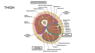
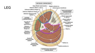
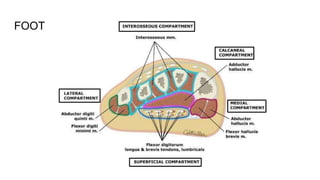
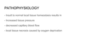
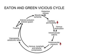
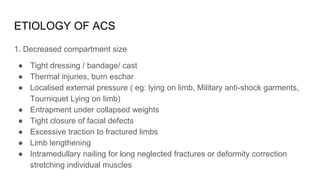
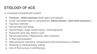
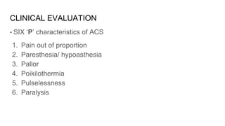
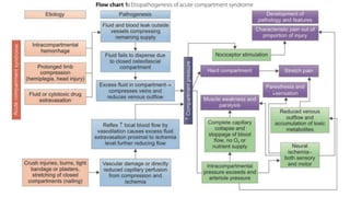
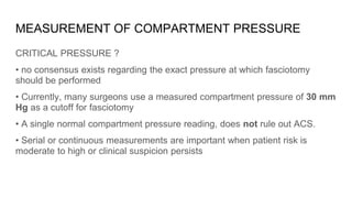
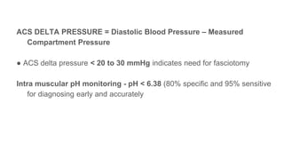
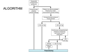
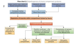
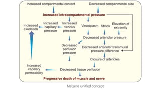
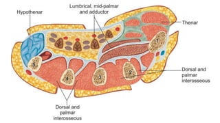
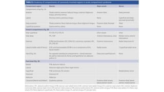
Orthopedics is a Reconstructive Surgery. Mangled extremity is an injury to at least three out of four systems (soft tissue, bone, nerves, and vessels). A Decision have to be made Amputation + Prosthesis Vs. Limb salvage procedure which includes Irrigation & Debridement, External fixation, Antibiotic bead spacers, Soft tissue coverage and finally Restoring Skeletal Stability by Salvage of Bone Defect
Hoffa's Fracture: Diagnosis, management & New Classification System by BAGARI...Vaibhav Bagaria
Hoffa's Fracture - coronal split fracture of distal femur, its diagnosis, management strategy, a new classification and tips and tricks of management. First described Hoffa, a new classification system by Bagaria et al helps plan the surgery for these tricky fracture. The most crucial step is not to miss these fractures in ER.
Spine and extremity injuries are common among people of all ages and can have a significant impact on mobility and quality of life. This PowerPoint presentation provides a comprehensive overview of spine and extremity injuries, including the causes, symptoms, and treatment options.
Through powerful images and personal stories, we showcase the impact of spine and extremity injuries on individuals, families, and communities. We highlight the challenges of accessing healthcare and rehabilitation services, particularly in low-resource settings, and the importance of early intervention and treatment.
The presentation provides detailed information about the various types of spine and extremity injuries, including fractures, dislocations, and soft tissue injuries. We also discuss the diagnostic procedures, including imaging tests and physical exams, and the treatment options, such as surgery, physical therapy, and pain management.
In addition, we explore the efforts being made to prevent and manage spine and extremity injuries. We highlight the importance of safety precautions, such as proper equipment use and ergonomic work practices, and the role of rehabilitation services in promoting recovery and restoring function.
Through this PowerPoint presentation, we aim to raise awareness about spine and extremity injuries and the importance of early diagnosis and treatment. We showcase the latest research and innovations in injury prevention and treatment, and the importance of collaboration and partnership to address the disease.
We urge the audience to take action in the fight against spine and extremity injuries, whether it be through spreading awareness, supporting organizations working on the ground, or advocating for policy change. Let us come together to create a world where everyone has access to the care and support they need to recover from spine and extremity injuries and live healthy, fulfilling lives.
2024.06.01 Introducing a competency framework for languag learning materials ...Sandy Millin
http://sandymillin.wordpress.com/iateflwebinar2024
Published classroom materials form the basis of syllabuses, drive teacher professional development, and have a potentially huge influence on learners, teachers and education systems. All teachers also create their own materials, whether a few sentences on a blackboard, a highly-structured fully-realised online course, or anything in between. Despite this, the knowledge and skills needed to create effective language learning materials are rarely part of teacher training, and are mostly learnt by trial and error.
Knowledge and skills frameworks, generally called competency frameworks, for ELT teachers, trainers and managers have existed for a few years now. However, until I created one for my MA dissertation, there wasn’t one drawing together what we need to know and do to be able to effectively produce language learning materials.
This webinar will introduce you to my framework, highlighting the key competencies I identified from my research. It will also show how anybody involved in language teaching (any language, not just English!), teacher training, managing schools or developing language learning materials can benefit from using the framework.
The French Revolution, which began in 1789, was a period of radical social and political upheaval in France. It marked the decline of absolute monarchies, the rise of secular and democratic republics, and the eventual rise of Napoleon Bonaparte. This revolutionary period is crucial in understanding the transition from feudalism to modernity in Europe.
For more information, visit-www.vavaclasses.com
Honest Reviews of Tim Han LMA Course Program.pptxtimhan337
Personal development courses are widely available today, with each one promising life-changing outcomes. Tim Han’s Life Mastery Achievers (LMA) Course has drawn a lot of interest. In addition to offering my frank assessment of Success Insider’s LMA Course, this piece examines the course’s effects via a variety of Tim Han LMA course reviews and Success Insider comments.
Synthetic Fiber Construction in lab .pptxPavel ( NSTU)
Synthetic fiber production is a fascinating and complex field that blends chemistry, engineering, and environmental science. By understanding these aspects, students can gain a comprehensive view of synthetic fiber production, its impact on society and the environment, and the potential for future innovations. Synthetic fibers play a crucial role in modern society, impacting various aspects of daily life, industry, and the environment. ynthetic fibers are integral to modern life, offering a range of benefits from cost-effectiveness and versatility to innovative applications and performance characteristics. While they pose environmental challenges, ongoing research and development aim to create more sustainable and eco-friendly alternatives. Understanding the importance of synthetic fibers helps in appreciating their role in the economy, industry, and daily life, while also emphasizing the need for sustainable practices and innovation.
Palestine last event orientationfvgnh .pptxRaedMohamed3
An EFL lesson about the current events in Palestine. It is intended to be for intermediate students who wish to increase their listening skills through a short lesson in power point.
Unit 8 - Information and Communication Technology (Paper I).pdfThiyagu K
This slides describes the basic concepts of ICT, basics of Email, Emerging Technology and Digital Initiatives in Education. This presentations aligns with the UGC Paper I syllabus.
Instructions for Submissions thorugh G- Classroom.pptxJheel Barad
This presentation provides a briefing on how to upload submissions and documents in Google Classroom. It was prepared as part of an orientation for new Sainik School in-service teacher trainees. As a training officer, my goal is to ensure that you are comfortable and proficient with this essential tool for managing assignments and fostering student engagement.
Introduction to AI for Nonprofits with Tapp NetworkTechSoup
Dive into the world of AI! Experts Jon Hill and Tareq Monaur will guide you through AI's role in enhancing nonprofit websites and basic marketing strategies, making it easy to understand and apply.
How to Make a Field invisible in Odoo 17Celine George
It is possible to hide or invisible some fields in odoo. Commonly using “invisible” attribute in the field definition to invisible the fields. This slide will show how to make a field invisible in odoo 17.
The Roman Empire A Historical Colossus.pdfkaushalkr1407
The Roman Empire, a vast and enduring power, stands as one of history's most remarkable civilizations, leaving an indelible imprint on the world. It emerged from the Roman Republic, transitioning into an imperial powerhouse under the leadership of Augustus Caesar in 27 BCE. This transformation marked the beginning of an era defined by unprecedented territorial expansion, architectural marvels, and profound cultural influence.
The empire's roots lie in the city of Rome, founded, according to legend, by Romulus in 753 BCE. Over centuries, Rome evolved from a small settlement to a formidable republic, characterized by a complex political system with elected officials and checks on power. However, internal strife, class conflicts, and military ambitions paved the way for the end of the Republic. Julius Caesar’s dictatorship and subsequent assassination in 44 BCE created a power vacuum, leading to a civil war. Octavian, later Augustus, emerged victorious, heralding the Roman Empire’s birth.
Under Augustus, the empire experienced the Pax Romana, a 200-year period of relative peace and stability. Augustus reformed the military, established efficient administrative systems, and initiated grand construction projects. The empire's borders expanded, encompassing territories from Britain to Egypt and from Spain to the Euphrates. Roman legions, renowned for their discipline and engineering prowess, secured and maintained these vast territories, building roads, fortifications, and cities that facilitated control and integration.
The Roman Empire’s society was hierarchical, with a rigid class system. At the top were the patricians, wealthy elites who held significant political power. Below them were the plebeians, free citizens with limited political influence, and the vast numbers of slaves who formed the backbone of the economy. The family unit was central, governed by the paterfamilias, the male head who held absolute authority.
Culturally, the Romans were eclectic, absorbing and adapting elements from the civilizations they encountered, particularly the Greeks. Roman art, literature, and philosophy reflected this synthesis, creating a rich cultural tapestry. Latin, the Roman language, became the lingua franca of the Western world, influencing numerous modern languages.
Roman architecture and engineering achievements were monumental. They perfected the arch, vault, and dome, constructing enduring structures like the Colosseum, Pantheon, and aqueducts. These engineering marvels not only showcased Roman ingenuity but also served practical purposes, from public entertainment to water supply.
Welcome to TechSoup New Member Orientation and Q&A (May 2024).pdfTechSoup
In this webinar you will learn how your organization can access TechSoup's wide variety of product discount and donation programs. From hardware to software, we'll give you a tour of the tools available to help your nonprofit with productivity, collaboration, financial management, donor tracking, security, and more.
3. DEFINITION
The muscle groups of the human limbs are divided into sections, or
compartments, formed by strong, unyielding fascial membranes.
"Elevation of the interstitial pressure in these closed osteofascial
compartment that result in microvascular compromise"
Most commonly involved - Compartments with relatively noncompliant fascial or
osseous structures (eg : anterior and deep posterior compartments of leg, volar
compartment of forearm)
4. TYPES
• Depending on the cause of the increased pressure and the duration of
symptoms
• A) ACUTE COMPARTMENT SYNDROME (surgical emergency)
• B) CHRONIC EXERTIONAL COMPARTMENT SYNDROME
5. ANATOMY OF COMPARTMENTS
Closed area of muscles, blood vessels and nerves surrounded by fascia
• ARM - 2 compartments
• FOREARM - 4 compartments
• HAND - 10 compartments
• THIGH - 3 compartments
• LEG - 4 compartments
• FOOT- 9 compartments
12. PATHOPHYSIOLOGY
- Insult to normal local tissue homeostasis results in
- Increased tissue pressure
- decreased capillary blood flow
- local tissue necrosis caused by oxygen deprivation
14. ETIOLOGY OF ACS
1. Decreased compartment size
● Tight dressing / bandage/ cast
● Thermal injuries, burn eschar
● Localised external pressure ( eg: lying on limb, Military anti-shock garments,
Tourniquet Lying on limb)
● Entrapment under collapsed weights
● Tight closure of facial defects
● Excessive traction to fractured limbs
● Limb lengthening
● Intramedullary nailing for long neglected fractures or deformity correction
stretching individual muscles
15. ETIOLOGY OF ACS
2. Increased compartment content
● Fracture - most common (both open and closed)
● Crush and direct blow to compartment (blunt trauma - 2nd most common )
● Vigorous exercise
● Animal bites and stings
● Hemorrhage ( large vessel injury) / anticoagulants
● Ruptured cysts (eg: baker’s cyst)
● Revascularization / Reperfusion after ischemia
● IV fluid extravasation
● High-pressure injections, Intraosseous fluid transfusion in children
● Reaming in intramedullary nailing
● Use of fluid pumps in arthroscopy
16. CLINICAL EVALUATION
• SIX ‘P’ characteristics of ACS
1. Pain out of proportion
2. Paresthesia/ hypoasthesia
3. Pallor
4. Poikilothermia
5. Pulselessness
6. Paralysis
17. PAIN
• Earliest symptom
• Classically out of proportion to injury
• Burning and deep aching in nature (symptom)
• Exaggerated with passive stretch of muscles (sign)
• Bony injury may cause pain(false positive)
• pain may be absent if nerves injured or patient has received efficient
analgesia/ anesthesia (postoperative)
• Can’t evaluate- head injury, drug abuse, intubated or sedated etc
18. PARESTHESIA
• Unreliable early complaint
• In acute anterior lower leg CS, the first sign to develop may be numbness
between the first 2 toes (superficial peroneal nerve),
• Test: pinprick, light touch , 2 point discrimination
• Decreased light touch is the best and first indicator
• Decreased 2-point discrimination is also a reliable early test
19. PARALYSIS
• Very late finding (being a function of thicker motor nerves)
• Irreversible nerve and muscle damage present
• Paresis may be present early but difficult to evaluate because of pain
• If objective evidence of a major sensory deficit, a motor deficit, or loss of
peripheral pulse is found, the syndrome is far advanced
20. PALLOR & PULSELESSNESS
• Rarely present
• Distal pulses are almost always present
• Indicates direct damage to vessels rather than compartment syndrome
• Vascular injury may be more of contributing factor to syndrome rather than
result
21.
22. DIAGNOSIS
Based on
● The history
● Clinical Examination findings
● The measurement of compartment pressures (not needed
always)
● Intramuscular pH monitoring ( rarely done)
23. MEASUREMENT OF COMPARTMENT PRESSURE
CRITICAL PRESSURE ?
• no consensus exists regarding the exact pressure at which fasciotomy
should be performed
• Currently, many surgeons use a measured compartment pressure of 30 mm
Hg as a cutoff for fasciotomy
• A single normal compartment pressure reading, does not rule out ACS.
• Serial or continuous measurements are important when patient risk is
moderate to high or clinical suspicion persists
24. ACS DELTA PRESSURE = Diastolic Blood Pressure ‒ Measured
Compartment Pressure
● ACS delta pressure < 20 to 30 mmHg indicates need for fasciotomy
Intra muscular pH monitoring - pH < 6.38 (80% specific and 95% sensitive
for diagnosing early and accurately
25. INTERPRETATION OF MEASUREMENTS
● The normal pressure of a tissue compartment falls between 0 and
8 mmHg .
● Compartment pressure > 30 mm of hg
● Delta pressure < 20-30 mm of hg
26. MEASUREMENT OF COMPARTMENT PRESSURE
INDICATIONS
● High clinical suspicion
● Significant tissue injury
● If clinical findings are equivocal or difficult to interpret (unresponsive
patients, uncooperative patients, Children, with multiple or distracting
injuries)
● Signs and symptoms not resolved after 60 min of appropriate
precaution
● Prophylactic with major corrective osteotomy of the leg and forearm
28. • TECHNIQUE - performed in each compartment as close to the fracture
site as possible (< 5 cm both proximally and distally) or maximal swelling
area
• Highest pressure is noted - is compared to diastolic blood pressure
• If Marginal readings - repeat physical examination and pressure
measurement
29. NON INVASIVE METHODS
• Ultrasonography: measures submicrometric displacement of fascia by
volume expansion
• Laser Doppler Flowmetry
• Infrared spectroscopy (NIRS) : determine temperature difference between
proximal and distal skin surface and tracking variations in the oxygenation of
muscle tissue
BECOMING POPULAR AND STANDARDISED
30. INVASIVE METHODS
Direct methods
● SOLID STATE TRANSDUCER TIPPED CATHETER SYSTEM
Indirect methods
● STRYKER HANDHELD SYSTEM
● ARTERIAL LINE MANOMETER
● WHITESIDES THREE WAY STOPCOCK APPARATUS
● WICK MONITOR
● STIC TECHNIQUE
31. STRYKER HANDHELD SYSTEM
The transducer is connected to a needle that has several side ports. These
side ports help eliminate muscle occlusion of the needle
32. WHITESIDES THREE WAY STOPCOCK APPARATUS
SANGWAN TECHNIQUE - saline manometer, cheap and easy to assemble
A simple arterial line setup (16–18 gauge needle) and a mercury manometer
-least accurate
-values consistently higher,
by an average of 5–19 mm Hg
33. WICK MONITOR
Dexon fibers that are fixed at the tip of a fluid filled polyethylene catheter (in
the form of a “wick”)
35. MANAGEMENT
EARLY MANAGEMENT- older concept , debatable
reserved only for patients presenting too late or missed (> 24–48 hours)
● remove cast or bandage
● positioning of the limb at the level of heart (do not elevate the affected
limb which decreases arterial pressure- further tissue injury)
● correct hypoperfusion- hydration with IV fluids, blood products (if acute
anemia)
● ensure patient is normotensive
● oxygen supplementation
● Role of mannitol - doubtful
36. MANAGEMENT
• NON-OPERATIVE
-if the stage is of impending compartment syndrome (serial clinical evaluation
and compartment pressure monitoring)
• OPERATIVE
-Emergency Fasciotomy + fracture stabilisation
37. FASCIOTOMY - prophylactic release of compartment pressure before
permanent damage occurs.
- Emergency procedure
- Will not reverse injury from trauma
- first suggested by Bardenheuer in 1906 and Murphy in 1914
FRACTURE CARE - stabilization ( Ex fix, IM nail )
38. INDICATIONS OF FASCIOTOMY
● Normotensive with positive clinical findings
● Compartment pressure > 30 mmHg
● Delta pressure < 25 mm hg
● If duration of increased pressure unknown or > 8 hours
● Worsening of clinical conditions
● Those who are uncooperative or unconscious, with pressure > 30 mm Hg
● Hypotensive and a compartment pressure > 20 mm Hg
● In hand > 15-20 mm Hg is a relative indication for release.
● Unequivocal clinical findings
39. CONTRAINDICATION OF FASCIOTOMY
Missed compartment syndrome - > 24-48 hours
- damage cannot be reversed
- significant infection rate when dead tissue exposed
40. FASCIOTOMY PRINCIPLES
• Make early diagnosis
• Long extensile incisions
• Release all fascial compartments
• Preserve neurovascular structures
• Debride necrotic tissues
• Coverage within 7-10 days
43. Volar curvilinear approach of Henry
● Skin incision proximal to the antecubital fossa on ulnar aspect of the arm
crossing the antecubital fossa horizontally in the flexion crease
● Extend incision - S-shape to the
wrist flexion crease and then cross
the crease into the palm to allow
release of the carpal tunnel.
44. ● Release the superficial fascia, and identify median nerve proximally and
release the lacertus fibrosus.
● Release the proximal edge of flexor pronator mass from humerus.
● Follow the median nerve between the FDS and FDP and decompress it
all the way to the carpal tunnel.
● Identify and preserve the palmar cutaneous branch. Release the carpal
tunnel.
● Inspect the deep volar muscles (FDP, FPL, PQ) and release fascial
coverings as needed.
45. Dorsal Approach
● Incision is similar to that used in the Thompson approach
● can extend from the lateral epicondyle to the mid-portion of the wrist
● Full incision may not be needed
● fascia of dorsal and mobile wad compartments is easily released.
46. DECOMPRESSION OF LEG
1) The two-incision techniques of Mubarak et al.
a) Anterolateral incision
b) Posteromedial incision
1) Single-incision technique of Davey et al.
47. Anterolateral incision
● To approach the anterior and lateral compartments
● extensive longitudinal skin incision, halfway between the fibular shaft
and tibial crest.
● Undermine the skin edges to widen exposure of the fascia.
● Identify anterior intermuscular septum
● Identify superficial peroneal nerve, which lies in the lateral
compartment next to the septum.
● Open the anterior compartment fascia and ensure complete fascial
release.
48. ● Make the lateral compartment fasciotomy in line with the fibular shaft.
● Direct the scissors proximally toward the fibular head and distally
toward the lateral malleolus.
● The fascial incision is posterior to
the superficial peroneal nerve.
49. Posteromedial incision
● longitudinal incision 2–3 cm posterior to the posterior tibial margin, to
approach the superficial and deep posterior compartments
● Retract saphenous vein and nerve anteriorly.
● Identify septum between the deep and superficial posterior
compartments
● Identify the tendon of the FDL in the deep post compartment and the
Achilles tendon in the superficial post compartment.
50. ● Decompress the superficial post compartment first by releasing fascia
proximally as far as possible and then distally behind the medial
malleolus.
● Release the deep post compartment distally and then proximally
under the soleus bridge.
● Release the soleus if it attaches to tibia distally more than halfway.
● If both compartments are anchored to the posteromedial edge of tibia,
the deep compartment is released from within the superficial
compartment by retracting soleus.
51.
52. SINGLE-INCISION PERIFIBULAR FASCIOTOMY
DAVEY ET AL
● Make a single longitudinal, lateral incision in line with the fibula,
extending from just distal to the head of the fibula to 3 to 4 cm proximal to
the lateral malleolus
● Undermine the skin anteriorly and avoid injuring the superficial peroneal
nerve.
● Perform a longitudinal fasciotomy of the anterior and lateral
compartments
● Undermine the skin posteriorly and perform a fasciotomy of the
superficial posterior compartment
53. ● Identify the interval between the superficial and lateral compartments
distally and develop this interval proximally by detaching the soleus from
the fibula.
● Subperiosteally dissect the flexor hallucis longus from the fibula.
● Retract the muscle and the peroneal vessels posteriorly.
● Identify the fascial attachment of the posterior tibial muscle to the fibula
and incise this fascia longitudinally
● Close only the skin over a suction drain or a negative pressure wound
device.
54.
55. FOOT DECOMPRESSION
● DORSAL—two incisions
overlying the second and fourth metatarsals is the gold standard.
Maintain the widest skin bridge and bluntly approach the metatarsals.
Continue blunt dissection into the web spaces, and decompress the
hematoma.
● MEDIAL—one incision
along the inferior border of first metatarsal, but superior to abductor muscle.
Enter the central compartment from superior aspect of abductor muscle.
56. ● CALCANEAL —one incision
uncommonly used
beginning medially, from the inferior border of the posterior tuberosity
extending toward the inferior surface of the first metatarsal.
DORSAL APPROACH. MEDIAL APPROACH
57. FASCIOTOMY OF THE THIGH
TARLOW ET AL.
● Prepare and drape the thigh exposing the limb from the iliac crest to
the knee joint.
● Make a lateral incision beginning just distal to the intertrochanteric line
and extending to the lateral epicondyle
● Use subcutaneous dissection to expose the iliotibial band and then
make a straight incision in line with the skin incision through the
iliotibial band
● reflect the vastus lateralis off the lateral intermuscular septum.
58. ● Make a 1.5-cm incision in the lateral intermuscular septum and, using
Metzenbaum scissors, extend it proximally and distally the length of
the incision.
● After the anterior and posterior compartments released, measure the
pressure of the medial compartment.
● If the pressure is elevated, make a separate medial incision to release
the adductor compartment.
59.
60. FRACTURE CARE
Stabilization of fracture is essential along with compartment decompression.
● For supracondylar fractures of humerus in children - K-wires are
preferred.
● metatarsal and metacarpal fractures - K-wires are preferred.
● Forearm fractures - compression or locked plates.
● Femur or tibia fractures - nailing is not good option (can worsen an
existing compartment syndrome) External fixator is better
61. POST OPERATIVE CARE AND REHABILITATION
● Pack the wounds loosely open (saline-dampened gauze)
● apply a bulky dressing + splinting in functional position
● Keep extremity above the level of heart to aid venous drainage.
● Shoe- lace technique of placing vessel loop suture and skin staple help in
gradual closure of skin wound.
● The sutures can be pulled even at bedside.
62. ● Return to OT for 2nd look in 2-5 days
- IV fluorescein and a Wood light - helpful in evaluating muscle viability
- If no muscle necrosis- then skin is loosely closed
- If closure is not accomplished
- Skin closure or SSG
●VAC (Vaccum Assisted Closure) highly
recommended immediate post -op
● Hyperbaric Oxygen
63. ● Begin active and active-assisted range-of-motion of the adjacent joints on
the second day after fasciotomy.
● After split-thickness skin grafting,
immobilize the limb for 3–5 days more.
64. Drugs that may help recovery from metabolic effects
by primarily scavenging free radicals
● Allopurinol
● Superoxide dismutase
● Deferoxamine
● Pentafraction of hydroxyethyl starch.
65. COMPLICATIONS
● Myonecrosis
● Nerve damage (Sensory and motor loss, Chronic pain)
● Volkmann ischemic contracture
● Reperfusion syndrome
● Recurrent ulceration and muscle herniation
● Infection
● Amputation
● Death
66. MYONECROSIS
● Functional impairement - after 2- 4 hr of ischemia
● Irreversible functional loss - after 4 -12 hr of ischemia
● CRUSH SYNDROME - sequelae of muscle necrosis (creatine
phosphokinase >20,000 IU) -
nonoliguria renal failure, myoglobinuria,
oliguria, shock, acidosis, hyperkalemia
and cardiac arrhythmias.
71. CHRONIC COMPARTMENT SYNDROME
● Also known as EXERTIONAL CS, RECURRENT CS, SUBACUTE CS
● Typical patients are young athelet (long distance runner) and military
recruits
● Occur mainly in lower limb
72. PATHOPHYSIOLOGY
Not yet fully understood
- Probably muscle volume can increase up to 20% of its resting size during
exercise
- increased muscle relaxation pressure during exercise
- Decreased muscle blood flow
- Ischemic pain and impaired muscle function
- Approximately 15% to 40% of patients treated for chronic exertional
compartment syndrome have been found to have a fascial hernia
74. PHYSICAL EXAM IN CCS
• Exercise/ Exertion induced pain
• Tenderness over the compartment
• Bilateral involvement is very common
• Fascial hernia common
76. WORK UP OF CCS
• Plain X ray- show 90% of stress fracture
• Bone scan - diffuse uptake - periostitis
- localized uptake - stress fracture
• Tinel test - +ve in nerve entrapment
• NCS - could be helpful
• MRI - promising results, fatty infiltration associated with chronic ischemia
• Near infrared spectroscopy
77. DIAGNOSIS OF CCS
• Intracompartmental testing is the hallmark of diagnosis
1) Pre-exercise resting pressure of 15 mm hg or more
2) After 1 minute of exercise pressure of 30 mm hg
3) After 5 minute of exercise pressure of 20 mm hg or more
4) An elevated pressure > 25mm hg after 15 minutes is a more reliable cutoff
78. TREATMENT OF CCS
• NONOPERATIVE-
- Analgesics (eg: NSAID)
- Electrostimulation
- Muscle relaxant
- Cessation or significant reduction of athletic activities
79. • OPERATIVE TREATMENT-
1. Single Incision Fasciotomy
2. Double Mini Incision Fasciotomy
3. Double Incision Fasciotomy
80. SINGLE INCISION FASCIOTOMY
● (FRONEK ET AL.)
● 5-cm longitudinal incision halfway btw the fibula and the tibial crest in the
midportion of the leg or over the fascial defect (if a muscular hernia is present
at the exit of the superficial peroneal nerve)
● Identify the nerve and the intermuscular septum
● pass a fasciotome into the anterior compartment in line with the anterior tibial
muscle
● In the lateral compartment, run the fasciotome posterior to the superficial
peroneal nerve in line with the fibular shaft
● Do not repair muscular hernias.
● Close the skin in the usual fashion and apply a sterile dressing.
82. DOUBLE MINI-INCISION FASCIOTOMY
● (MOUHSINE ET AL.)
● Without the use of a tourniquet, make two vertical 2-cm skin incisions
over the anterior compartment 15 cm apart
● Identify the fascia and carefully develop a subcutaneous flap with blunt
dissection of the bridge of skin and subcutaneous tissue.
● Identify the anterior intermuscular septum and the superficial peroneal
nerve through the distal incision (10 to 12 cm proximal to the lateral
malleolus).
83. ● Retract the skin anteriorly and posteriorly to allow anterior and/or lateral
fasciotomy under direct vision
● If needed, use a gloved finger to complete the release.
● Close the two incisions with 3.0 monofilament and sterile adhesive strips
and apply a firm bandage from midfoot to the knee.
85. DOUBLE INCISION FASCIOTOMY
● (RORABECK)
● Make two incisions in the leg 1 cm behind the posteromedial border of the
tibia
● Identify the saphenous vein in the proximal incision and retract it anteriorly
along with the nerve.
● Enter and release the superficial compartment
● Incise the deep fascia
● Expose the deep compartment, including the flexor digitorum longus and
posterior tibial muscles, by detaching the soleal bridge.
86. ● Identify the neurovascular bundle and posterior tibial tendon
● incise proximally and distally the fascia overlying the tendon. The posterior
tibial tendon is the key to posterior compartment decompression, and it
usually is constricted proximally between the two origins of the flexor hallucis
longus; enlarge the opening between these two structures to check for
constriction.
● Release the tourniquet and obtain meticulous hemostasis.
● Close the wound over a suction drain to minimize the risk of hematoma
formation
88. POST FASCIOTOMY CARE
- Close the wound with an interdermal running stitch
- Apply light dressing
- Early ROM encouraged
- Weight bearing on crutches is allowed on POD1
- After suture removal at 2-3 weeks Light jogging is allowed
- exercises as tolerated over the next 3–6 weeks according to the
patient’s abilities and pain tolerance.
89. COMPLICATIONS
● Hemorrhage
● Skin breakdown
● Altered sensation over the fasciotomy site
● Recurrence (6–11%)—more with subcutaneous and endoscopic
technique
● Infection
● Deep vein thrombosis
● Vascular injury
● Nerve damage / Nerve entrapment
● Lymphocele
● Complex regional pain syndrome.
90. REFERENCE
● Campbell's Operative Orthopaedics - 14th Edition
● Handbook of Fractures 6th Edition 2019 By Kenneth Egol, Kenneth J. Koval &
Joseph Zuckerman
● UpToDate: Evidence-based Clinical Decision Support
● Essential Orthopedics Principles & Practice by Manish Kumar Varshney