This document summarizes pelvic anatomy, classification of pelvic ring injuries, and radiographic evaluation. It describes:
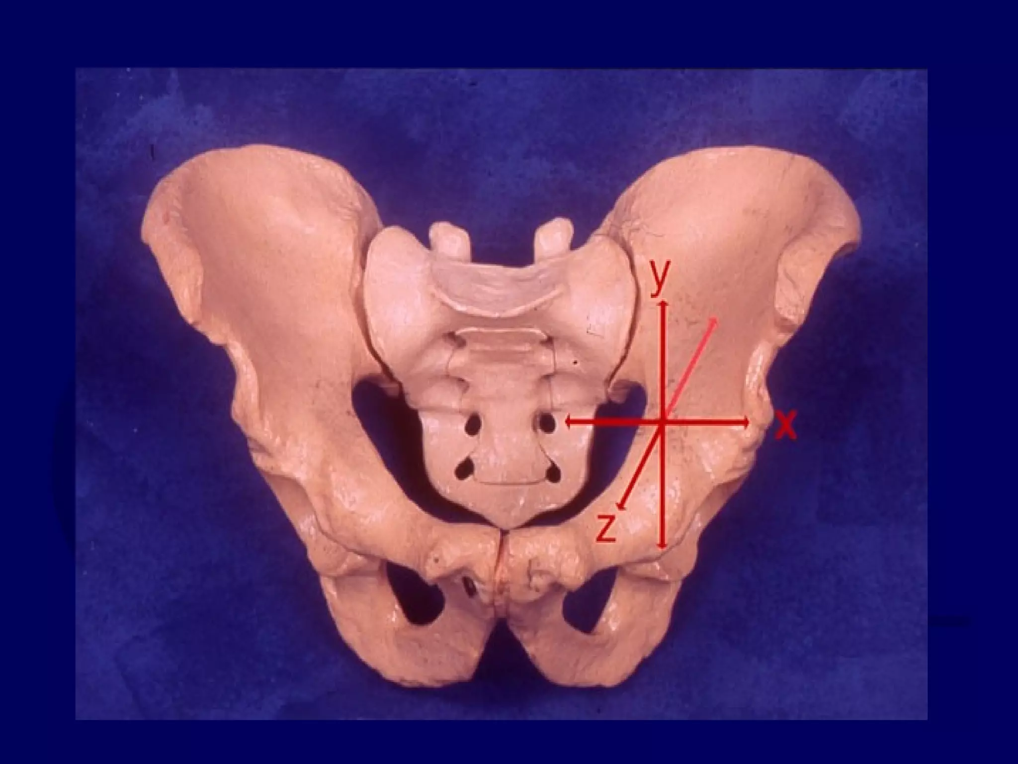
1) Important pelvic landmarks, ligaments, muscles, and arterial bleeders. Neurologic damage most commonly involves L5 and S1 nerves.
2) Classification systems include the anatomical Letournel system and the stability-focused systems of Pennal, Bucholz, and Tile.
3) The Young-Burgess system predicts associated injuries based on the vector force and classifies injuries as lateral compression, anteroposterior compression, or vertical shear.
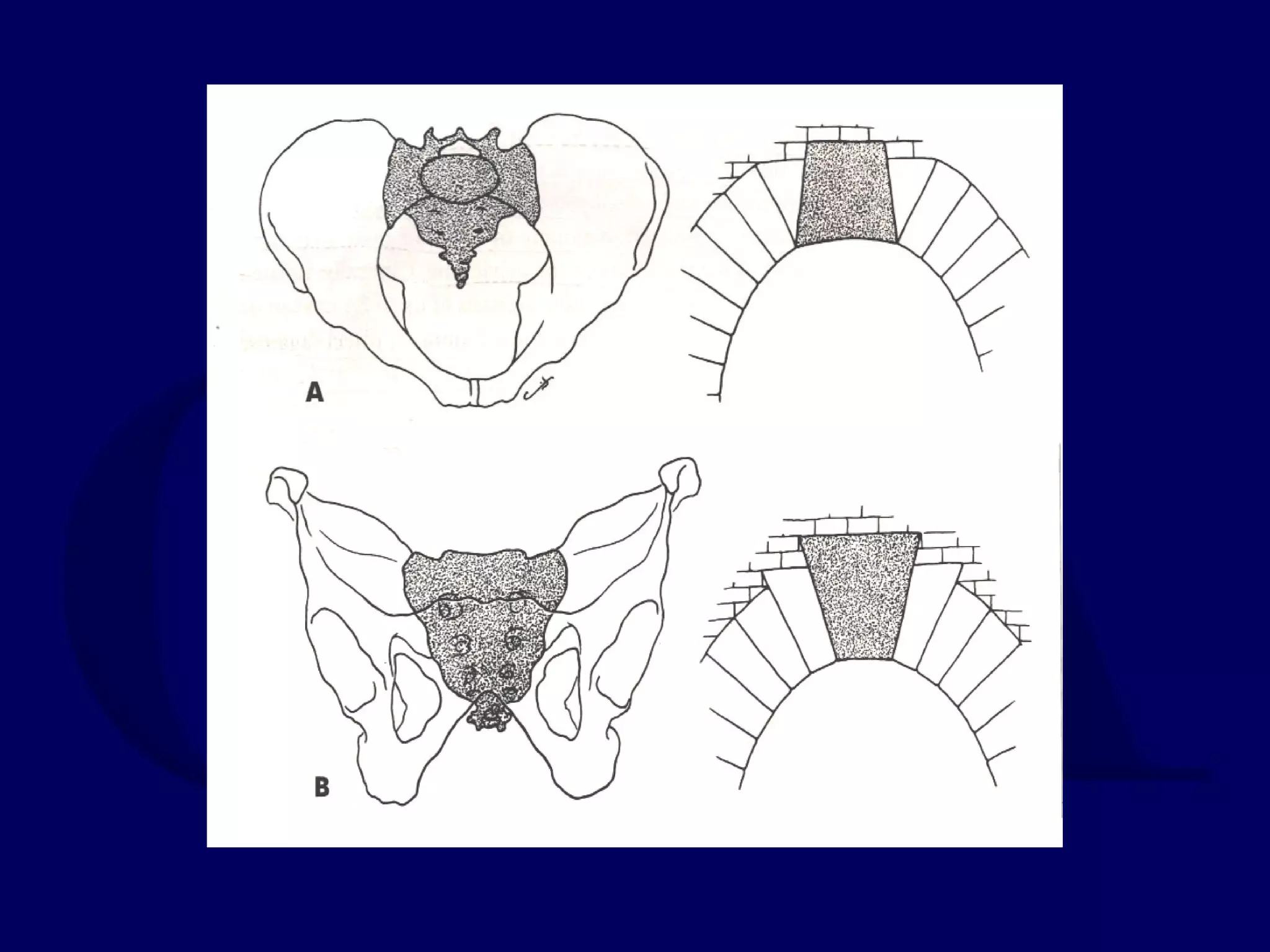
4) Radiographic evaluation involves anteroposterior, inlet, and outlet views on radiographs
Disorders of the Great toe (hallux) are very important as they are very painful, causes many clinical symptoms,and very difficult to treat.The presentation compiled from various important orthopedic textbooks and international journals.
HUMAN ANATOMY
regional anatomy
regional anatomy of pelvic
changsha medical university lecture
csmu lecture by an chen
uploaded by Prabesh raj jamkatel
pelvics
Trauma Image Interpretation of the Pelvis and Hip Radiographs: Using ABCSuk121chris
An informative presentation describing basics of image interpretation for the Pelvis and Proximal Hip by utilising ABCS; a step-by-step method described by Otto Chan's book entitled ABC to Emergency Radiology. This presentation includes local and external image examples of traumatic abnormalities of the pelvis and hip. Radiographers, Nurses and Emergency Doctors may find this useful to enhance their image interpretation skills. This presentation was developed for a In-service CPD session in 2013. Questions and/or feedback are welcome by email: abigheadache [at] gmail.com
All about Spondyloarthropaties also known as Seronegative Arthritis in a nutshell....includes Pathology,signs and symptoms, investigations, and latest approved treatment of all subtypes....compiled from Turek and Harrisons textbook.
Radiological evaluation of Lower Limb in acute ED setting !!Runal Shah
Radiological evaluation of Lower Limb in acute ED setting !!
How to evaluate lower limb injuries in ED by primary look out... How to assess simple bony injuries ! A simple radiological approach for ED physicians..
Basic spine anatomy is the first step in understanding the spine profession. Being familiar with spine anatomy makes you spine-minded, understand pathological spine diseases, correlate symptoms and signs, and facilitate your surgical skills.
This is a teaching lecture given twice by Prof. Dr. Mohamed Mohi Eldin, professor of neurosurgery, in the Basic Spine Course, Egyptian Medical Syndicate, Cairo, March 2009 and in 2010.
Ethanol (CH3CH2OH), or beverage alcohol, is a two-carbon alcohol
that is rapidly distributed in the body and brain. Ethanol alters many
neurochemical systems and has rewarding and addictive properties. It
is the oldest recreational drug and likely contributes to more morbidity,
mortality, and public health costs than all illicit drugs combined. The
5th edition of the Diagnostic and Statistical Manual of Mental Disorders
(DSM-5) integrates alcohol abuse and alcohol dependence into a single
disorder called alcohol use disorder (AUD), with mild, moderate,
and severe subclassifications (American Psychiatric Association, 2013).
In the DSM-5, all types of substance abuse and dependence have been
combined into a single substance use disorder (SUD) on a continuum
from mild to severe. A diagnosis of AUD requires that at least two of
the 11 DSM-5 behaviors be present within a 12-month period (mild
AUD: 2–3 criteria; moderate AUD: 4–5 criteria; severe AUD: 6–11 criteria).
The four main behavioral effects of AUD are impaired control over
drinking, negative social consequences, risky use, and altered physiological
effects (tolerance, withdrawal). This chapter presents an overview
of the prevalence and harmful consequences of AUD in the U.S.,
the systemic nature of the disease, neurocircuitry and stages of AUD,
comorbidities, fetal alcohol spectrum disorders, genetic risk factors, and
pharmacotherapies for AUD.
Flu Vaccine Alert in Bangalore Karnatakaaddon Scans
As flu season approaches, health officials in Bangalore, Karnataka, are urging residents to get their flu vaccinations. The seasonal flu, while common, can lead to severe health complications, particularly for vulnerable populations such as young children, the elderly, and those with underlying health conditions.
Dr. Vidisha Kumari, a leading epidemiologist in Bangalore, emphasizes the importance of getting vaccinated. "The flu vaccine is our best defense against the influenza virus. It not only protects individuals but also helps prevent the spread of the virus in our communities," he says.
This year, the flu season is expected to coincide with a potential increase in other respiratory illnesses. The Karnataka Health Department has launched an awareness campaign highlighting the significance of flu vaccinations. They have set up multiple vaccination centers across Bangalore, making it convenient for residents to receive their shots.
To encourage widespread vaccination, the government is also collaborating with local schools, workplaces, and community centers to facilitate vaccination drives. Special attention is being given to ensuring that the vaccine is accessible to all, including marginalized communities who may have limited access to healthcare.
Residents are reminded that the flu vaccine is safe and effective. Common side effects are mild and may include soreness at the injection site, mild fever, or muscle aches. These side effects are generally short-lived and far less severe than the flu itself.
Healthcare providers are also stressing the importance of continuing COVID-19 precautions. Wearing masks, practicing good hand hygiene, and maintaining social distancing are still crucial, especially in crowded places.
Protect yourself and your loved ones by getting vaccinated. Together, we can help keep Bangalore healthy and safe this flu season. For more information on vaccination centers and schedules, residents can visit the Karnataka Health Department’s official website or follow their social media pages.
Stay informed, stay safe, and get your flu shot today!
Lung Cancer: Artificial Intelligence, Synergetics, Complex System Analysis, S...Oleg Kshivets
RESULTS: Overall life span (LS) was 2252.1±1742.5 days and cumulative 5-year survival (5YS) reached 73.2%, 10 years – 64.8%, 20 years – 42.5%. 513 LCP lived more than 5 years (LS=3124.6±1525.6 days), 148 LCP – more than 10 years (LS=5054.4±1504.1 days).199 LCP died because of LC (LS=562.7±374.5 days). 5YS of LCP after bi/lobectomies was significantly superior in comparison with LCP after pneumonectomies (78.1% vs.63.7%, P=0.00001 by log-rank test). AT significantly improved 5YS (66.3% vs. 34.8%) (P=0.00000 by log-rank test) only for LCP with N1-2. Cox modeling displayed that 5YS of LCP significantly depended on: phase transition (PT) early-invasive LC in terms of synergetics, PT N0—N12, cell ratio factors (ratio between cancer cells- CC and blood cells subpopulations), G1-3, histology, glucose, AT, blood cell circuit, prothrombin index, heparin tolerance, recalcification time (P=0.000-0.038). Neural networks, genetic algorithm selection and bootstrap simulation revealed relationships between 5YS and PT early-invasive LC (rank=1), PT N0—N12 (rank=2), thrombocytes/CC (3), erythrocytes/CC (4), eosinophils/CC (5), healthy cells/CC (6), lymphocytes/CC (7), segmented neutrophils/CC (8), stick neutrophils/CC (9), monocytes/CC (10); leucocytes/CC (11). Correct prediction of 5YS was 100% by neural networks computing (area under ROC curve=1.0; error=0.0).
CONCLUSIONS: 5YS of LCP after radical procedures significantly depended on: 1) PT early-invasive cancer; 2) PT N0--N12; 3) cell ratio factors; 4) blood cell circuit; 5) biochemical factors; 6) hemostasis system; 7) AT; 8) LC characteristics; 9) LC cell dynamics; 10) surgery type: lobectomy/pneumonectomy; 11) anthropometric data. Optimal diagnosis and treatment strategies for LC are: 1) screening and early detection of LC; 2) availability of experienced thoracic surgeons because of complexity of radical procedures; 3) aggressive en block surgery and adequate lymph node dissection for completeness; 4) precise prediction; 5) adjuvant chemoimmunoradiotherapy for LCP with unfavorable prognosis.
Recomendações da OMS sobre cuidados maternos e neonatais para uma experiência pós-natal positiva.
Em consonância com os ODS – Objetivos do Desenvolvimento Sustentável e a Estratégia Global para a Saúde das Mulheres, Crianças e Adolescentes, e aplicando uma abordagem baseada nos direitos humanos, os esforços de cuidados pós-natais devem expandir-se para além da cobertura e da simples sobrevivência, de modo a incluir cuidados de qualidade.
Estas diretrizes visam melhorar a qualidade dos cuidados pós-natais essenciais e de rotina prestados às mulheres e aos recém-nascidos, com o objetivo final de melhorar a saúde e o bem-estar materno e neonatal.
Uma “experiência pós-natal positiva” é um resultado importante para todas as mulheres que dão à luz e para os seus recém-nascidos, estabelecendo as bases para a melhoria da saúde e do bem-estar a curto e longo prazo. Uma experiência pós-natal positiva é definida como aquela em que as mulheres, pessoas que gestam, os recém-nascidos, os casais, os pais, os cuidadores e as famílias recebem informação consistente, garantia e apoio de profissionais de saúde motivados; e onde um sistema de saúde flexível e com recursos reconheça as necessidades das mulheres e dos bebês e respeite o seu contexto cultural.
Estas diretrizes consolidadas apresentam algumas recomendações novas e já bem fundamentadas sobre cuidados pós-natais de rotina para mulheres e neonatos que recebem cuidados no pós-parto em unidades de saúde ou na comunidade, independentemente dos recursos disponíveis.
É fornecido um conjunto abrangente de recomendações para cuidados durante o período puerperal, com ênfase nos cuidados essenciais que todas as mulheres e recém-nascidos devem receber, e com a devida atenção à qualidade dos cuidados; isto é, a entrega e a experiência do cuidado recebido. Estas diretrizes atualizam e ampliam as recomendações da OMS de 2014 sobre cuidados pós-natais da mãe e do recém-nascido e complementam as atuais diretrizes da OMS sobre a gestão de complicações pós-natais.
O estabelecimento da amamentação e o manejo das principais intercorrências é contemplada.
Recomendamos muito.
Vamos discutir essas recomendações no nosso curso de pós-graduação em Aleitamento no Instituto Ciclos.
Esta publicação só está disponível em inglês até o momento.
Prof. Marcus Renato de Carvalho
www.agostodourado.com
Ozempic: Preoperative Management of Patients on GLP-1 Receptor Agonists Saeid Safari
Preoperative Management of Patients on GLP-1 Receptor Agonists like Ozempic and Semiglutide
ASA GUIDELINE
NYSORA Guideline
2 Case Reports of Gastric Ultrasound
These simplified slides by Dr. Sidra Arshad present an overview of the non-respiratory functions of the respiratory tract.
Learning objectives:
1. Enlist the non-respiratory functions of the respiratory tract
2. Briefly explain how these functions are carried out
3. Discuss the significance of dead space
4. Differentiate between minute ventilation and alveolar ventilation
5. Describe the cough and sneeze reflexes
Study Resources:
1. Chapter 39, Guyton and Hall Textbook of Medical Physiology, 14th edition
2. Chapter 34, Ganong’s Review of Medical Physiology, 26th edition
3. Chapter 17, Human Physiology by Lauralee Sherwood, 9th edition
4. Non-respiratory functions of the lungs https://academic.oup.com/bjaed/article/13/3/98/278874
New Drug Discovery and Development .....NEHA GUPTA
The "New Drug Discovery and Development" process involves the identification, design, testing, and manufacturing of novel pharmaceutical compounds with the aim of introducing new and improved treatments for various medical conditions. This comprehensive endeavor encompasses various stages, including target identification, preclinical studies, clinical trials, regulatory approval, and post-market surveillance. It involves multidisciplinary collaboration among scientists, researchers, clinicians, regulatory experts, and pharmaceutical companies to bring innovative therapies to market and address unmet medical needs.
Report Back from SGO 2024: What’s the Latest in Cervical Cancer?bkling
Are you curious about what’s new in cervical cancer research or unsure what the findings mean? Join Dr. Emily Ko, a gynecologic oncologist at Penn Medicine, to learn about the latest updates from the Society of Gynecologic Oncology (SGO) 2024 Annual Meeting on Women’s Cancer. Dr. Ko will discuss what the research presented at the conference means for you and answer your questions about the new developments.
1. Radiographic Evaluation,
Anatomy, and Classification of
Pelvic Ring Injuries
Kyle F. Dickson, MD
Chief of Orthopaedics, Charity Hospital
Director of Orthopaedic Trauma
Tulane University
Created March 2004
Revised April 2007
23. Pelvic Ring
• No inherent stability
• Ligaments give the pelvis stability
24.
25. Symphyseal Ligaments
• Resist external rotation in double-leg stance
• Rami act as struts to resist compressive and
internal rotation in single leg stance
• Sectioning causes little pelvic instability
26. Ghanayem, J Trauma 1995
• Abdominal wall contributes to pelvic
stability (laparotomy increased pelvic
displacement in cadaveric model)
29. Sacrum
• Inlet View Reverse keystone where
compression forces displace sacrum
anteriorly
• Outlet View True keystone compression
locks sacrum into pelvic ring
• Small rotating movements during gait
30. Posterior Ligaments
• Ant. SI Joint – resist external rotation
• Post. SI and Interosseous – posterior
stability by tension band (strongest in body)
• Iliolumbar ligaments augments posterior
complex
31. Sacrotuberous (sacrum behind sacro-
spinous into ischial tuberosily vertically)
Resists shear and flexion of SI joint
Sacrospinous – (anterior sacral body to
ischial spine horizontally) resists external
rotation
32. Normal SI Joint Motion with Gait
• < 6 mm of translation
• < 6° rotation
• Intact cadaver resist 5,837 N (1,212 lbs)
33. Nachemson, Acta Orthop Scand
1966
• Sitting 710 N (160 lbs) at each Si joint
• Lying 196 N (44 lbs)
• Lateral decubitus 686 N (154 lbs)
• Standing 980 N (220 lbs)
34. Sitting or Double Leg Stance
• Pubic rami tension and compression
posteriorly
• External rotation injury – displaces in
sitting or double leg stance
35.
36. Single Leg Stance
• Tension shear posteriorly and compression
of rami
• Will displace internal rotation injury
38. Stability – ability of pelvic ring
to withstand physiologic forces
without abnormal deformation
39.
40. Translational Deformities
• X axis – Diastasis or impaction
• Y axis – Caudad or cephalad displacement
• Z axis – Anterior or posterior displacement
41. Rotational Deformities
• X axis – Flexion or extension
• Y axis – Internal rotation or external
rotation
• Z axis – Abduction or adduction
42. Deformity of Pelvis
• Defined from an anatomically positioned
pelvis in space
• Deformity a combination of rotational &
translational deformities
43. Deformity of Pelvis (cont.)
• Does not deform around a single point but
can be represented as a vector from a
normally positioned pelvis
• Acute deformity difficult to measure but
direction often able to be determined
44. Pelvic Instability
• These injuries which will have worsening
deformity
• Physical exam and radiographic evaluation
45. Determining Stability
• Integrity of posterior bone and ligament,
unstable = vertical plane displacement
• Some partial instability in rotation
46. Physical Exam
• Symmetrical palpable ASIS, iliac wing, and
symphysis
• ASIS compression test
• Iliac wing compression test
57. Placement of Wires Show
• Ant. SI joint lateral to post. SI
• Radiographic brim does not always
correlate with anatomical brim
58.
59.
60.
61.
62.
63. CT Scan
• Better defines posterior injury
• Amount of displacement versus impaction
• Rotation of fragments
• Amount of comminution
• Assess neural foramina
64. Radiographic Signs of Instability
• Sacroiliac displacement of 5 mm in any
plane
• Posterior fracture gap (rather than
impaction)
• Avulsion of fifth lumbar transverse process,
lateral border of sacrum (sacrotuberous
ligament), or ischial spine (sacrospinous
ligament)
65. Classification
• Aids in predicting hemodynamic instability
• Aids in predicting visceral and g.u. injuries
• Aids in predicting pelvic instability
• Aids in understanding mechanism of injury,
force vector of injury, and surgical tactic for
reduction
66. Classification Systems
• Anatomical (Letournel)
• Stability & Deformity (Pennal, Bucholz,
Tile)
• Vector force and associated injuries (Young
& Burgess)
73. OTA/AO – Pelvic Injury
Classification
• 61A – Lesion sparing (or with no
displacement of ) posterior arch
• B – Incomplete disruption at posterior arch;
partially stable
• C – Complete disruption of posterior arch;
unstable
74. A Fractures – Ring Intact
• A-1 – Fracture of innominate bone;
avulsion
• A-2 – Fracture of innominate bone; direct
blow
• A-3 – Transverse fracture of sacrum and
coccyx
84. Young and Burgess, Rad 1986
• Increases clinicians diagnosis of frequently
missed lesions
• Predictive index for associated injuries
• Helps clinicians to select treatment based on
probable pathology and hemodynamic
status
85. Lateral Compression
• LC-1 – Ant. superior inf. rami or symphysis
and compression of sacrum same side
• LC-2 - LC-1 – anteriorly and posteriorly
crescent fracture near anterior border at SI
joint → Ileum rotated internally
88. Patient WH
• Progressive IR deformity that became fixed
• Required anterior release & post sacral
osteotomy followed by external rotation
• Pre-& postop, AP and inlet, and 2 year
follow-up
101. LC (cont.)
• LC-3 – Windswept pelvis – LCI or II on
one side of the pelvis and open book (APC)
on contralateral side (roll over mechanism
by IR on LC side and ER on contralateral
side)
104. Anteroposterior Compression
• Diastasis anteriorly through symphysis
pubis or vertical Rami fractures
• Posteriorly usually through SI joint –
amount of displacement defines subset
105. Anteroposterior (cont.)
• APC-1 – 1-2 cm symphysis diastasis and
minimal SI diastasis anteriorly (external
rotation of hemipelvis – stable pelvis).
120. Patient NJ
• VS initially attempted to be treated with
anterior plate and ex-fix with hardware
failure
• 3 stage pelvic reconstruction ( ant. →
post→ ant. 2 yr follow-up – Auburn
football player)
136. Acknowledgment
Return to
Pelvis
Index
E-mail OTA
about
Questions/Comments
If you would like to volunteer as an author for
the Resident Slide Project or recommend updates
to any of the following slides, please send an e-
mail to ota@aaos.org
Joel Matta, Phil Kregor, and Mark
Vrahas for the use of their slides