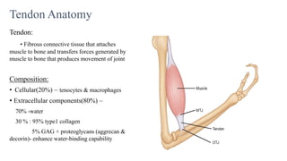
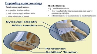
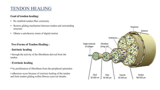
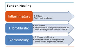
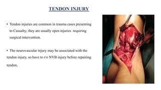
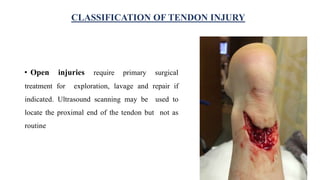
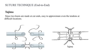
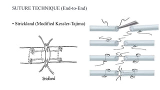
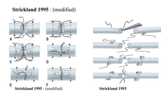
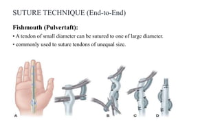
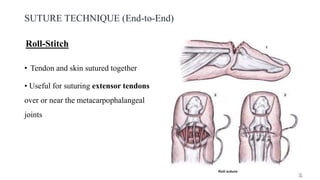
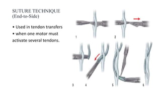
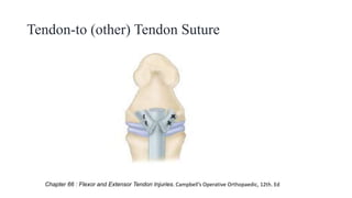
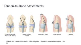
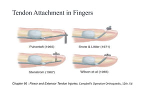
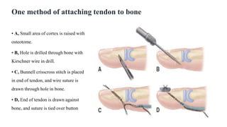
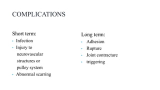
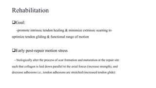
This document discusses tendon anatomy, injury, and repair. It describes the composition and vascular supply of tendons. Common tendon injuries include open wounds requiring surgical repair and closed injuries causing deformities. The goals of repair are to reestablish tendon continuity and gliding function. Various suture techniques are discussed for end-to-end, end-to-side, and tendon-to-bone repairs. Post-operative rehabilitation aims to promote intrinsic healing while minimizing scarring through early controlled motion to optimize tendon gliding and range of motion recovery.
This is a lecture presentation on applying external fixator on open fracture specially on tibia. This method is a classical method. Various new and dynamic fixators are there but the basics are the same.
Arthroscopic ACL Reconstruction By Dr Shekhar ShrivastavDelhiArthroscopy
Arthroscopic Acl Reconstruction By Dr Shekhar Shrivastav.
HOW NORMAL KNEE WORKS ?
The knee is the largest joint in the body, and one of the most easily injured. It is made up of the lower end of the thigh bone(femur), the upper end of the shin bone (tibia), and the knee cap (patella), which slides in a groove on the end of the femur. Four bands of tissue, the anterior and posterior cruciate ligaments, and the medial and lateral collateral ligaments connect the femur and the tibia and provide joint stability. The surfaces where the femur, tibia and patella touch are covered with articular cartilage, a smooth substance that cushions the bones and enables them to glide freely. Semicircular rings of tough fibrous-cartilage tissue called the lateral and medial menisci act as shock absorbers and stabilizers.
WHAT IS THE ROLE OF ACL ?
ACL along with other ligaments of the knee joint and meniscus provides stability to the knee joint.
WHAT IS LIGAMENT RECONSTRUCTION ( ACL ) ?
Ligament reconstruction involves replacing the torn ligament with a tendon (graft) from your knee and fixing the graft in place with screws. This procedure is performed with the use of the arthroscope. The anterior cruciate ligament (ACL) is the most common ligament requiring reconstruction procedures. The torn ligament is excised arthroscopically and new ligament is prepared by ligament grafts taken from your own body. Bony tunnels are prepared in femur and tibia using specialized instruments through which the new ligament is passed and fixed with special screws. This procedure requires relative rest or leave from your work or studies for about 2-3 weeks after which you will be allowed normal day to day activities.
WHEN CAN THE PATIENT BE AMBULATED AFTER SURGERY ?
The patient can walk from the same evening of the surgery. Initially the patient is advised to walk with a brace and a walking cane. Strengthening and range of motion exercises for the knee are started from the next day. The patient is discharged from the hospital 2nd or 3rd day after surgery. The patient can walk without support by 10-14 days depending on muscle strengthening. Slow Jogging and other strenuous activities are permitted after 3 months and the patient can return to active sports only 8-9 months after surgery.
Torn ACL Reconstructed ACL
For Further Queries contact your Orthopedic Surgeon at
+ 91 9971192233
New Directions in Targeted Therapeutic Approaches for Older Adults With Mantl...i3 Health
i3 Health is pleased to make the speaker slides from this activity available for use as a non-accredited self-study or teaching resource.
This slide deck presented by Dr. Kami Maddocks, Professor-Clinical in the Division of Hematology and
Associate Division Director for Ambulatory Operations
The Ohio State University Comprehensive Cancer Center, will provide insight into new directions in targeted therapeutic approaches for older adults with mantle cell lymphoma.
STATEMENT OF NEED
Mantle cell lymphoma (MCL) is a rare, aggressive B-cell non-Hodgkin lymphoma (NHL) accounting for 5% to 7% of all lymphomas. Its prognosis ranges from indolent disease that does not require treatment for years to very aggressive disease, which is associated with poor survival (Silkenstedt et al, 2021). Typically, MCL is diagnosed at advanced stage and in older patients who cannot tolerate intensive therapy (NCCN, 2022). Although recent advances have slightly increased remission rates, recurrence and relapse remain very common, leading to a median overall survival between 3 and 6 years (LLS, 2021). Though there are several effective options, progress is still needed towards establishing an accepted frontline approach for MCL (Castellino et al, 2022). Treatment selection and management of MCL are complicated by the heterogeneity of prognosis, advanced age and comorbidities of patients, and lack of an established standard approach for treatment, making it vital that clinicians be familiar with the latest research and advances in this area. In this activity chaired by Michael Wang, MD, Professor in the Department of Lymphoma & Myeloma at MD Anderson Cancer Center, expert faculty will discuss prognostic factors informing treatment, the promising results of recent trials in new therapeutic approaches, and the implications of treatment resistance in therapeutic selection for MCL.
Target Audience
Hematology/oncology fellows, attending faculty, and other health care professionals involved in the treatment of patients with mantle cell lymphoma (MCL).
Learning Objectives
1.) Identify clinical and biological prognostic factors that can guide treatment decision making for older adults with MCL
2.) Evaluate emerging data on targeted therapeutic approaches for treatment-naive and relapsed/refractory MCL and their applicability to older adults
3.) Assess mechanisms of resistance to targeted therapies for MCL and their implications for treatment selection
These lecture slides, by Dr Sidra Arshad, offer a quick overview of physiological basis of a normal electrocardiogram.
Learning objectives:
1. Define an electrocardiogram (ECG) and electrocardiography
2. Describe how dipoles generated by the heart produce the waveforms of the ECG
3. Describe the components of a normal electrocardiogram of a typical bipolar leads (limb II)
4. Differentiate between intervals and segments
5. Enlist some common indications for obtaining an ECG
Study Resources:
1. Chapter 11, Guyton and Hall Textbook of Medical Physiology, 14th edition
2. Chapter 9, Human Physiology - From Cells to Systems, Lauralee Sherwood, 9th edition
3. Chapter 29, Ganong’s Review of Medical Physiology, 26th edition
4. Electrocardiogram, StatPearls - https://www.ncbi.nlm.nih.gov/books/NBK549803/
5. ECG in Medical Practice by ABM Abdullah, 4th edition
6. ECG Basics, http://www.nataliescasebook.com/tag/e-c-g-basics
Explore natural remedies for syphilis treatment in Singapore. Discover alternative therapies, herbal remedies, and lifestyle changes that may complement conventional treatments. Learn about holistic approaches to managing syphilis symptoms and supporting overall health.
Factory Supply Best Quality Pmk Oil CAS 28578–16–7 PMK Powder in Stockrebeccabio
Factory Supply Best Quality Pmk Oil CAS 28578–16–7 PMK Powder in Stock
Telegram: bmksupplier
signal: +85264872720
threema: TUD4A6YC
You can contact me on Telegram or Threema
Communicate promptly and reply
Free of customs clearance, Double Clearance 100% pass delivery to USA, Canada, Spain, Germany, Netherland, Poland, Italy, Sweden, UK, Czech Republic, Australia, Mexico, Russia, Ukraine, Kazakhstan.Door to door service
Hot Selling Organic intermediates
Anti ulcer drugs and their Advance pharmacology ||
Anti-ulcer drugs are medications used to prevent and treat ulcers in the stomach and upper part of the small intestine (duodenal ulcers). These ulcers are often caused by an imbalance between stomach acid and the mucosal lining, which protects the stomach lining.
||Scope: Overview of various classes of anti-ulcer drugs, their mechanisms of action, indications, side effects, and clinical considerations.
Ozempic: Preoperative Management of Patients on GLP-1 Receptor Agonists Saeid Safari
Preoperative Management of Patients on GLP-1 Receptor Agonists like Ozempic and Semiglutide
ASA GUIDELINE
NYSORA Guideline
2 Case Reports of Gastric Ultrasound
Report Back from SGO 2024: What’s the Latest in Cervical Cancer?bkling
Are you curious about what’s new in cervical cancer research or unsure what the findings mean? Join Dr. Emily Ko, a gynecologic oncologist at Penn Medicine, to learn about the latest updates from the Society of Gynecologic Oncology (SGO) 2024 Annual Meeting on Women’s Cancer. Dr. Ko will discuss what the research presented at the conference means for you and answer your questions about the new developments.
Title: Sense of Taste
Presenter: Dr. Faiza, Assistant Professor of Physiology
Qualifications:
MBBS (Best Graduate, AIMC Lahore)
FCPS Physiology
ICMT, CHPE, DHPE (STMU)
MPH (GC University, Faisalabad)
MBA (Virtual University of Pakistan)
Learning Objectives:
Describe the structure and function of taste buds.
Describe the relationship between the taste threshold and taste index of common substances.
Explain the chemical basis and signal transduction of taste perception for each type of primary taste sensation.
Recognize different abnormalities of taste perception and their causes.
Key Topics:
Significance of Taste Sensation:
Differentiation between pleasant and harmful food
Influence on behavior
Selection of food based on metabolic needs
Receptors of Taste:
Taste buds on the tongue
Influence of sense of smell, texture of food, and pain stimulation (e.g., by pepper)
Primary and Secondary Taste Sensations:
Primary taste sensations: Sweet, Sour, Salty, Bitter, Umami
Chemical basis and signal transduction mechanisms for each taste
Taste Threshold and Index:
Taste threshold values for Sweet (sucrose), Salty (NaCl), Sour (HCl), and Bitter (Quinine)
Taste index relationship: Inversely proportional to taste threshold
Taste Blindness:
Inability to taste certain substances, particularly thiourea compounds
Example: Phenylthiocarbamide
Structure and Function of Taste Buds:
Composition: Epithelial cells, Sustentacular/Supporting cells, Taste cells, Basal cells
Features: Taste pores, Taste hairs/microvilli, and Taste nerve fibers
Location of Taste Buds:
Found in papillae of the tongue (Fungiform, Circumvallate, Foliate)
Also present on the palate, tonsillar pillars, epiglottis, and proximal esophagus
Mechanism of Taste Stimulation:
Interaction of taste substances with receptors on microvilli
Signal transduction pathways for Umami, Sweet, Bitter, Sour, and Salty tastes
Taste Sensitivity and Adaptation:
Decrease in sensitivity with age
Rapid adaptation of taste sensation
Role of Saliva in Taste:
Dissolution of tastants to reach receptors
Washing away the stimulus
Taste Preferences and Aversions:
Mechanisms behind taste preference and aversion
Influence of receptors and neural pathways
Impact of Sensory Nerve Damage:
Degeneration of taste buds if the sensory nerve fiber is cut
Abnormalities of Taste Detection:
Conditions: Ageusia, Hypogeusia, Dysgeusia (parageusia)
Causes: Nerve damage, neurological disorders, infections, poor oral hygiene, adverse drug effects, deficiencies, aging, tobacco use, altered neurotransmitter levels
Neurotransmitters and Taste Threshold:
Effects of serotonin (5-HT) and norepinephrine (NE) on taste sensitivity
Supertasters:
25% of the population with heightened sensitivity to taste, especially bitterness
Increased number of fungiform papillae
Title: Sense of Smell
Presenter: Dr. Faiza, Assistant Professor of Physiology
Qualifications:
MBBS (Best Graduate, AIMC Lahore)
FCPS Physiology
ICMT, CHPE, DHPE (STMU)
MPH (GC University, Faisalabad)
MBA (Virtual University of Pakistan)
Learning Objectives:
Describe the primary categories of smells and the concept of odor blindness.
Explain the structure and location of the olfactory membrane and mucosa, including the types and roles of cells involved in olfaction.
Describe the pathway and mechanisms of olfactory signal transmission from the olfactory receptors to the brain.
Illustrate the biochemical cascade triggered by odorant binding to olfactory receptors, including the role of G-proteins and second messengers in generating an action potential.
Identify different types of olfactory disorders such as anosmia, hyposmia, hyperosmia, and dysosmia, including their potential causes.
Key Topics:
Olfactory Genes:
3% of the human genome accounts for olfactory genes.
400 genes for odorant receptors.
Olfactory Membrane:
Located in the superior part of the nasal cavity.
Medially: Folds downward along the superior septum.
Laterally: Folds over the superior turbinate and upper surface of the middle turbinate.
Total surface area: 5-10 square centimeters.
Olfactory Mucosa:
Olfactory Cells: Bipolar nerve cells derived from the CNS (100 million), with 4-25 olfactory cilia per cell.
Sustentacular Cells: Produce mucus and maintain ionic and molecular environment.
Basal Cells: Replace worn-out olfactory cells with an average lifespan of 1-2 months.
Bowman’s Gland: Secretes mucus.
Stimulation of Olfactory Cells:
Odorant dissolves in mucus and attaches to receptors on olfactory cilia.
Involves a cascade effect through G-proteins and second messengers, leading to depolarization and action potential generation in the olfactory nerve.
Quality of a Good Odorant:
Small (3-20 Carbon atoms), volatile, water-soluble, and lipid-soluble.
Facilitated by odorant-binding proteins in mucus.
Membrane Potential and Action Potential:
Resting membrane potential: -55mV.
Action potential frequency in the olfactory nerve increases with odorant strength.
Adaptation Towards the Sense of Smell:
Rapid adaptation within the first second, with further slow adaptation.
Psychological adaptation greater than receptor adaptation, involving feedback inhibition from the central nervous system.
Primary Sensations of Smell:
Camphoraceous, Musky, Floral, Pepperminty, Ethereal, Pungent, Putrid.
Odor Detection Threshold:
Examples: Hydrogen sulfide (0.0005 ppm), Methyl-mercaptan (0.002 ppm).
Some toxic substances are odorless at lethal concentrations.
Characteristics of Smell:
Odor blindness for single substances due to lack of appropriate receptor protein.
Behavioral and emotional influences of smell.
Transmission of Olfactory Signals:
From olfactory cells to glomeruli in the olfactory bulb, involving lateral inhibition.
Primitive, less old, and new olfactory systems with different path
These simplified slides by Dr. Sidra Arshad present an overview of the non-respiratory functions of the respiratory tract.
Learning objectives:
1. Enlist the non-respiratory functions of the respiratory tract
2. Briefly explain how these functions are carried out
3. Discuss the significance of dead space
4. Differentiate between minute ventilation and alveolar ventilation
5. Describe the cough and sneeze reflexes
Study Resources:
1. Chapter 39, Guyton and Hall Textbook of Medical Physiology, 14th edition
2. Chapter 34, Ganong’s Review of Medical Physiology, 26th edition
3. Chapter 17, Human Physiology by Lauralee Sherwood, 9th edition
4. Non-respiratory functions of the lungs https://academic.oup.com/bjaed/article/13/3/98/278874
Prix Galien International 2024 Forum ProgramLevi Shapiro
June 20, 2024, Prix Galien International and Jerusalem Ethics Forum in ROME. Detailed agenda including panels:
- ADVANCES IN CARDIOLOGY: A NEW PARADIGM IS COMING
- WOMEN’S HEALTH: FERTILITY PRESERVATION
- WHAT’S NEW IN THE TREATMENT OF INFECTIOUS,
ONCOLOGICAL AND INFLAMMATORY SKIN DISEASES?
- ARTIFICIAL INTELLIGENCE AND ETHICS
- GENE THERAPY
- BEYOND BORDERS: GLOBAL INITIATIVES FOR DEMOCRATIZING LIFE SCIENCE TECHNOLOGIES AND PROMOTING ACCESS TO HEALTHCARE
- ETHICAL CHALLENGES IN LIFE SCIENCES
- Prix Galien International Awards Ceremony
3. Tendon Anatomy
Tendon:
• Fibrous connective tissue that attaches
muscle to bone and transfers forces generated by
muscle to bone that produces movement of joint
Composition:
• Cellular(20%) – tenocytes & macrophages
• Extracellular components(80%) –
70% -water
30 % : 95% type1 collagen
5% GAG + proteoglycans (aggrecan &
decorin)- enhance water-binding capability
4. Paratenon covered tendons
e.g., patellar, Achilles tendons
• rich vascular supply so heals better
• often injured due to trauma
Sheathed tendons
e.g., hand flexor tendons
• less vascularized with avascular areas that receive
nutrition by diffusion
• often injured due to laceration and at risk for adhesions
Depending upon coverings
6. Pulley system: (flexor compartment of digits)
Intermittent fibrous condensations that exist along the tendon sheath to
secure the tendon to the adjacent bone
• Digits 1-4 contain:
5 annular pulleys (A1 to A5)
3 cruciate pulleys (C1 to C3)
(A2 and A4 are the most important pulleys to
prevent flexor tendon bowstringing)
• Thumb contains
2 annular pulley
7. Tendon nutrition
1. Blood supply
From segmental vessels arising from surrounding
vessels
- In digits, flexor tendons through vincula; these are
folds of mesotenon through which run the small
vessels that penetrate the tendons
2. Synovial fluid: Supplies sheathed tendons
Produced within tenosynovial sheath
9. FLEXOR ZONES OF HAND (Verdan’s
zones): 5 zones
ZONE I: contains only FDP tendons
Extends from just distal to insertion of FDS tendon to site of
insertion of FDP tendon.
ZONE II: (bunnell’s“no man’s land”) critical area of pulleys
• Contains both FDP & FDS tendons
• Between distal palmar crease & insertion of superficialis tendon
10. ZONE III: Comprises area of lumbrical origin
Between distal margin of transverse carpal ligament &
beginning of critical area of pulleys or 1st annulus.
ZONE IV:
Zone covered by transverse carpal ligament
ZONE V:
Zone proximal to transverse carpal ligament and include
forearm
12. Goal of tendon healing:
• Re establish tendon fiber continuity
• Restore gliding mechanism between tendon and surrounding
structure
• Obtain a satisfactory return of digital motion
Two Forms of Tendon Healing :
–Intrinsic healing
• through the activity of the fibroblasts derived from the
tendon.
–Extrinsic healing
• by proliferation of fibroblasts from the peripheral epitendon
• adhesions occur because of extrinsic healing of the tendon
and limit tendon gliding within fibrous synovial sheaths
TENDON HEALING
13.
14. • Tendon injuries are common in trauma cases presenting
to Casualty; they are usually open injuries requiring
surgical intervention.
• The neurovascular injury may be associated with the
tendon injury, so have to r/o NVB injury before repairing
tendon.
TENDON INJURY
15. Tendon injury may be classified as:
Open or closed, sharp or blunt, and traumatic or degenerative,
based on nature and etiology of injury
Extensor or flexor, based on the tendons are involved
CLASSIFICATION OF TENDON INJURY
16. • Open injuries require primary surgical
treatment for exploration, lavage and repair if
indicated. Ultrasound scanning may be used to
locate the proximal end of the tendon but not as
routine
CLASSIFICATION OF TENDON INJURY
17. • Closed tendon injuries:
Shearing stress to the tendon may result in
closed tendon injuries
eg.: mallet fingers, Boutonniere deformities
and avulsions, ultrasound scanning is useful to
know level of injury, also for measurement of
gap between the tendon ends and to identify
pulley lesions and inflammatory processes.
CLASSIFICATION OF TENDON INJURY
mallet fingers
Boutonniere
deformities
18. • Open or closed tendon injuries may be partial or
complete
• When movement is present but painful this can
indicate a partial tendon injury
• In complete tendon injury, generally
movement is restricted
COMPLETE AND PARTIAL TENDON INJURY
19. 1.Primary repair:
Golden period
With in 24hrs in a clean wound
best results
2.Delayed primary repair
1-10 days
Done: suspicion of infection , viability questionable or came late
3.Secondary repair
10-14days up to 4wks
4.Late secondary
After 4 wks
Delay several days if wound infected
TENDON REPAIR
21. TENDON REPAIR
Ideal
• Gap resistant
• Strong enough to tolerate forces generated by early controlled active motion protocols
• 10-50% decrease in repair strength from day 5-21 post repair in immobilized tendons
• This is effect is minimized (possibly eliminated) through application of early motion stress
• Minimal bulk
• Minimal interference with tendon vascularity
22. Strickland stresses six characteristics of an ideal tendon
repair:
(1) easy placement of sutures in the tendon,
(2) secure suture knots,
(3) smooth juncture of tendon ends,
(4) minimal gapping at the repair site,
(5) minimal interference with tendon vascularity, and
(6) sufficient strength throughout healing to permit application of early
motion stress to the tendon.
TENDON REPAIR
23. Direct repair:
• if laceration is more than 1 cm
from insertion
Tendon advancement:
• if the laceration is less then 1 cm
from insertion.
TENDON REPAIR
24. Ideal Suture material:
Non reactive
Pliable
Small calibre (4-0) for core sutures and (6-0) for epitendon
Strong
Easy to handle
Common material: Polyester (Ethibond), Nylon, prolene
TENDON REPAIR
25.
26. Suture configuration
3 Groups
Group 1 (e.g. : simple sutures)
• the suture pull is parallel to the tendon collagen bundles, transmitting the stress of the repair directly
to the opposing tendon ends.
• Weakest
27. Group 2 (e.g. : Bunnell suture)
• stress is transmitted directly across the juncture by the suture material and depends on the strength of the
suture itself.
Group 3 e.g. : Pulvertaft technique (fish-mouth weave);
• sutures are placed perpendicular to the tendon collagen bundles and the applied stress
• Strongest & most suitable
28. Suture configuration
Modified suture configs.
Multiple-strand modifications
• Savage (six strands)
• Lee (four strands)
The Tang and Cruciate repairs
• better tensile strength and elastic properties
A four-strand adaptation of the Kessler repair
• significantly stronger than the Kessler technique
29. DEPENDING UPON APPROACHES
• Epitenon-first technique
22% stronger than the modified Kessler
technique
• Circumferential suture :
Interlocking horizontal mattress suture
• greatest resistance to gap formation,
• highest stiffness
best overall
Suture configuration
33. SUTURE TECHNIQUE (End-to-End)
Kessler
• Mainly used for tendon repair in the fingers and palm.
• disadvantage: knots being left exposed on the tendon surface
Modified Kessler (Smith-Evans
modification)
Advantage:
• A single piece of suture material is used.
• knot is left in the cut surface of the tendon.
• minimize the problem of exposed suture material
Disadvantage: difficulty to achieve satisfactory approximation
of the tendon ends.
40. SUTURE TECHNIQUE (End-to-End)
Fishmouth (Pulvertaft):
• A tendon of small diameter can be sutured to one of large diameter.
• commonly used to suture tendons of unequal size.
41. SUTURE TECHNIQUE (End-to-End)
• Tendon and skin sutured together
• Useful for suturing extensor tendons
over or near the metacarpophalangeal
joints
Roll-Stitch
45. Tendon Attachment in Fingers
Chapter 66 : Flexor and Extensor Tendon Injuries. Campbell’s Operative Orthopaedic, 12th. Ed
46. One method of attaching tendon to bone
• A, Small area of cortex is raised with
osteotome.
• B, Hole is drilled through bone with
Kirschner wire in drill.
• C, Bunnell crisscross stitch is placed
in end of tendon, and wire suture is
drawn through hole in bone.
• D, End of tendon is drawn against
bone, and suture is tied over button
47. COMPLICATIONS
Short term:
• Infection
• Injury to
neurovascular
structures or
pulley system
• Abnormal scarring
Long term:
• Adhesion
• Rupture
• Joint contracture
• triggering
48. Rehabilitation
Goal:
-promote intrinsic tendon healing & minimize extrinsic scarring to
optimize tendon gliding & functional range of motion
Early post-repair motion stress
– biologically alter the process of scar formation and maturation at the repair site
such that collagen is laid down parallel to the axial forces (increase strength), and
decrease adhesions i.e., tendon adhesions are stretched (increased tendon glide)
49. Methods of post-op tendon management are:
• Immobilization: Complete immobilization of tendon for 3 ½ weeks after Surgery; but
greater chances of production of scar adherence; greater incidence of tendon rupture
• Controlled passive motion: passive flexion and extension followed by active
extension and passive flexion
• Early active motion: early active extension but passive flexion, Method minimizes
scar adhesions while enhancing tendon nutrition and blood flow