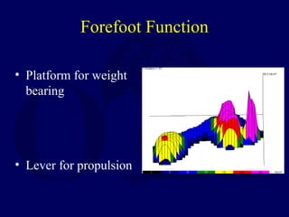
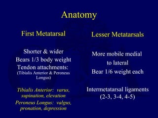
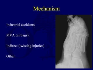
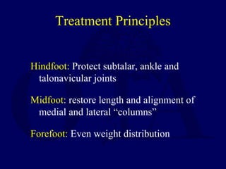
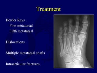
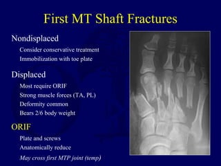
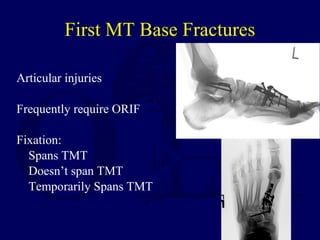
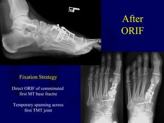
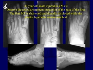
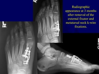
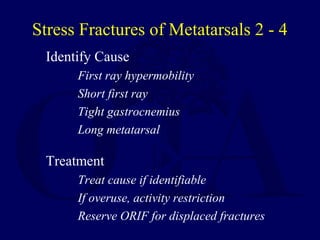
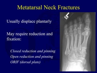
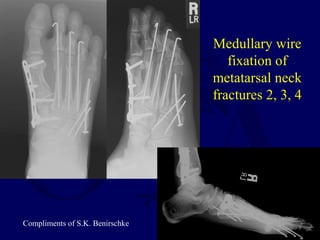
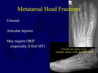
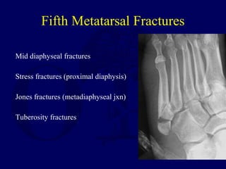
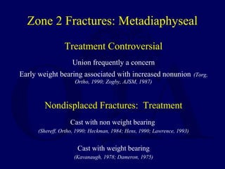
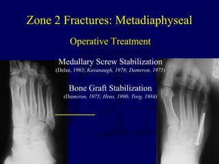
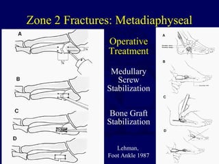
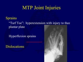
Forefoot Fractures
Sean E. Nork, MD
Harborview Medical Center
Foot Trauma and Outcomes
Turchin et al, JOT, 1999
28 patients: Polytrauma +/- foot injury
Age, gender, ISS matched
Results SF-36 5/8 components worse with foot injury
WOMAC All 3 components worse with foot injury
Jurkovich et al, JT, 1995
Highest Sickness Impact Profile (SIP) @ 6 & 12 months
Patients with foot trauma (compared to other lower extremity injuries)
Foot Function
Hindfoot: Shock absorption, propulsion,
deceleration
Midfoot: Controls relationship between
hindfoot and forefoot
Forefoot: Platform for standing and lever for
push off
Forefoot Function
• Platform for weight
bearing
• Lever for propulsion
Anatomy
First Metatarsal
Shorter & wider
Bears 1/3 body weight
Tendon attachments:
(Tibialis Anterior & Peroneus
Longus)
Tibialis Anterior: varus,
supination, elevation
Peroneus Longus: valgus,
pronation, depression
Lesser Metatarsals
More mobile medial
to lateral
Bear 1/6 weight each
Intermetatarsal ligaments
(2-3, 3-4, 4-5)
Sesamoids
Medial (tibial) & Lateral (fibular)
Within FHB tendons
Articulate with 1st MT head
Weight bearing through sesamoids
Tibial Sesamoid: Tibial FHB
Abductor Hallucis
Fibular Sesamoid: Fibular FHB
Adductor Hallucis
Deep Tverse MT ligament
Phalanges
Great toe (2)
Lesser toes (3)
FDB attaches @
intermediate
FDL/EDL attaches @ distal
Biomechanics
Metatarsal heads in
contact with floor 60-
80% of stance phase
Toes in contact with
floor 75% of stance
phase
Cavanagh, PR, F&A, 1987
Hughes, J, JBJS[Br], 1990
Mechanism
Industrial accidents
MVA (airbags)
Indirect (twisting injuries)
Other
Physical Examination
Gross deformity
Dislocations
Sensation
Capillary refill
Foot Compartments
Radiographs
Foot trauma series
AP/lat/oblique
Don’t forget oblique
Sesamoid view
Tangential view (MT heads)
Contralateral foot films (comparison)
CT Scan (occasionally)
Treatment Principles
Hindfoot: Protect subtalar, ankle and
talonavicular joints
Midfoot: restore length and alignment of
medial and lateral “columns”
Forefoot: Even weight distribution
Treatment
Border Rays
First metatarsal
Fifth metatarsal
Dislocations
Multiple metatarsal shafts
Intraarticular fractures
First MT Shaft Fractures
Nondisplaced
Consider conservative treatment
Immobilization with toe plate
Displaced
Most require ORIF
Strong muscle forces (TA, PL)
Deformity common
Bears 2/6 body weight
ORIF
Plate and screws
Anatomically reduce
May cross first MTP joint (temp)
Articular injuries
Frequently require ORIF
Fixation:
Spans TMT
Doesn’t span TMT
Temporarily Spans TMT
First MT Base Fractures
36 year old male
s/p MVC
Active
Note articular
comminution
After
ORIF
Fixation Strategy
Direct ORIF of comminuted
first MT base fractre
Temporary spanning across
first TMT joint
43 year old male injured in a MVC
Observe the articular segment impaction of the base of the first.
The first MT is shortened and dorsally displaced while the
plantar ligaments remain attached.
The patient underwent ORIF of the
base of the first metatarsal with
spanning of the first TMT, given
the level of comminution observed.
Additionally, temporary spanning
external fixation was used.
Radiographic
appearance at 3 months
after removal of the
external fixator and
metatarsal neck k-wire
fixations.
Non-displaced Metatarsal Fractures 2-4
Single metatarsal fractures
Treatment usually nonoperative
Symptomatic: hard shoe vs AFO vs cast
Multiple metatarsal fractures
Usually symptomatic treatment (as above)
May require ORIF if other associated injuries
Displaced Metatarsal Shaft Fractures
Sagittal plane displacement & angulation is most important.
Reestablish length, rotation, & declination
Dorsal deformity can produce transfer metatarsalgia
Plantar deformity can produce increased load at affected metatarsal
Treatment Options
Closed Reduction
Intramedullary pinning with k-wire (0.054” or 0.062”)
Pinning of distal segment to adjacent metatarsal
ORIF with dorsal plate fixation
This patient sustained an open
second metatarsal fracture in a
crush injury. Given the soft
tissue injury and continued
pressure on the dorsal skin,
operative fixation was elected.
Fixation consisted of a dorsal
2.0 mm plate application after
appropriate irrigation of the
open fracture.
This patient was
treated with ORIF
of multiple
metatarsal
fractures (3,4,5)
through a dorsal
approach.
Fixation consisted
of a 2.7 mm DCP
on the fifth and
2.0 mm plates on
the third and
fourth
metatarsals.
Medullary K-wires
in Lesser MTs
Exit wire distally through
the proximal phalanx
Plantar wire exit may
produce a hyperextension
deformity of the MTP
ST Hansen,
Skeletal Trauma
Stress Fractures of Metatarsals 2 - 4
Identify Cause
First ray hypermobility
Short first ray
Tight gastrocnemius
Long metatarsal
Treatment
Treat cause if identifiable
If overuse, activity restriction
Reserve ORIF for displaced fractures
Usually displace plantarly
May require reduction and
fixation:
Closed reduction and pinning
Open reduction and pinning
ORIF (dorsal plate)
Metatarsal Neck Fractures
This patient sustained
multiple metatarsal neck
fractures after an MVA.
Note additional fractures at
the first and fifth metatarsals
Medullary wire
fixation of
metatarsal neck
fractures 2, 3, 4
Compliments of S.K. Benirschke
Unusual
Articular injuries
May require ORIF
(especially if first MT)
Metatarsal Head Fractures
Circular saw injury to the
articular surface of the first MT head
Fifth Metatarsal Fractures
Mid diaphyseal fractures
Stress fractures (proximal diaphysis)
Jones fractures (metadiaphyseal jxn)
Tuberosity fractures
Proximal Fifth Metatarsal Fractures
Dameron, TB, JAAOS, 1995
Zone 1 cancellous tuberosity
insertion of PB & plantar fascia
involve metatarsocuboid joint
Zone 2 distal to tuberosity
extend to 4/5 articulation
Zone 3 distal to proximal ligaments
usually stress fractures
extend to diaphysis for 1.5 cm
Proximal Fifth Metatarsal Fractures
Dameron, TB, JAAOS, 1995
Relative Frequency
Zone 1 93%
Zone 2 4%
Zone 3 3%
Fifth Metatarsal Blood Supply
Smith, J et al, F&A, 1992
Cadaver Arterial Injection
Study (n = 10)
Nutrient artery with intramedullary
branches (retrograde flow to
proximal fifth metatarsal)
Multiple metaphyseal arteries
Conclusions: Fracture distal to the
tuberosity disrupts the nutrient
arterial supply and creates relative
avascularity
Shereff, M et al, F&A, 1991
Fresh leg specimens (after
BKA) (n = 15)
Extraosseus circulation:
dorsal metatarsal artery
plantar metatarsal artery
fibular plantar marginal artery
Intraosseus circulation:
Nutrient artery
Metaphyseal vessels
Periosteal complex
Fifth Metatarsal Blood Supply
Smith et al, Foot Ankle 1993
Zone 1 Fractures: Tuberosity
Etiology
Avulsion from lateral plantar aponeurosis
(Richli & Rosenthal, AJR, 1984)
Treatment
Symptomatic
Hard shoe
Healing usually uneventful
(Dameron, T, JBJS, 1975)
Lawrence, SL, Foot
Ankle, 1993
Zone 1 Fractures: Tuberosity
Weiner, et al, F & A Int, 1997
60 patients
Randomized to short leg cast vs soft dressing only
Weight bearing in hard shoe in all
Healing in 44(average) - 65(all) days
Soft dressing only: shorter recuperation (33 vs 46
days) and similar foot score (92 vs 86)
Conclusions: Faster return to function without
compromising radiographic union or clinical
outcome in patients treated without casting.
Zone 2 Fractures: Metadiaphyseal
Treatment Controversial
Union frequently a concern
Early weight bearing associated with increased nonunion (Torg,
Ortho, 1990; Zogby, AJSM, 1987)
Nondisplaced Fractures: Treatment
Cast with non weight bearing
(Shereff, Ortho, 1990; Heckman, 1984; Hens, 1990; Lawrence, 1993)
Cast with weight bearing
(Kavanaugh, 1978; Dameron, 1975)
Zone 2 Fractures: Metadiaphyseal
Operative Treatment
Medullary Screw Stabilization
(Delee, 1983; Kavanaugh, 1978; Dameron, 1975)
Bone Graft Stabilization
(Dameron, 1975; Hens, 1990; Torg, 1984)
Zone 2 Fractures: Metadiaphyseal
Operative
Treatment
Medullary
Screw
Stabilization
Bone Graft
Stabilization
Lehman,
Foot Ankle 1987
Comminuted fracture
of the base of the fifth
metatarsal
After ORIF of the
fifth metatarsal
MTP Joint Injuries
Sprains
“Turf Toe”: hyperextension with injury to thee
plantar plate
Hyperflexion sprains
Dislocations
First MTP Dislocations
Jahss, F&A, 1980
Type I: Hallux dislocation without disrupting sesamoid
Irreducible closed!
MT incarcerated by conjoined tendons and intact
sesamoid
Open reduction required (dorsal, plantar, or medial
approach)
Type II: Disruption of intersesamoid ligament (type A)
Transverse fracture of one of the sesamoids (type B)
Usually stable after reduction
Treatment usually conservative and symptomatic
(hard shoe for 4-6 weeks)
Lesser MTP Dislocations
Uncommon
Dorsal vs Lateral
Usually stable post reduction
Rarely require open reduction
If unstable post reduction, consider k-wire fixation
Proximal Phalanx Fractures
ORIF for transverse & displaced (?)
ORIF intraarticular fractures (?)
Interphalangeal Joint Fractures
Nonoperative treatment usually
Distal Phalanx Fractures
Taping usually adequate
Hard shoe
Fractures of the
Great Toe
Sesamoid Injuries
Sesamoiditis
Acute fractures
Stress fractures in dancers and runners
Treatment
Acute: padding
strap MTP @ neutral or slight flexion
immobilization in cast/shoe
Chronic: consider bone grafting
sesamoidectomy: not a simple procedure, assoc
with hallux drift and transfer lesions, requires tendon
(FHB) repair.
Fractures of the LesserToes
Correct alignment & rotation
Attempt taping to adjacent
toe
May require open reduction
and pinning if adequate
reduction not obtained
ST Hansen,
Skeletal Trauma
The Crushed Foot
Soft Tissue Evaluation
Assess whether salvageable
sensate, perfused, adequate plantar tissue
Wash open wounds
Reposition bone deformity that
threatens the skin
Reduce dislocations
Release compartments as needed
Recommended Readings
Cavanaugh, PR, et al. Pressure Distribution Patterns under Symptom-free
Feet during barefoot standing. Foot Ankle, 7:262-276, 1987
Dameron, TB, Fractures of the Proximal Fifth Metatarsal: Selecting the Best
Treatment Option. J Acad Orthop Surg, 3(2): 110-114, 1995.
Holmes, James. AAOS Monograph “The Traumatized Foot”, pages 55-75,
2002.
Lawrence, SJ, and Botte, MJ. Foot Fellow’s Review: Jones’ Fractures and
Related Fractures of the Proximal Fifth Metatarsal. Foot & Ankle, 14(6),
358-365, 1987.
Smith, JW, et al. The Intraosseus Blood Supply of the Fifth Metatarsal:
Implications for Proximal Fracture Healing. Foot & Ankle, 13(3), 143-
152, 1992
Thank You
Sean E. Nork, MD
Harborview Medical Center
University of Washington
Return to
Lower Extremity
Index

L17 forefoot fxs

  • 1.
    Forefoot Fractures Sean E.Nork, MD Harborview Medical Center
  • 2.
    Foot Trauma andOutcomes Turchin et al, JOT, 1999 28 patients: Polytrauma +/- foot injury Age, gender, ISS matched Results SF-36 5/8 components worse with foot injury WOMAC All 3 components worse with foot injury Jurkovich et al, JT, 1995 Highest Sickness Impact Profile (SIP) @ 6 & 12 months Patients with foot trauma (compared to other lower extremity injuries)
  • 3.
    Foot Function Hindfoot: Shockabsorption, propulsion, deceleration Midfoot: Controls relationship between hindfoot and forefoot Forefoot: Platform for standing and lever for push off
  • 4.
    Forefoot Function • Platformfor weight bearing • Lever for propulsion
  • 5.
    Anatomy First Metatarsal Shorter &wider Bears 1/3 body weight Tendon attachments: (Tibialis Anterior & Peroneus Longus) Tibialis Anterior: varus, supination, elevation Peroneus Longus: valgus, pronation, depression Lesser Metatarsals More mobile medial to lateral Bear 1/6 weight each Intermetatarsal ligaments (2-3, 3-4, 4-5)
  • 6.
    Sesamoids Medial (tibial) &Lateral (fibular) Within FHB tendons Articulate with 1st MT head Weight bearing through sesamoids Tibial Sesamoid: Tibial FHB Abductor Hallucis Fibular Sesamoid: Fibular FHB Adductor Hallucis Deep Tverse MT ligament
  • 7.
    Phalanges Great toe (2) Lessertoes (3) FDB attaches @ intermediate FDL/EDL attaches @ distal
  • 8.
    Biomechanics Metatarsal heads in contactwith floor 60- 80% of stance phase Toes in contact with floor 75% of stance phase Cavanagh, PR, F&A, 1987 Hughes, J, JBJS[Br], 1990
  • 9.
  • 10.
  • 11.
    Radiographs Foot trauma series AP/lat/oblique Don’tforget oblique Sesamoid view Tangential view (MT heads) Contralateral foot films (comparison) CT Scan (occasionally)
  • 12.
    Treatment Principles Hindfoot: Protectsubtalar, ankle and talonavicular joints Midfoot: restore length and alignment of medial and lateral “columns” Forefoot: Even weight distribution
  • 13.
    Treatment Border Rays First metatarsal Fifthmetatarsal Dislocations Multiple metatarsal shafts Intraarticular fractures
  • 14.
    First MT ShaftFractures Nondisplaced Consider conservative treatment Immobilization with toe plate Displaced Most require ORIF Strong muscle forces (TA, PL) Deformity common Bears 2/6 body weight ORIF Plate and screws Anatomically reduce May cross first MTP joint (temp)
  • 15.
    Articular injuries Frequently requireORIF Fixation: Spans TMT Doesn’t span TMT Temporarily Spans TMT First MT Base Fractures
  • 16.
    36 year oldmale s/p MVC Active Note articular comminution
  • 17.
    After ORIF Fixation Strategy Direct ORIFof comminuted first MT base fractre Temporary spanning across first TMT joint
  • 18.
    43 year oldmale injured in a MVC Observe the articular segment impaction of the base of the first. The first MT is shortened and dorsally displaced while the plantar ligaments remain attached.
  • 19.
    The patient underwentORIF of the base of the first metatarsal with spanning of the first TMT, given the level of comminution observed. Additionally, temporary spanning external fixation was used.
  • 20.
    Radiographic appearance at 3months after removal of the external fixator and metatarsal neck k-wire fixations.
  • 21.
    Non-displaced Metatarsal Fractures2-4 Single metatarsal fractures Treatment usually nonoperative Symptomatic: hard shoe vs AFO vs cast Multiple metatarsal fractures Usually symptomatic treatment (as above) May require ORIF if other associated injuries
  • 22.
    Displaced Metatarsal ShaftFractures Sagittal plane displacement & angulation is most important. Reestablish length, rotation, & declination Dorsal deformity can produce transfer metatarsalgia Plantar deformity can produce increased load at affected metatarsal Treatment Options Closed Reduction Intramedullary pinning with k-wire (0.054” or 0.062”) Pinning of distal segment to adjacent metatarsal ORIF with dorsal plate fixation
  • 23.
    This patient sustainedan open second metatarsal fracture in a crush injury. Given the soft tissue injury and continued pressure on the dorsal skin, operative fixation was elected.
  • 24.
    Fixation consisted ofa dorsal 2.0 mm plate application after appropriate irrigation of the open fracture.
  • 25.
    This patient was treatedwith ORIF of multiple metatarsal fractures (3,4,5) through a dorsal approach. Fixation consisted of a 2.7 mm DCP on the fifth and 2.0 mm plates on the third and fourth metatarsals.
  • 26.
    Medullary K-wires in LesserMTs Exit wire distally through the proximal phalanx Plantar wire exit may produce a hyperextension deformity of the MTP ST Hansen, Skeletal Trauma
  • 27.
    Stress Fractures ofMetatarsals 2 - 4 Identify Cause First ray hypermobility Short first ray Tight gastrocnemius Long metatarsal Treatment Treat cause if identifiable If overuse, activity restriction Reserve ORIF for displaced fractures
  • 28.
    Usually displace plantarly Mayrequire reduction and fixation: Closed reduction and pinning Open reduction and pinning ORIF (dorsal plate) Metatarsal Neck Fractures
  • 29.
    This patient sustained multiplemetatarsal neck fractures after an MVA. Note additional fractures at the first and fifth metatarsals
  • 30.
    Medullary wire fixation of metatarsalneck fractures 2, 3, 4 Compliments of S.K. Benirschke
  • 31.
    Unusual Articular injuries May requireORIF (especially if first MT) Metatarsal Head Fractures Circular saw injury to the articular surface of the first MT head
  • 32.
    Fifth Metatarsal Fractures Middiaphyseal fractures Stress fractures (proximal diaphysis) Jones fractures (metadiaphyseal jxn) Tuberosity fractures
  • 33.
    Proximal Fifth MetatarsalFractures Dameron, TB, JAAOS, 1995 Zone 1 cancellous tuberosity insertion of PB & plantar fascia involve metatarsocuboid joint Zone 2 distal to tuberosity extend to 4/5 articulation Zone 3 distal to proximal ligaments usually stress fractures extend to diaphysis for 1.5 cm
  • 34.
    Proximal Fifth MetatarsalFractures Dameron, TB, JAAOS, 1995 Relative Frequency Zone 1 93% Zone 2 4% Zone 3 3%
  • 35.
    Fifth Metatarsal BloodSupply Smith, J et al, F&A, 1992 Cadaver Arterial Injection Study (n = 10) Nutrient artery with intramedullary branches (retrograde flow to proximal fifth metatarsal) Multiple metaphyseal arteries Conclusions: Fracture distal to the tuberosity disrupts the nutrient arterial supply and creates relative avascularity Shereff, M et al, F&A, 1991 Fresh leg specimens (after BKA) (n = 15) Extraosseus circulation: dorsal metatarsal artery plantar metatarsal artery fibular plantar marginal artery Intraosseus circulation: Nutrient artery Metaphyseal vessels Periosteal complex
  • 36.
    Fifth Metatarsal BloodSupply Smith et al, Foot Ankle 1993
  • 37.
    Zone 1 Fractures:Tuberosity Etiology Avulsion from lateral plantar aponeurosis (Richli & Rosenthal, AJR, 1984) Treatment Symptomatic Hard shoe Healing usually uneventful (Dameron, T, JBJS, 1975) Lawrence, SL, Foot Ankle, 1993
  • 38.
    Zone 1 Fractures:Tuberosity Weiner, et al, F & A Int, 1997 60 patients Randomized to short leg cast vs soft dressing only Weight bearing in hard shoe in all Healing in 44(average) - 65(all) days Soft dressing only: shorter recuperation (33 vs 46 days) and similar foot score (92 vs 86) Conclusions: Faster return to function without compromising radiographic union or clinical outcome in patients treated without casting.
  • 39.
    Zone 2 Fractures:Metadiaphyseal
  • 40.
    Treatment Controversial Union frequentlya concern Early weight bearing associated with increased nonunion (Torg, Ortho, 1990; Zogby, AJSM, 1987) Nondisplaced Fractures: Treatment Cast with non weight bearing (Shereff, Ortho, 1990; Heckman, 1984; Hens, 1990; Lawrence, 1993) Cast with weight bearing (Kavanaugh, 1978; Dameron, 1975) Zone 2 Fractures: Metadiaphyseal
  • 41.
    Operative Treatment Medullary ScrewStabilization (Delee, 1983; Kavanaugh, 1978; Dameron, 1975) Bone Graft Stabilization (Dameron, 1975; Hens, 1990; Torg, 1984) Zone 2 Fractures: Metadiaphyseal
  • 42.
    Zone 2 Fractures:Metadiaphyseal Operative Treatment Medullary Screw Stabilization Bone Graft Stabilization Lehman, Foot Ankle 1987
  • 43.
    Comminuted fracture of thebase of the fifth metatarsal
  • 44.
    After ORIF ofthe fifth metatarsal
  • 45.
    MTP Joint Injuries Sprains “TurfToe”: hyperextension with injury to thee plantar plate Hyperflexion sprains Dislocations
  • 46.
    First MTP Dislocations Jahss,F&A, 1980 Type I: Hallux dislocation without disrupting sesamoid Irreducible closed! MT incarcerated by conjoined tendons and intact sesamoid Open reduction required (dorsal, plantar, or medial approach) Type II: Disruption of intersesamoid ligament (type A) Transverse fracture of one of the sesamoids (type B) Usually stable after reduction Treatment usually conservative and symptomatic (hard shoe for 4-6 weeks)
  • 47.
    Lesser MTP Dislocations Uncommon Dorsalvs Lateral Usually stable post reduction Rarely require open reduction If unstable post reduction, consider k-wire fixation
  • 48.
    Proximal Phalanx Fractures ORIFfor transverse & displaced (?) ORIF intraarticular fractures (?) Interphalangeal Joint Fractures Nonoperative treatment usually Distal Phalanx Fractures Taping usually adequate Hard shoe Fractures of the Great Toe
  • 49.
    Sesamoid Injuries Sesamoiditis Acute fractures Stressfractures in dancers and runners Treatment Acute: padding strap MTP @ neutral or slight flexion immobilization in cast/shoe Chronic: consider bone grafting sesamoidectomy: not a simple procedure, assoc with hallux drift and transfer lesions, requires tendon (FHB) repair.
  • 50.
    Fractures of theLesserToes Correct alignment & rotation Attempt taping to adjacent toe May require open reduction and pinning if adequate reduction not obtained ST Hansen, Skeletal Trauma
  • 51.
    The Crushed Foot SoftTissue Evaluation Assess whether salvageable sensate, perfused, adequate plantar tissue Wash open wounds Reposition bone deformity that threatens the skin Reduce dislocations Release compartments as needed
  • 52.
    Recommended Readings Cavanaugh, PR,et al. Pressure Distribution Patterns under Symptom-free Feet during barefoot standing. Foot Ankle, 7:262-276, 1987 Dameron, TB, Fractures of the Proximal Fifth Metatarsal: Selecting the Best Treatment Option. J Acad Orthop Surg, 3(2): 110-114, 1995. Holmes, James. AAOS Monograph “The Traumatized Foot”, pages 55-75, 2002. Lawrence, SJ, and Botte, MJ. Foot Fellow’s Review: Jones’ Fractures and Related Fractures of the Proximal Fifth Metatarsal. Foot & Ankle, 14(6), 358-365, 1987. Smith, JW, et al. The Intraosseus Blood Supply of the Fifth Metatarsal: Implications for Proximal Fracture Healing. Foot & Ankle, 13(3), 143- 152, 1992
  • 53.
    Thank You Sean E.Nork, MD Harborview Medical Center University of Washington Return to Lower Extremity Index