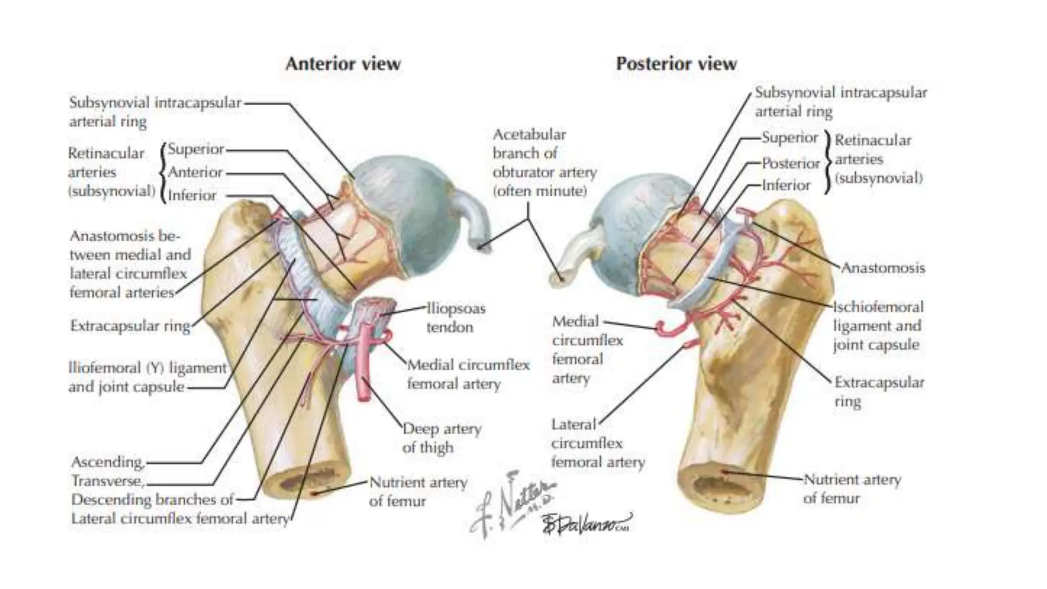
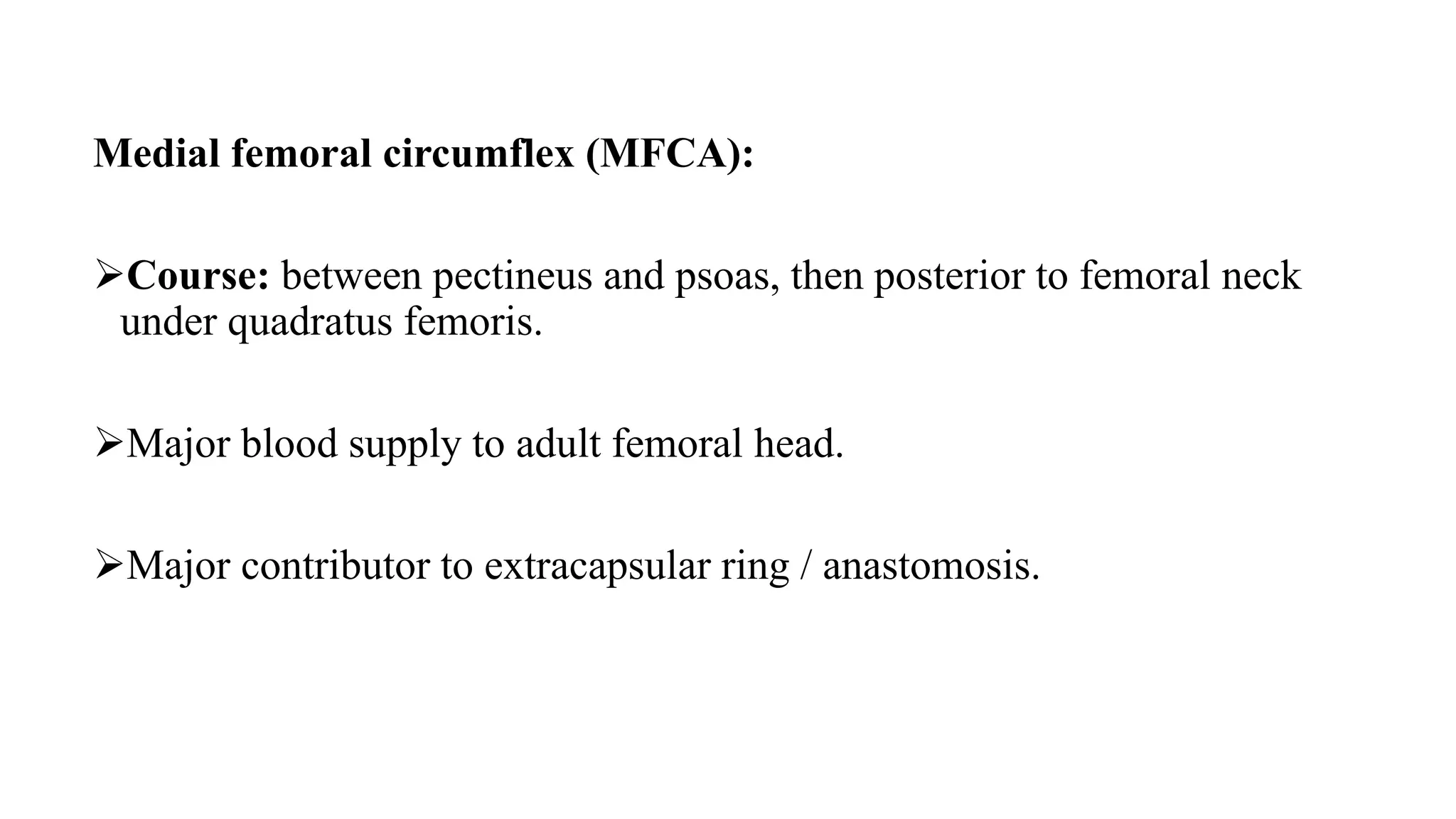
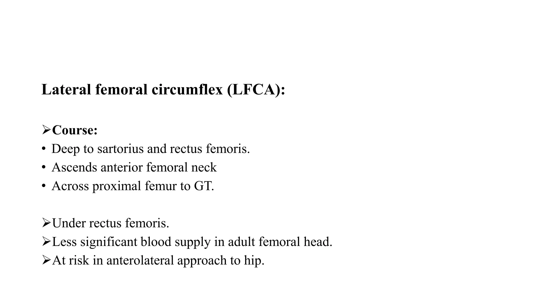
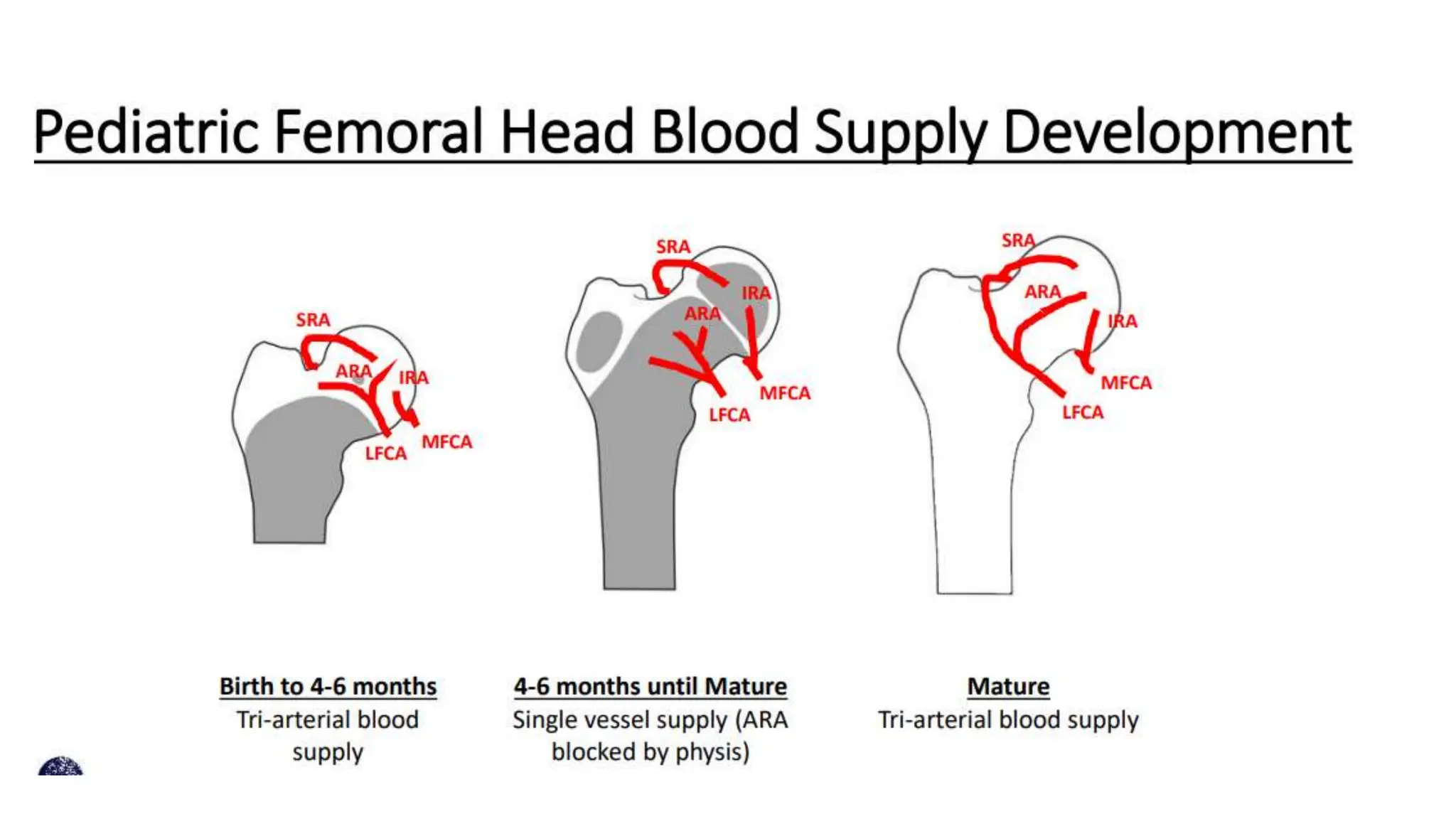
Blood supply of head of
femur
Aorta
Abdominal aorta Internal iliac artery
External iliac artery
Profunda femoris artery
Femoral artery
Lateral circumflex femoral artery
Obturator artery
Extracapsular anastomosis
Medial circumflex femoral artery
Superior
Anterior
Inferior
Retinacular artery
Artery to ligament of teres
Intracapsular anastomosis
Supply head of femur
Medial femoral circumflex (MFCA):
Course: between pectineus and psoas, then posterior to femoral neck
under quadratus femoris.
Major blood supply to adult femoral head.
Major contributor to extracapsular ring / anastomosis.
Lateral femoral circumflex (LFCA):
Course:
• Deep to sartorius and rectus femoris.
• Ascends anterior femoral neck
• Across proximal femur to GT.
Under rectus femoris.
Less significant blood supply in adult femoral head.
At risk in anterolateral approach to hip.
Extracapsular ring:
Formed at the base of femoral neck primarily from branches of MFCA and LFCA.
Lateral branch  from ring laterally toward GT  supply greater trochanter.
Ascending cervical arteries  along extracapsular femoral neck branch from
extracapsular ring.
Retinacular arteries  along intracapsular femoral neck  intracapsular continuation
of cervical arteries from a 2nd intracapsular ring at base of head.
Subsynovial intracapsular arterial ring:
Formed at the base of the femoral head.
Epiphyseal arteries and lateral epiphyseal artery:
Enter bone at border of articular surface in posterosuperior neck.
Will form intraosseous anastomoses.
Lat. Epiphyseal supplies most of WB femoral head.
Obturator Artery:
Artery of ligamentum teres  Through ligamentum teres to fovea  Minimal
supply to the adult femoral head.
Medial epiphyseal artery  Interosseous terminal branches  Anastomose with
lateral epiphyseal arteries.
Paediatric femoral head blood supply:
• 0-4yr  MFCA, LFCA, and ligamentum teres artery;
• 4-8yr: mostly MFCA, minimal LFCA and ligamentum teres artery;
• 8yrs: MFCA is predominant.
Paediatric femoral head blood supply:

Blood supply of head of femur. ,

  • 1.
    Blood supply ofhead of femur
  • 2.
    Aorta Abdominal aorta Internaliliac artery External iliac artery Profunda femoris artery Femoral artery Lateral circumflex femoral artery Obturator artery Extracapsular anastomosis Medial circumflex femoral artery Superior Anterior Inferior Retinacular artery Artery to ligament of teres Intracapsular anastomosis Supply head of femur
  • 4.
    Medial femoral circumflex(MFCA): Course: between pectineus and psoas, then posterior to femoral neck under quadratus femoris. Major blood supply to adult femoral head. Major contributor to extracapsular ring / anastomosis.
  • 6.
    Lateral femoral circumflex(LFCA): Course: • Deep to sartorius and rectus femoris. • Ascends anterior femoral neck • Across proximal femur to GT. Under rectus femoris. Less significant blood supply in adult femoral head. At risk in anterolateral approach to hip.
  • 7.
    Extracapsular ring: Formed atthe base of femoral neck primarily from branches of MFCA and LFCA. Lateral branch  from ring laterally toward GT  supply greater trochanter. Ascending cervical arteries  along extracapsular femoral neck branch from extracapsular ring. Retinacular arteries  along intracapsular femoral neck  intracapsular continuation of cervical arteries from a 2nd intracapsular ring at base of head.
  • 8.
    Subsynovial intracapsular arterialring: Formed at the base of the femoral head. Epiphyseal arteries and lateral epiphyseal artery: Enter bone at border of articular surface in posterosuperior neck. Will form intraosseous anastomoses. Lat. Epiphyseal supplies most of WB femoral head.
  • 9.
    Obturator Artery: Artery ofligamentum teres  Through ligamentum teres to fovea  Minimal supply to the adult femoral head. Medial epiphyseal artery  Interosseous terminal branches  Anastomose with lateral epiphyseal arteries.
  • 10.
    Paediatric femoral headblood supply: • 0-4yr  MFCA, LFCA, and ligamentum teres artery; • 4-8yr: mostly MFCA, minimal LFCA and ligamentum teres artery; • 8yrs: MFCA is predominant.
  • 12.
  • 13.
  • 14.
  • 15.