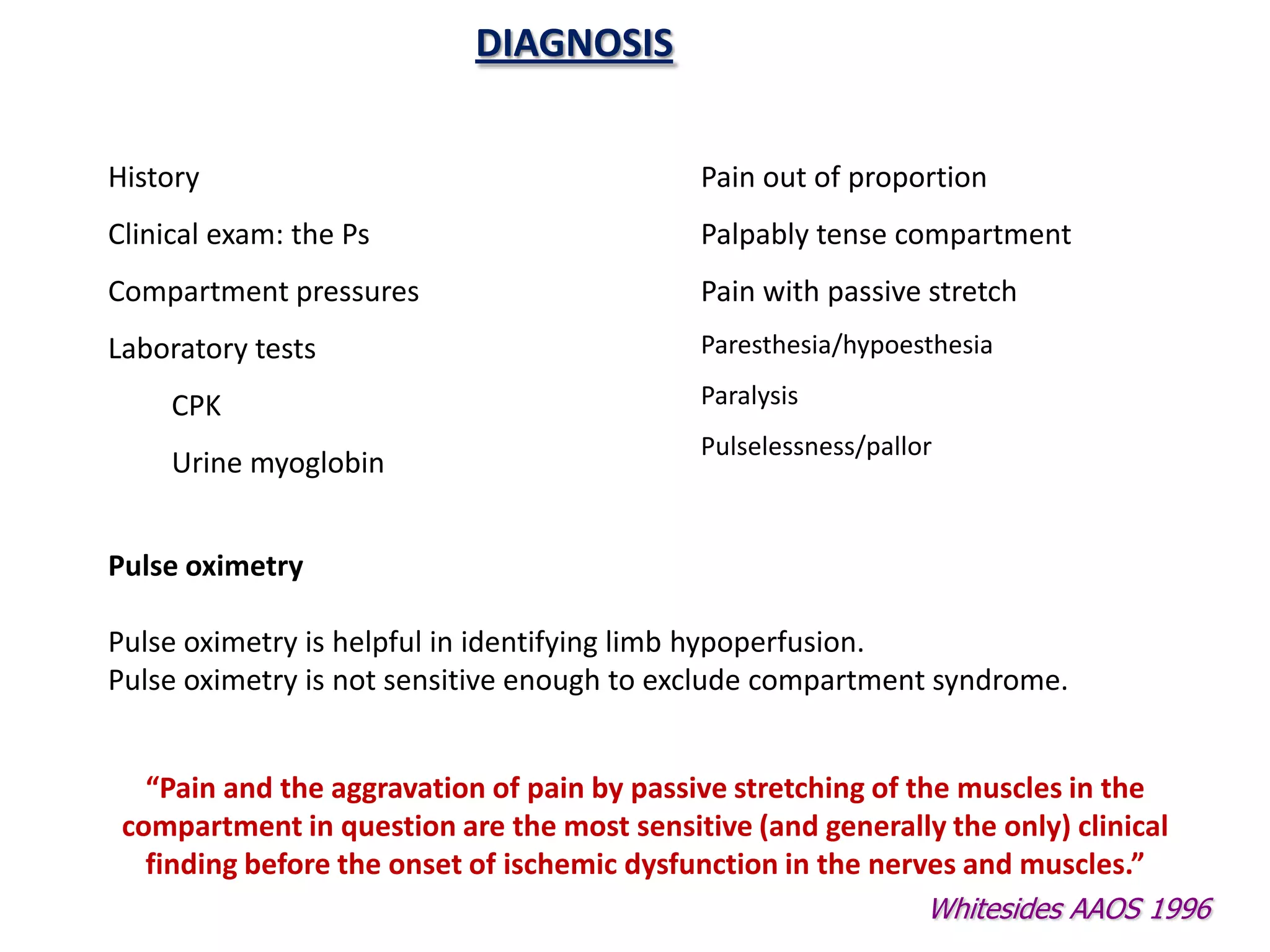
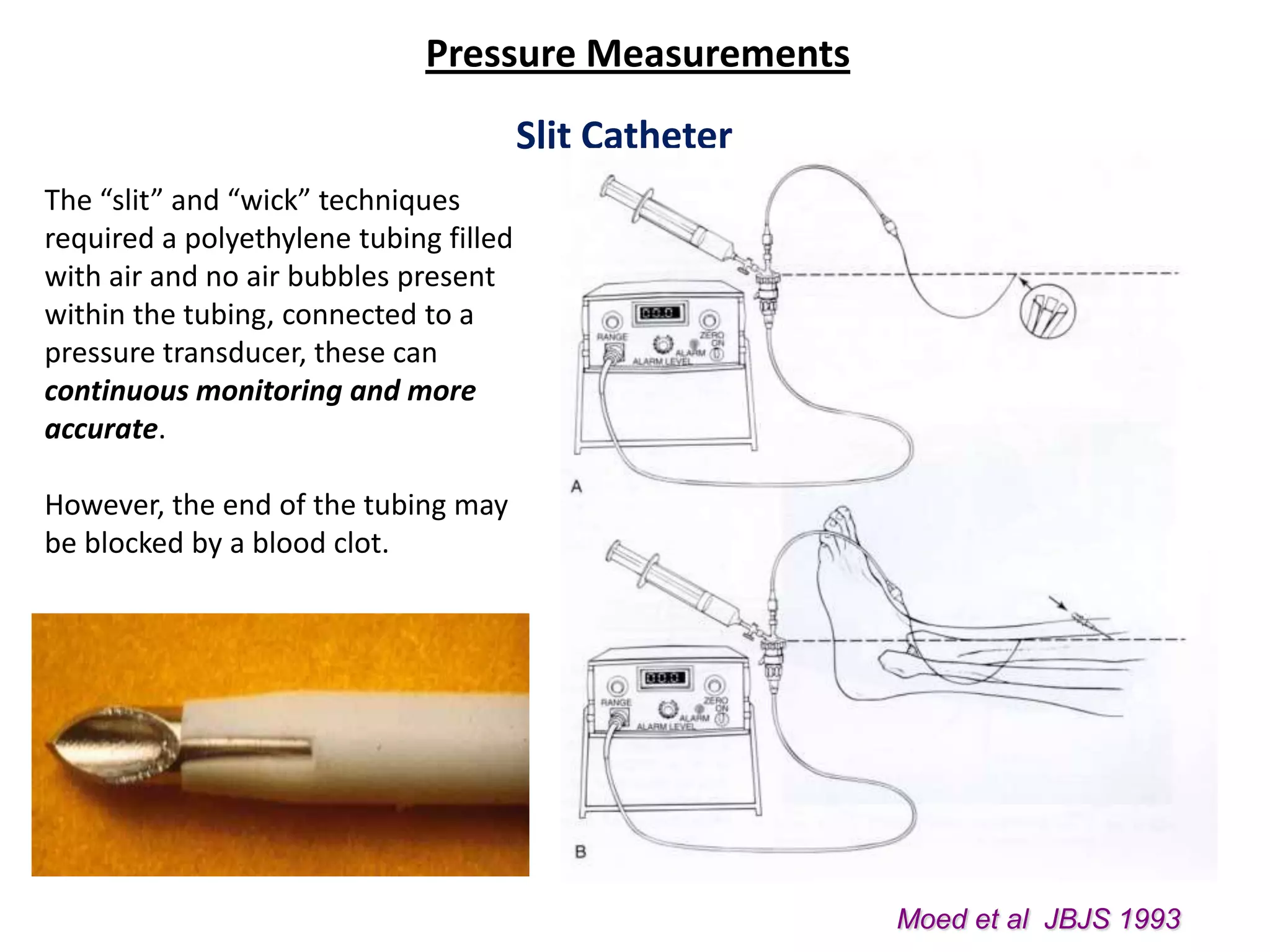
This document discusses compartment syndrome, beginning with a definition and classification. It then covers the history of compartment syndrome, notable contributors, and key events in understanding the condition. Etiology, incidence rates, effects of patient positioning, traction, and intramedullary nailing are examined. The pathophysiology and timeline of tissue damage are described. Diagnosis is discussed, highlighting indicators such as pain, tense compartments, pressure measurements, and laboratory tests. Clinical parameters like pain, paresthesia, paralysis and pallor are also outlined. The document emphasizes that pain, especially with passive stretching, is the most important sign and should not be waited on before intervention.
Seminar presentation by 4th year medical student of Lincoln University College, supervised by HRPZ Orthopedic's specialist.
Reference were from reliable medical websites and also from texttbook; Apley and Solomon's Concise System of Orthopaedics and Trauma, 4th Ed.
This is a lecture presentation on applying external fixator on open fracture specially on tibia. This method is a classical method. Various new and dynamic fixators are there but the basics are the same.
compartment syndrome, causes, compartments of legs,compartments of forearm,compartments of hand,compartments of foot, compartments of arm,compartments of thigh,fasciotomy of leg,fasciotomy of forearm, fasciotomy of hand,fasciotomy of foot, fasciotomy of thigh, fasciotomy of arm
Acute Transverse Myelitis
Blockage of the Spinal Cord’s Blood Supply
Cervical Spondylosis
Compression of the Spinal Cord
Hereditary Spastic Paraparesis
Subacute Combined Degeneration
Syrinx of the Spinal Cord and Brain Stem
Ethanol (CH3CH2OH), or beverage alcohol, is a two-carbon alcohol
that is rapidly distributed in the body and brain. Ethanol alters many
neurochemical systems and has rewarding and addictive properties. It
is the oldest recreational drug and likely contributes to more morbidity,
mortality, and public health costs than all illicit drugs combined. The
5th edition of the Diagnostic and Statistical Manual of Mental Disorders
(DSM-5) integrates alcohol abuse and alcohol dependence into a single
disorder called alcohol use disorder (AUD), with mild, moderate,
and severe subclassifications (American Psychiatric Association, 2013).
In the DSM-5, all types of substance abuse and dependence have been
combined into a single substance use disorder (SUD) on a continuum
from mild to severe. A diagnosis of AUD requires that at least two of
the 11 DSM-5 behaviors be present within a 12-month period (mild
AUD: 2–3 criteria; moderate AUD: 4–5 criteria; severe AUD: 6–11 criteria).
The four main behavioral effects of AUD are impaired control over
drinking, negative social consequences, risky use, and altered physiological
effects (tolerance, withdrawal). This chapter presents an overview
of the prevalence and harmful consequences of AUD in the U.S.,
the systemic nature of the disease, neurocircuitry and stages of AUD,
comorbidities, fetal alcohol spectrum disorders, genetic risk factors, and
pharmacotherapies for AUD.
ARTIFICIAL INTELLIGENCE IN HEALTHCARE.pdfAnujkumaranit
Artificial intelligence (AI) refers to the simulation of human intelligence processes by machines, especially computer systems. It encompasses tasks such as learning, reasoning, problem-solving, perception, and language understanding. AI technologies are revolutionizing various fields, from healthcare to finance, by enabling machines to perform tasks that typically require human intelligence.
HOT NEW PRODUCT! BIG SALES FAST SHIPPING NOW FROM CHINA!! EU KU DB BK substit...GL Anaacs
Contact us if you are interested:
Email / Skype : kefaya1771@gmail.com
Threema: PXHY5PDH
New BATCH Ku !!! MUCH IN DEMAND FAST SALE EVERY BATCH HAPPY GOOD EFFECT BIG BATCH !
Contact me on Threema or skype to start big business!!
Hot-sale products:
NEW HOT EUTYLONE WHITE CRYSTAL!!
5cl-adba precursor (semi finished )
5cl-adba raw materials
ADBB precursor (semi finished )
ADBB raw materials
APVP powder
5fadb/4f-adb
Jwh018 / Jwh210
Eutylone crystal
Protonitazene (hydrochloride) CAS: 119276-01-6
Flubrotizolam CAS: 57801-95-3
Metonitazene CAS: 14680-51-4
Payment terms: Western Union,MoneyGram,Bitcoin or USDT.
Deliver Time: Usually 7-15days
Shipping method: FedEx, TNT, DHL,UPS etc.Our deliveries are 100% safe, fast, reliable and discreet.
Samples will be sent for your evaluation!If you are interested in, please contact me, let's talk details.
We specializes in exporting high quality Research chemical, medical intermediate, Pharmaceutical chemicals and so on. Products are exported to USA, Canada, France, Korea, Japan,Russia, Southeast Asia and other countries.
Tom Selleck Health: A Comprehensive Look at the Iconic Actor’s Wellness Journeygreendigital
Tom Selleck, an enduring figure in Hollywood. has captivated audiences for decades with his rugged charm, iconic moustache. and memorable roles in television and film. From his breakout role as Thomas Magnum in Magnum P.I. to his current portrayal of Frank Reagan in Blue Bloods. Selleck's career has spanned over 50 years. But beyond his professional achievements. fans have often been curious about Tom Selleck Health. especially as he has aged in the public eye.
Follow us on: Pinterest
Introduction
Many have been interested in Tom Selleck health. not only because of his enduring presence on screen but also because of the challenges. and lifestyle choices he has faced and made over the years. This article delves into the various aspects of Tom Selleck health. exploring his fitness regimen, diet, mental health. and the challenges he has encountered as he ages. We'll look at how he maintains his well-being. the health issues he has faced, and his approach to ageing .
Early Life and Career
Childhood and Athletic Beginnings
Tom Selleck was born on January 29, 1945, in Detroit, Michigan, and grew up in Sherman Oaks, California. From an early age, he was involved in sports, particularly basketball. which played a significant role in his physical development. His athletic pursuits continued into college. where he attended the University of Southern California (USC) on a basketball scholarship. This early involvement in sports laid a strong foundation for his physical health and disciplined lifestyle.
Transition to Acting
Selleck's transition from an athlete to an actor came with its physical demands. His first significant role in "Magnum P.I." required him to perform various stunts and maintain a fit appearance. This role, which he played from 1980 to 1988. necessitated a rigorous fitness routine to meet the show's demands. setting the stage for his long-term commitment to health and wellness.
Fitness Regimen
Workout Routine
Tom Selleck health and fitness regimen has evolved. adapting to his changing roles and age. During his "Magnum, P.I." days. Selleck's workouts were intense and focused on building and maintaining muscle mass. His routine included weightlifting, cardiovascular exercises. and specific training for the stunts he performed on the show.
Selleck adjusted his fitness routine as he aged to suit his body's needs. Today, his workouts focus on maintaining flexibility, strength, and cardiovascular health. He incorporates low-impact exercises such as swimming, walking, and light weightlifting. This balanced approach helps him stay fit without putting undue strain on his joints and muscles.
Importance of Flexibility and Mobility
In recent years, Selleck has emphasized the importance of flexibility and mobility in his fitness regimen. Understanding the natural decline in muscle mass and joint flexibility with age. he includes stretching and yoga in his routine. These practices help prevent injuries, improve posture, and maintain mobilit
Lung Cancer: Artificial Intelligence, Synergetics, Complex System Analysis, S...Oleg Kshivets
RESULTS: Overall life span (LS) was 2252.1±1742.5 days and cumulative 5-year survival (5YS) reached 73.2%, 10 years – 64.8%, 20 years – 42.5%. 513 LCP lived more than 5 years (LS=3124.6±1525.6 days), 148 LCP – more than 10 years (LS=5054.4±1504.1 days).199 LCP died because of LC (LS=562.7±374.5 days). 5YS of LCP after bi/lobectomies was significantly superior in comparison with LCP after pneumonectomies (78.1% vs.63.7%, P=0.00001 by log-rank test). AT significantly improved 5YS (66.3% vs. 34.8%) (P=0.00000 by log-rank test) only for LCP with N1-2. Cox modeling displayed that 5YS of LCP significantly depended on: phase transition (PT) early-invasive LC in terms of synergetics, PT N0—N12, cell ratio factors (ratio between cancer cells- CC and blood cells subpopulations), G1-3, histology, glucose, AT, blood cell circuit, prothrombin index, heparin tolerance, recalcification time (P=0.000-0.038). Neural networks, genetic algorithm selection and bootstrap simulation revealed relationships between 5YS and PT early-invasive LC (rank=1), PT N0—N12 (rank=2), thrombocytes/CC (3), erythrocytes/CC (4), eosinophils/CC (5), healthy cells/CC (6), lymphocytes/CC (7), segmented neutrophils/CC (8), stick neutrophils/CC (9), monocytes/CC (10); leucocytes/CC (11). Correct prediction of 5YS was 100% by neural networks computing (area under ROC curve=1.0; error=0.0).
CONCLUSIONS: 5YS of LCP after radical procedures significantly depended on: 1) PT early-invasive cancer; 2) PT N0--N12; 3) cell ratio factors; 4) blood cell circuit; 5) biochemical factors; 6) hemostasis system; 7) AT; 8) LC characteristics; 9) LC cell dynamics; 10) surgery type: lobectomy/pneumonectomy; 11) anthropometric data. Optimal diagnosis and treatment strategies for LC are: 1) screening and early detection of LC; 2) availability of experienced thoracic surgeons because of complexity of radical procedures; 3) aggressive en block surgery and adequate lymph node dissection for completeness; 4) precise prediction; 5) adjuvant chemoimmunoradiotherapy for LCP with unfavorable prognosis.
- Video recording of this lecture in English language: https://youtu.be/lK81BzxMqdo
- Video recording of this lecture in Arabic language: https://youtu.be/Ve4P0COk9OI
- Link to download the book free: https://nephrotube.blogspot.com/p/nephrotube-nephrology-books.html
- Link to NephroTube website: www.NephroTube.com
- Link to NephroTube social media accounts: https://nephrotube.blogspot.com/p/join-nephrotube-on-social-media.html
The prostate is an exocrine gland of the male mammalian reproductive system
It is a walnut-sized gland that forms part of the male reproductive system and is located in front of the rectum and just below the urinary bladder
Function is to store and secrete a clear, slightly alkaline fluid that constitutes 10-30% of the volume of the seminal fluid that along with the spermatozoa, constitutes semen
A healthy human prostate measures (4cm-vertical, by 3cm-horizontal, 2cm ant-post ).
It surrounds the urethra just below the urinary bladder. It has anterior, median, posterior and two lateral lobes
It’s work is regulated by androgens which are responsible for male sex characteristics
Generalised disease of the prostate due to hormonal derangement which leads to non malignant enlargement of the gland (increase in the number of epithelial cells and stromal tissue)to cause compression of the urethra leading to symptoms (LUTS
micro teaching on communication m.sc nursing.pdfAnurag Sharma
Microteaching is a unique model of practice teaching. It is a viable instrument for the. desired change in the teaching behavior or the behavior potential which, in specified types of real. classroom situations, tends to facilitate the achievement of specified types of objectives.
Factory Supply Best Quality Pmk Oil CAS 28578–16–7 PMK Powder in Stockrebeccabio
Factory Supply Best Quality Pmk Oil CAS 28578–16–7 PMK Powder in Stock
Telegram: bmksupplier
signal: +85264872720
threema: TUD4A6YC
You can contact me on Telegram or Threema
Communicate promptly and reply
Free of customs clearance, Double Clearance 100% pass delivery to USA, Canada, Spain, Germany, Netherland, Poland, Italy, Sweden, UK, Czech Republic, Australia, Mexico, Russia, Ukraine, Kazakhstan.Door to door service
Hot Selling Organic intermediates
Anti ulcer drugs and their Advance pharmacology ||
Anti-ulcer drugs are medications used to prevent and treat ulcers in the stomach and upper part of the small intestine (duodenal ulcers). These ulcers are often caused by an imbalance between stomach acid and the mucosal lining, which protects the stomach lining.
||Scope: Overview of various classes of anti-ulcer drugs, their mechanisms of action, indications, side effects, and clinical considerations.
3. COMPARTMENT SYNDROME
Elevated tissue pressure within a closed fascial space
Reduces tissue perfusion - ischemia
Results in cell death - necrosis
True Orthopaedic Emergency
Limb Compartment Syndrome
Acute
Chronic
Abdominal Compartment Syndrome
4. HISTORY
Richard von Volkmann
1881
Volkmann published an article in which he
attempted to describe the condition of irreversible
contractures of the flexor muscles of the hand to
ischemic processes occurring in the forearm
Application of restrictive dressing to an injured limb
German surgeon, Halle
1830 - 1889, Volkmann's Ischæmic Contracture. When the wrist is flexed
born in Leipzig, died in Jena. to a right angle it is possible to extend the fingers.
5. HISTORY: TIMELINE
Hildebrand 1906
First used the term Volkmann ischemic
contracture to describe the final result of any
untreated compartment syndrome, and was
the first to suggest that elevated tissue
pressure may be related to ischemic
contracture.
Thomas 1909
Reviewed the 112 published cases of
Volkmann ischemic contracture and found
fractures to be the predominant cause. Also,
noted that tight bandages, an arterial
embolus, or arterial insufficiency could also
lead to the problem.
Murphy 1914
First to suggest that fasciotomy might prevent
the contracture. Also, suggested that tissue
pressure and fasciotomy were related to the
development of contracture.
6. HISTORY: TIMELINE
Ellis 1958
Reported a 2% incidence of
compartment syndrome with tibia
fractures, and increased attention
was paid to contractures involving the
lower extremities
Seddon, Kelly, and Whitesides 1967
Demonstrated the existence of 4
compartments in the leg and to the
need to decompress more than just
the anterior compartment. Since
then, compartment syndrome has
been shown to affect many areas of
the body, including the hand, foot,
thigh, and buttocks
8. AETIOLOGY
Fractures-closed and open Exertional states
Blunt trauma GSW
Temp vascular occlusion IV/A-lines
Cast/dressing Hemophiliac/coag
Closure of fascial defects Intraosseous IV(infant)
Burns/electrical Snake bite
Arterial injury
Patients with a coagulopathy are at particular risk of compartment syndrome.
9. FRACTURE
The most common cause
Incidence of accompanying compartment syndrome of 9.1%
The incidence is directly proportional to the degree of injury to soft
tissue and bone
ACS may be more prevalent after a low energy injury (lack of
compartment disruption)
Tibial diaphyseal #
Distal radius #
Forearm #
Blick et al JBJS 1986
10. BLUNT TRAUMA
2nd most common cause
About 23% of CS
25% due to direct blow
McQueen et al; JBJS Br 2000
11. Hematoma after arterial puncture resulting in
compartment syndrome
Mannitol extravasation during partial nephrectomy
leading to forearm compartment syndrome
12. INCIDENCE
McQueen et al; JBJS Br 2000
164 pts with CS,
149 male, 15 female
Most pts were usually under 35 y
69% with associated fx, about half were tibial shaft
23% soft tissue injury without fx
Ranges of 2-12% have been published
Type of Fx % of Incidence Incidence
ACS all ages <35
Tibial 36% 4.3% 5.9%(3 fold)
diaphysis
Distal 9.8% 0.25% 1.4%(30 fold)
radius
Forearm 7.9% 3.1% 3.2%
diaphysis
McQueen et al; JBJS Br 2000
13. Patient Positioning
Leaving the calf free when the leg is
placed in the hemilithotomy position
instead of using a standard well-leg
holder
Increases the difference between the
diastolic blood pressure and the
intramuscular pressure
May decrease the risk of compartment
syndrome
Elevation of leg
Pressure on posterior compartment
Circumferential inflated devices
Wraps
Meyer, Mubarak JBJS 2002
14. Role of Traction
• Pressure increases linear
with increasing weight
– Posterior compartment
of leg most effected
– 1 kg added weight
• 5% increase in
posterior
compartment
• <2% increase in
anterior
compartment
• Calcaneal traction increases
dorsiflexion
15. Compartment Syndrome and Intramedullary Nailing
Nassif et al, J Orthop Trauma, 2000
Effect of acute reamed vs unreamed intramedullary nailing on compartment
pressure when treating closed tibial shaft fractures: a randomised prospective
study.
Highest pressures occurred during reaming in reamed group and during nail insertion
in unreamend group.
However no significant difference in pressures between the two groups
SPRINT Trial
No significant difference in rates of fasciotomy following reamed and unreamed tibial
nailing.
16. PATHOPHYSIOLOGY
Normal tissue pressure
0-4 mm Hg
8-10 with exertion
Absolute pressure theory
30 mm Hg - Mubarak
45 mm Hg – Matsen
Pressure gradient theory
< 30 mm Hg of diastolic
pressure
– Whitesides
McQueen, et al
Reperfusion Injury
18. HOW DO WE DIAGNOSE PRESENCE OF
COMPARTMENT SYNDROME IN A TRAUMATISED LIMB?
19. DIAGNOSIS
History Pain out of proportion
Clinical exam: the Ps Palpably tense compartment
Compartment pressures Pain with passive stretch
Laboratory tests Paresthesia/hypoesthesia
CPK Paralysis
Pulselessness/pallor
Urine myoglobin
Pulse oximetry
Pulse oximetry is helpful in identifying limb hypoperfusion.
Pulse oximetry is not sensitive enough to exclude compartment syndrome.
“Pain and the aggravation of pain by passive stretching of the muscles in the
compartment in question are the most sensitive (and generally the only) clinical
finding before the onset of ischemic dysfunction in the nerves and muscles.”
Whitesides AAOS 1996
20. CLINICAL PARAMETERS
Pain
First symptom
Classically out of portion to injury, ischemic character
Exaggerated with passive stretch of the involved muscles in compartment
Earliest symptom but inconsistent, minimal in deep post compartment.
Not available in obtunded patient
Pressure
Early finding
Only objective finding
Refers to palpation of compartment and its tension or firmness
Paresthesia
Also early sign
Peripheral nerve tissue is more sensitive than muscle to ischemia
Permanent damage may occur in 75 minutes
Difficult to interpret
Will progress to anesthesia if pressure not relieved
21. CLINICAL PARAMETERS
Paralysis
Very late finding
Irreversible nerve and muscle damage present
Paresis may be present early
Difficult to evaluate because of pain
If motor deficit develops, full recovery is rare
Pallor & Pulselessness
Rarely present
Indicates direct damage to vessels rather than compartment syndrome
(therefore arteriography indicated)
Vascular injury may be more of contributing factor to syndrome rather than
result
22. CLINICAL PARAMETERS
Pain – most important.
Especially pain out of proportion to
the injury (child becoming more and
more restless /needing more
analgesia)
Most reliable signs are pain on
passive stretching and pain on
palpation of the involved
compartment
Other features like pallor,
pulselessness, paralysis, paraesthesia
etc. appear very late and we should
not wait for these things.
Missing the boat
Pale
Willis &Rorabeck OCNA 1990 Pulseless
Paralyzed
23. VALUE OF THESE CLINICAL PARAMETRS
IN DIAGNOSIS OF COMPARTMENT SYNDROME ?
Ulmer T:
The clinical diagnosis of compartment syndrome of the lower leg: are clinical findings
predictive of the disorder?
J Orthop Trauma 2002
24. Clinical Sensitivity (Ulmer T 02)
Pain Paresthesia PPS Paresis
Sensitivity 0.19 0.13 0.19 0.13
Specificity 0.97 0.98 0.97 0.97
PPV 0.14 0.15 0.14 0.11
NPV 0.98 0.98 0.98 0.98
Do these numbers reflect anything more than a low incidence? MISSED CASES
To achieve a probability of over 90% of ACS being present 3 clinical findings must be
present. The third clinical finding is paresis, thus to achieve an accurate clinical diagnosis
of ACS the condition must be allowed to progress until late which is clearly unacceptable.
25. CLINICAL EVALUATION
Beware of epidural analgesia
Strecker JBJS 1986
Morrow J. Trauma 1994
Beware long acting nerve blocks
Hyder JBJS Br 1995
Beware controlled intravenous opiate analgesia
26. COMPARTMENT PRESSURE MONITORING
Raised tissue pressure is primary event in ACS, changes in ICP will
precede the clinical signs and symptoms
When to monitor?
How to monitor – continuous or single measurement?
Threshold for diagnosis of Compartment syndrome and Fasciotomy?
28. Compartment Pressure Monitoring
Harris et al, J Trauma, 2006:
Continuous compartment pressure monitoring for tibia fractures: does it
influence outcome?
Randomized 200 Extraarticular tibial shaft fractures
Monitored (36 h continuous pressure monitoring) and unmonitored groups
Level of Evidence : 1
Results:
05 cases of CS in nonmonitored group, 0 cases in monitored group
Monitored group 18 pts had ∆P(DBP-ICP) of < 30 mm Hg, none developed CS or
late sequelae.
In awake and alert pt, diagnosis of CS using clinical signs in appropriate time
possible and continuous pressure monitoring in these pt not necessary.
29. Pressure Measurements
Whitesides Technique
Simple
Disadv - injection of saline into the compartment and this way aggravates an
impending syndrome
(Whitesides, CORR 1975)
30. Pressure Measurements
Slit Catheter
The “slit” and “wick” techniques
required a polyethylene tubing filled
with air and no air bubbles present
within the tubing, connected to a
pressure transducer, these can
continuous monitoring and more
accurate.
However, the end of the tubing may
be blocked by a blood clot.
Moed et al JBJS 1993
31. Pressure Measurements
Stryker STIC Monitor.
A solid-state transducer intracompartmental
catheter (STIC) may be used, which is more
accurate and reliable.
Can monitor ICP for up to 16 hours.
32. Pressure Measurements
Near-Infrared Spectroscopy (NIRS)
A technique that allows tracking of variations in the oxygenation of muscle tissue.
It can be useful for chronic compartment syndrome in adults, but it is of little value in
acute CS as changes in the relative oxygenation may have already occurred, at the
moment of measurement.
NIRS measures soft tissue oxygenation (StO 2 ) noninvasively, is potentially a new
noninvasive technique for the early detection of acute compartment syndrome (ACS).
Animal models of ACS have shown that StO 2 correlates with perfusion pressure in
the compartment. The StO2 difference (measured noninvasively) was significantly
lower among patients with an ACS, suggesting that NIRS can detect decreasing tissue
oxygenation in trauma patients who are developing an ACS.
33. Pressure Measurements
Laser Doppler Flowmetry
Laser Doppler Flowmetry (LDF) is a
non-invasive method to estimate
the blood perfusion in the
microcirculation.
Uses a flexible fibre optic wire
which is introduced into the
muscle compartment and the
signals from this wire are recorded
on a computer.
It can be used as an adjuvant
diagnostic tool for chronic CS.
34. Pressure Measurements
Measurements must be made in all
compartments
Anterior and deep posterior are usually
highest
Measurement made within 5 cm of fx
Distance From Fracture Effects Pressure
Marginal readings must be followed with
repeat physical exam and repeat
compartment pressure measurement
Heckman, Whitesides JBJS 1994
35. What is Critical Pressure?
Significant individual variations in tolerance to raised ICP is largely
because of variation in Systemic BP
>30 mm Hg as absolute number (Roraback)
>45 mm Hg as absolute number (Matsen)
<30 mm Hg for ∆p (where ∆p =diastolic pressure – compartment pressure, McQueen)
<40 mm Hg for ∆P (where ∆P mean arterial pressure* – compartment pressure,
Heppenstall)
*mean arterial pressure is diastolic pressure plus 1/3 of pulse pressure
Whether the Absolute pressure or ∆P should be used to diagnose
Compartment Syndrome ?
36. Threshold for fasciotomy
McQueen, Court-Brown JBJS Br 1996
116 pts with tibial diaphyseal fx had continuous monitoring of anterior
compartment pressure for 24 hours
53 pts had ICP over 30 mmHg
30 pts had ICP over 40 mmHg
04 pts had ICP over 50 mmHg
Only 03 had ∆P(DBP-ICP) of < 30, they had fasciotomy
None of the patients had any sequelae of the compartment syndrome
Decompression should be performed if the differential
pressure level drops to under 30 mmHg (∆P)
37. SUSPECTED COMPARTMENT SYNDROME
Unequivocal + Findings Pt. not alert/polytrauma/inconc.
Comp. pressure measurement
w/i 30 mm Hg >30 mm Hg of DBP
Serial exams
FASCIOTOMY FASCIOTOMY
McQueen JBJSB 1996
38. Management
• High index of Clinical Suspicion
• Ensure patient is normotensive, as hypotension reduces prefusion pressure
and facilitates further tissue injury.
• Remove cicumferential bandages and cast
• Maintain the limb at level of the heart as elevation reduces the arterial inflow
and the arterio-venous pressure gradient on which perfusion depends.
Perfusion pressure = A pr(30-35mmHg) – V pr(10-15mmHg)
• Supplemental oxygen administration.
39. Removal of Circumferential Casts
Compartmental pressure falls by 30% when cast is split on one side
Falls by 65% when the cast is spread after splitting.
Splitting the padding reduces it by a further 10% and complete removal of cast by
another 15%
Total of 85-90% reduction by just taking off the plaster!
Garfin, Mubarak JBJS 1981
42. Indications for Fasciotomy
Unequivocal clinical findings
Pressure within 15-20 mm hg of DBP
Rising tissue pressure
Significant tissue injury or high risk pt
> 6 hours of total limb ischemia
Injury at high risk of compartment syndrome
CONTRAINDICATION -
Missed compartment syndrome (>24-48 hrs)
43. Fasciotomy Principles
Make early diagnosis
Long extensile incisions
Release all fascial compartments
Preserve neurovascular structures
Debride necrotic tissues
Coverage within 7-10 days
44. Lower Leg
4 compartments
Lateral
Peroneus longus and brevis
Anterior
EHL, EDC, Tibialis anterior,
Peroneus tertius
Supeficial posterior
Gastrocnemius, Soleus
Deep posterior
Tibialis posterior, FHL, FDL
47. Lower Leg
Double Incision (Recommended)
In most instances it affords better
exposure of the four compartments
2 vertical incisions separated by minimum
8 cm
One incision over anterior and lateral
compartments
Superficial peroneal nerve
One incision located 1-2 cm behind
postero
-medial aspect of tibia
Saphenous nerve and vein
Mubarak et al JBJS 1977
51. Lower Leg
Look for Superficial Peroneal Nerve
Superficial peroneal nerve exits from lateral
compartment about 10 cm above lateral
malleolus and courses into the anterior
compartment
Risk of injury
Use a Generous Incision
Lengthening the skin incisions to an average of 16 cm
decreases intracompartmental pressures significantly.
The skin envelope is a contributing factor in acute
compartment syndromes of the leg and The use of
generous skin incisions is supported
Cohen, Mubarak JBJS Br 1991
52. Forearm
3 compartments
Mobile wad
BR,ECRL,ECRB
Volar
Superficial and deep flexors
Dorsal
Extensors
Pronator quadratus described as a
separate compartment
53. Forearm
Volar-Henry approach
Include a carpal tunnel release
Release lacertus fibrosus and fascia
Protect median nerve, brachial artery
and tendons after release
Consider dorsal release
54. Foot
9 compartments
Medial,
Superficial,
Lateral,
Calcaneal
Interossei(4),
Adductor
Careful exam with any swelling
Clinical suspicion with certain mechanisms of injury
Lisfranc fracture dislocation
Calcaneus fracture
55. Foot
Dorsal incision
To release the interosseous and
adductor
Medial incision
To release the medial, superficial
lateral and calcaneal compartments
56. Hand
10 separate osteofascial compartments
dorsal interossei (4)
palmar interossei (3)
thenar and hypothenar (2)
adductor pollicis (1)
non specific aching of the hand
disproportionate pain
loss of digital motion & continued swelling
MP extension and PIP flexion
difficult to measure tissue pressure
57. Thigh
Lateral to release anterior and
posterior compartments
May require medial incision
for adductor compartment
Vastus lateralis
Lateral septum
58. Delayed Fasciotomy - Is it Safe?
Sheridan, Matsen.JBJS 1976
Infection rate of 46% and amputation rate of 21% after a delay of 12 hours
4.5 % complications for early fasciotomies and 54% for delayed ones
Recommendations
If the CS has existed for more than 8-10 hrs, supportive treatment of acute renal
failure should be considered.
Skin is left intact and late reconstructions maybe planned.
59. Delayed Fasciotomy - Is it Safe?
Finkelstein et al. J Trauma 1996
5 pts, nine fasciotomies in lower limbs
Avg delay 56 h. (35-96 hrs).
1 pt died of septicaemia and multi organ failure, the others required amputations
Recommendations:
In delayed cases, routine fasciotomy may not be successful
Should we do Fasciotomy in delayed cases?
If there is no likelihood of any surviving muscles and ICP is low, withhold
Fasciotomy.
If any possibility of viable muscle or if ICP more than critical levels, Fasciotomy
should be done.
60. Wound Management
After the fasciotomy, a bulky compression dressing and a splint are applied.
“VAC” (Vacuum Assisted Closure) can be used
Foot should be placed in neutral to prevent equinus contracture.
Incision for the fasciotomy usually can be closed after 3 - 5 days
62. Wound Management
Wound is not closed at initial surgery
Second look debridement with consideration for coverage after 48-72 hrs
Limb should not be at risk for further swelling
Pt should be adequately stabilized
Usually requires skin graft
DPC possible if residual swelling is minimal
Flap coverage needed if nerves, vessels, or bone exposed
Goal is to obtain definitive coverage within 7-10 days
64. Complications Related to Fasciotomies
• Altered sensation within the margins of the wound (77%)
• Dry, scaly skin (40%)
• Pruritus (33%)
• Discolored wounds (30%)
• Swollen limbs (25%)
• Tethered scars (26%)
• Recurrent ulceration (13%)
• Muscle herniation (13%)
• Pain related to the wound (10%)
• Tethered tendons (7%)
Fitzgerald, McQueen Br J Plast Surg 2000
65. Complications related to CS
Late Sequelae
Volkmann’s contracture
Weak dorsiflexors
Claw toes
Sensory loss
Chronic pain
Amputation
66. Chronic Compartment Syndrome
Chronic CS usually occurs in young active patients after
intense muscular activity.
It is usually detected in the tibial shaft (anterior or
posterior deep compartment).
Main symptoms involve pain, parasthesia of the muscle
within the affected compartment, after intense and
continuing (over than 20-30 min) streching of muscle
groups.
The symptoms recess progressively by interrupting any
kind of exercise (15-20min).
Differential diagnosis include stress fracture, superficial
fibular nerve entrapment syndrome, posterior tibial
muscle tendonitis.
67. CASE
A 27 year-old man sustained an undisplaced
midshaft fracture of his left tibia.
Following an orthopedic consult, he was put in a
long leg cast and sent home, with orthopedics
follow up arranged for the next day.
Overnight he re-presented to the emergency
department with increased pain in his leg and
paraesthesiae in his toes.
The cast was removed.
Peripheral pulses were intact, but the anterior
compartment of the leg, in particular, was tense
and tender to palpation.
He was taken to the operating theatre in the
middle of the night after an anterior compartment
pressure measurement of 60 mmHg was obtained.
68. CASE REPORTS
Simvastatin-induced bilateral leg compartment syndrome and myonecrosis
associated with hypothyroidism
A 54-year-old hypothyroid male
taking thyroxine and simvastatin
presented with bilateral leg
compartment syndrome and
myonecrosis.
Urgent fasciotomies were
performed and the patient
made an uneventful recovery
with the withdrawal of
simvastatin.
It is likely that this complication
will be seen more often with
the increased worldwide use of
this drug and its approval for all
arteriopathic patients.
Postgrad Med J 2007;83:152-153 doi:10.1136/pgmj.2006.051334
69. CASE REPORTS
Bilateral compartment syndrome in thighs and legs by methanol intoxication
Methanol intoxication is infrequent even
though it is easily obtainable. One of the
complications in locomotor apparatus is the
development of a compartment syndrome of
the lower extremities.
A 49-year-old man with a compartment
syndrome in all compartments of both legs
and the anterior compartment of both thighs
due to methanol intoxication.
The patient underwent a bilateral fasciotomy
of the legs and thighs.
He also had haemodialysis sessions because
of acute renal insufficiency.
After 4 weeks of haemodialysis, covering of
the fasciotomies with cutaneous autograft
and rehabilitation treatment, the patient was
Emerg Med J 2008;25:540-541 doi:10.1136/emj.2008.058461
able to walk on his own again.
70. CASE REPORTS
65 y M, H/o a street fight 5 days back
Presented with tachycardia,
hypotension, fever, confusion.
O/E - secretion through a small injury
on the dorsal aspect of the proximal
phalanx of 3 finger in left hand;
increase of local temperature, slow
capillary reflux, edema on the
forearm, flictenas on dorsum of hand
and forearm, exacerbated pain at
finger mobilization, and decrease in
distal sensibility (median nerve
territory), no crepitation was
detected.
X Rays – No fractures, No gas, No FB
TLC - 22000 The Internet Journal of Emergency Medicine 2003 :
Volume 1 Number 2
71. CASE REPORTS
Diagnosis - Hand and forearm compartmental
syndrome secondary to hand infection.
Rx-
Antitetanic immunization
Antibiotics
Pressure in dorsal compartment was (20mm
Hg) and in palmar compartment (42 mm Hg).
A Henry approach was performed,
decompressing palmar spaces including carpal
tunnel, observing edema in muscles and
venous congestion, with no infection signs.
Dorsal approach on hand and forearm was
done, purulent material (about 120 ml)
drained from preretinacular space, carefully
cleaning was performed, leaving the wound
open and material was sent to culture and
pathology.
72. CASE REPORTS
The upper limb splinted and remained
elevated.
Sensibility and capillary reflux improve
immediately surgery, and systemic symptoms
disappear after 6 hours.
A beta hemolytic streptococcus was isolated.
At 24/48 and 72 hours new toilettes were
performed, with daily evaluation.
The patient was discharged after a week of
treatment, and started hand rehabilitation,
and occupational therapy.
After 3 weeks the wound was closed by second
intention and mobility was acceptable.