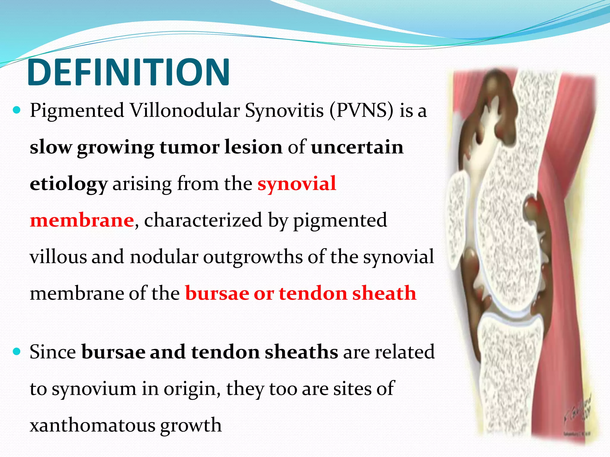
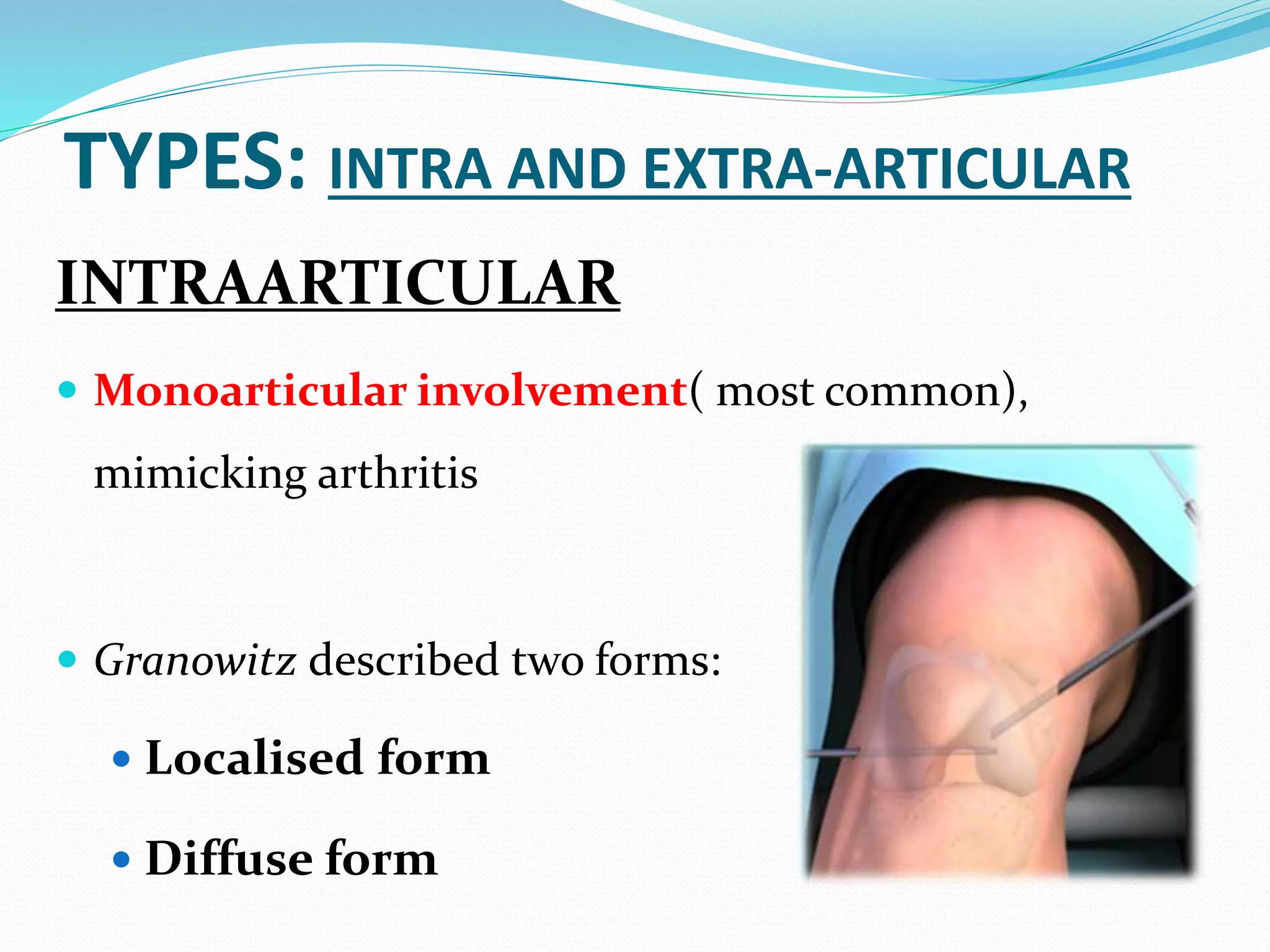
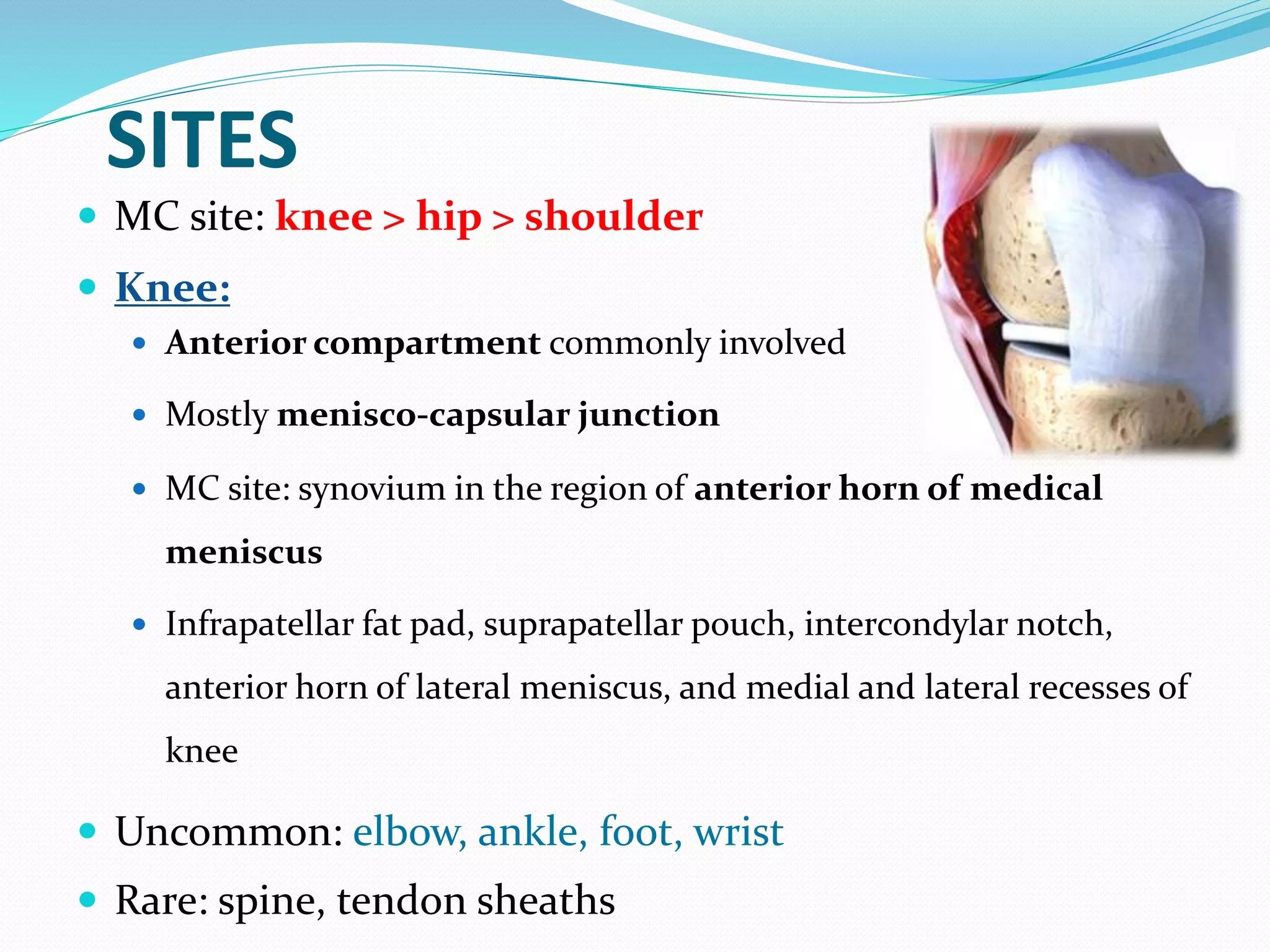
By: Dr. Bipul Borthakur
Professor,
Dept of Orthopaedics, SMCH
DEFINITION
 Pigmented Villonodular Synovitis (PVNS) is a
slow growing tumor lesion of uncertain
etiology arising from the synovial
membrane, characterized by pigmented
villous and nodular outgrowths of the synovial
membrane of the bursae or tendon sheath
 Since bursae and tendon sheaths are related
to synovium in origin, they too are sites of
xanthomatous growth
PREVALENCE
 Age: 3rd to 4th decade of life, rare in children
 Sex: no sex based predilection
 Incidence: 1.8 per million
 Appendicular skeleton, especially large joints
such as knee and hip joints are frequently
involved
 No predilection for any laterality
ETIOPATHOGENESIS
Repetitive trauma (50% cases)
Recurrent local hemorrhage to
affected joint
Proliferation of synovium of
joints, tendon sheaths or bursae
Progressive erosive arthropathies
(especially in haemophilics)
ETIOPATHOGENESIS
 It is a reactive condition, and not a true
neoplasm
 Recurrent atraumatic haemarthrosis is a
characteristic feature
 Often aggressive, with marked extra-articular
extension
TYPES: INTRA AND EXTRA-ARTICULAR
INTRAARTICULAR
 Monoarticular involvement( most common),
mimicking arthritis
 Granowitz described two forms:
 Localised form
 Diffuse form
 Localized form
 Focal involvement of the synovium
 Nodular/ sessile or pedunculated well circumscribed masses
 Diffuse form
 More common
 Affect virtually the entire synovium
EXTRAARTICULAR
 Tendon sheath and bursae PVNS
SITES
 MC site: knee > hip > shoulder
 Knee:
 Anterior compartment commonly involved
 Mostly menisco-capsular junction
 MC site: synovium in the region of anterior horn of medical
meniscus
 Infrapatellar fat pad, suprapatellar pouch, intercondylar notch,
anterior horn of lateral meniscus, and medial and lateral recesses of
knee
 Uncommon: elbow, ankle, foot, wrist
 Rare: spine, tendon sheaths
CLINICAL FEATURES
Symptoms Signs
 Pain: 80%
 Swelling: 75%
 Stiffness
 Locking
 catching
 Instability
 Palpable mass: 12%
 Effusion – floating
patella
 Tenderness
 Decreased ROM
 Usually not palpable;
In localised variety,
can show a joint
mouse
TYPE SPECIFIC FEATURES
LOCALIZED PVNS
 Initially painless
 If untreated, caused continuous pain and
discomfort, limiting activities of daily living
 At knee, mostly presents with signs and
symptoms of meniscal pathology
 Locking
 Catching
 Instability
 Well circumscribed soft tissue mass
DIFFUSE PVNS
 Slow, insidious onset pain
 Swelling
 Stiffness
 Most of or all of the joint involved
 Decreased ROM
 Poorly localised/ ill defined mass
 May encroach on nearby neurovascular structures
 Osteoarthritis: continued inflammation and joint erosions
lead to articular cartilage destruction
INVESTIGATION : X-RAY
 Soft tissue swelling
 Cysts, bony erosions in joint-
mimicks gout.
 Osteoporosis is characteristically
absent
 Can affect epiphysis in children
 Reciprocal bony lesions on
opposite sides of joint, despite
articular preservation- highly
suggestive
 Degenerative changes,
osteoarthritic changes
MRI
 Investigation of choice
 LPVNS:
 Nodular mass (periarticular or
synovial)
 bone erosion
 DPVNS:
 extensive mass and thickening of
joint lining
 destruction of bone and cartilage
ULTRASONOGRAPHY
 Loculated joint effusion
 Complex heterogenous echogenic
masses with markedly thickened
synovium
ASPIRATION
 Blood tinged thick orange brown
fluid containing cholesterol in large
amounts
ARTHROSCOPY
 Diagnostic and
therapeutic value
 Direct visualisation
of synovium
 Normal arthroscopic
findings however does
not exclude PVNS
HISTOPATHOLOGY
 Synovium looks like a “shaggy carpet”
 Histiocytes
 lipid laden macrophages
 hemosiderin containing cells
 frequent giant cells
 Subsynovial nodular proliferation of
large round, polyhedral or spindle cells
with prominent cytoplasm and pale
nuclei
DIFFERENTIAL DIAGNOSIS
 Hemophilic lobular synovitis
 Menisceal tear or other ligamentous injury
 Synovial chondromatosis
 Osteoarthritis
 Gouty arthritis
 Rheumatoid arthritis
TREATMENT
 Local excision- for nodular form
 Total Synovectomy- arthroscopic or open
 Radiotherapy
 Advanced cases: Arthrodesis/ arthroplasty +
extensive synovectomy
SYNOVECTOMY
Open :
 Anterior approach or medial
parapatellar approach: for the
diffuse form
 Posterior approach done
subsequently for extensions into
the popliteal fossa
SYNOVECTOMY
Arthroscopic:
 Gained popularity because of
several advantages over open
technique
 Preferred for LPVNS
 Higher recurrence rate in DPVNS
 Vascular or neurological injury:
especially in posterior extra-
articular extension or fibrosis after
irradiation
RADIOTHERAPY
 3500- 4000 cGy
 Radiation induced synovectomy
 Intra-articular radiation
synovectomy with Yttrium-90
 Use: Recurrence
PROGNOSIS
LPVNS: DPVNS:
 Excellent prognosis
 Low recurrence rate: 8%
 Complete surgical
excision is difficult
 Recurrence rate: upto 46%
THANK YOU
“yajñārthātkarmaṇo’nyatra loko’yaṃ karmabandhanaḥ
tadarthaṃ karma kaunteya muktasaṅgaḥ samācara”
“Man is bound by his own action except when it is performed for the sake of sacrifice.
Therefore, Arjuna, efficiently perform your duty, free from attachment, for the sake of
sacrifice alone.”

Pigmented villonodular synovitis

  • 1.
    By: Dr. BipulBorthakur Professor, Dept of Orthopaedics, SMCH
  • 2.
    DEFINITION  Pigmented VillonodularSynovitis (PVNS) is a slow growing tumor lesion of uncertain etiology arising from the synovial membrane, characterized by pigmented villous and nodular outgrowths of the synovial membrane of the bursae or tendon sheath  Since bursae and tendon sheaths are related to synovium in origin, they too are sites of xanthomatous growth
  • 3.
    PREVALENCE  Age: 3rdto 4th decade of life, rare in children  Sex: no sex based predilection  Incidence: 1.8 per million  Appendicular skeleton, especially large joints such as knee and hip joints are frequently involved  No predilection for any laterality
  • 4.
    ETIOPATHOGENESIS Repetitive trauma (50%cases) Recurrent local hemorrhage to affected joint Proliferation of synovium of joints, tendon sheaths or bursae Progressive erosive arthropathies (especially in haemophilics)
  • 5.
    ETIOPATHOGENESIS  It isa reactive condition, and not a true neoplasm  Recurrent atraumatic haemarthrosis is a characteristic feature  Often aggressive, with marked extra-articular extension
  • 6.
    TYPES: INTRA ANDEXTRA-ARTICULAR INTRAARTICULAR  Monoarticular involvement( most common), mimicking arthritis  Granowitz described two forms:  Localised form  Diffuse form
  • 7.
     Localized form Focal involvement of the synovium  Nodular/ sessile or pedunculated well circumscribed masses  Diffuse form  More common  Affect virtually the entire synovium EXTRAARTICULAR  Tendon sheath and bursae PVNS
  • 8.
    SITES  MC site:knee > hip > shoulder  Knee:  Anterior compartment commonly involved  Mostly menisco-capsular junction  MC site: synovium in the region of anterior horn of medical meniscus  Infrapatellar fat pad, suprapatellar pouch, intercondylar notch, anterior horn of lateral meniscus, and medial and lateral recesses of knee  Uncommon: elbow, ankle, foot, wrist  Rare: spine, tendon sheaths
  • 9.
    CLINICAL FEATURES Symptoms Signs Pain: 80%  Swelling: 75%  Stiffness  Locking  catching  Instability  Palpable mass: 12%  Effusion – floating patella  Tenderness  Decreased ROM  Usually not palpable; In localised variety, can show a joint mouse
  • 10.
    TYPE SPECIFIC FEATURES LOCALIZEDPVNS  Initially painless  If untreated, caused continuous pain and discomfort, limiting activities of daily living  At knee, mostly presents with signs and symptoms of meniscal pathology  Locking  Catching  Instability  Well circumscribed soft tissue mass
  • 11.
    DIFFUSE PVNS  Slow,insidious onset pain  Swelling  Stiffness  Most of or all of the joint involved  Decreased ROM  Poorly localised/ ill defined mass  May encroach on nearby neurovascular structures  Osteoarthritis: continued inflammation and joint erosions lead to articular cartilage destruction
  • 12.
    INVESTIGATION : X-RAY Soft tissue swelling  Cysts, bony erosions in joint- mimicks gout.  Osteoporosis is characteristically absent  Can affect epiphysis in children
  • 13.
     Reciprocal bonylesions on opposite sides of joint, despite articular preservation- highly suggestive  Degenerative changes, osteoarthritic changes
  • 14.
    MRI  Investigation ofchoice  LPVNS:  Nodular mass (periarticular or synovial)  bone erosion  DPVNS:  extensive mass and thickening of joint lining  destruction of bone and cartilage
  • 15.
    ULTRASONOGRAPHY  Loculated jointeffusion  Complex heterogenous echogenic masses with markedly thickened synovium ASPIRATION  Blood tinged thick orange brown fluid containing cholesterol in large amounts
  • 16.
    ARTHROSCOPY  Diagnostic and therapeuticvalue  Direct visualisation of synovium  Normal arthroscopic findings however does not exclude PVNS
  • 17.
    HISTOPATHOLOGY  Synovium lookslike a “shaggy carpet”  Histiocytes  lipid laden macrophages  hemosiderin containing cells  frequent giant cells  Subsynovial nodular proliferation of large round, polyhedral or spindle cells with prominent cytoplasm and pale nuclei
  • 18.
    DIFFERENTIAL DIAGNOSIS  Hemophiliclobular synovitis  Menisceal tear or other ligamentous injury  Synovial chondromatosis  Osteoarthritis  Gouty arthritis  Rheumatoid arthritis
  • 19.
    TREATMENT  Local excision-for nodular form  Total Synovectomy- arthroscopic or open  Radiotherapy  Advanced cases: Arthrodesis/ arthroplasty + extensive synovectomy
  • 20.
    SYNOVECTOMY Open :  Anteriorapproach or medial parapatellar approach: for the diffuse form  Posterior approach done subsequently for extensions into the popliteal fossa
  • 21.
    SYNOVECTOMY Arthroscopic:  Gained popularitybecause of several advantages over open technique  Preferred for LPVNS  Higher recurrence rate in DPVNS  Vascular or neurological injury: especially in posterior extra- articular extension or fibrosis after irradiation
  • 22.
    RADIOTHERAPY  3500- 4000cGy  Radiation induced synovectomy  Intra-articular radiation synovectomy with Yttrium-90  Use: Recurrence
  • 23.
    PROGNOSIS LPVNS: DPVNS:  Excellentprognosis  Low recurrence rate: 8%  Complete surgical excision is difficult  Recurrence rate: upto 46%
  • 24.
    THANK YOU “yajñārthātkarmaṇo’nyatra loko’yaṃkarmabandhanaḥ tadarthaṃ karma kaunteya muktasaṅgaḥ samācara” “Man is bound by his own action except when it is performed for the sake of sacrifice. Therefore, Arjuna, efficiently perform your duty, free from attachment, for the sake of sacrifice alone.”