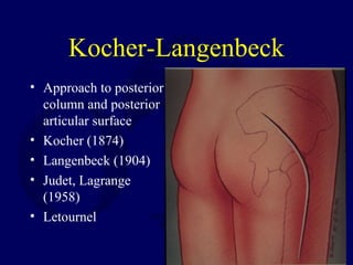
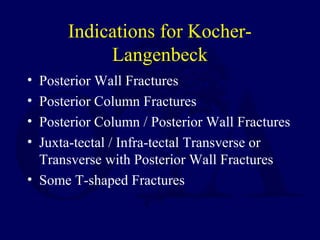
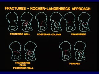
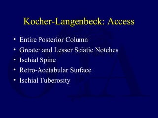
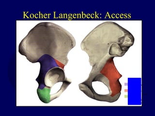
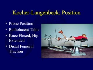
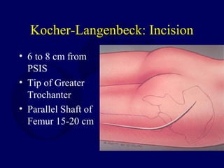
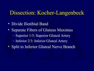
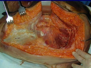
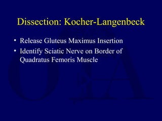
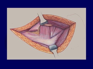
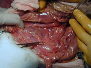
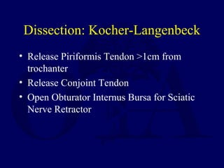
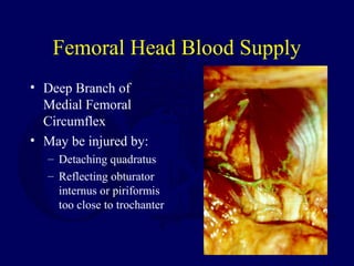
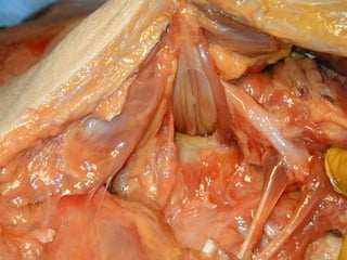
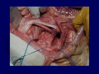
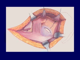
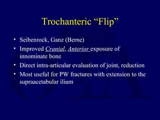
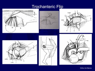
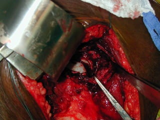
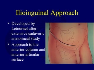
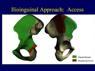
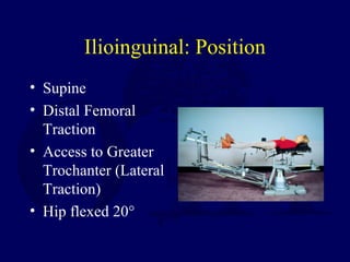
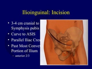
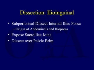
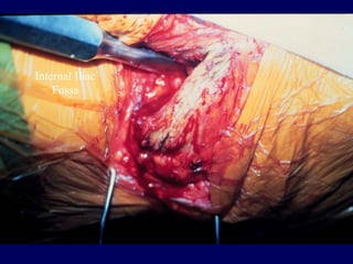
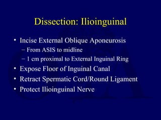
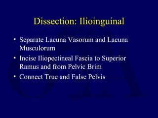
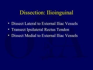
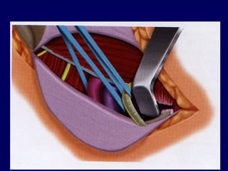
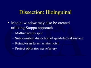
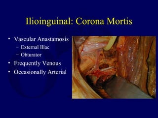
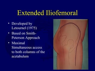
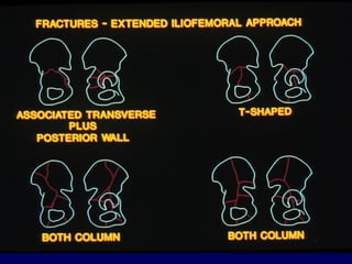
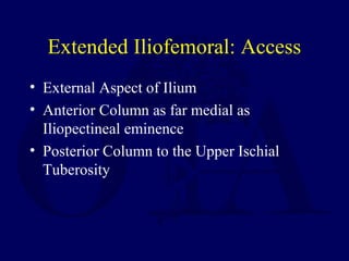
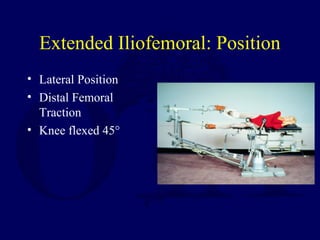
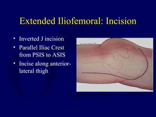
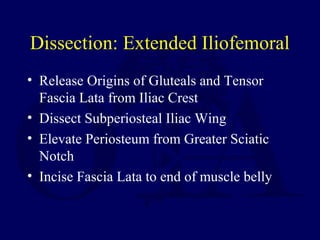
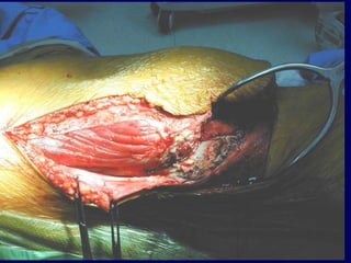
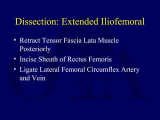
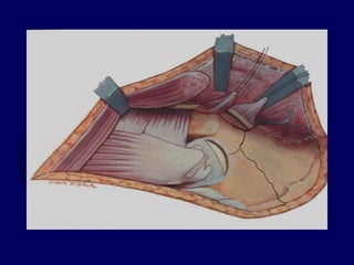
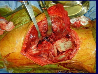
Surgical Approaches for
Fractures of the Acetabulum
Original Author: Mark Reilly, MD
Created February 2004, Updated February 2007
Treatment Protocol
• Radiographs Allow Proper Fracture
Classification
• Fracture Location and Displacement
Determine Need for Surgery
• Fracture Pattern Determines Approach
Surgical Approach
• Single Approach Preferred
– Kocher Langenbeck
– Ilioinguinal
– Extended Iliofemoral
Kocher-Langenbeck
• Approach to posterior
column and posterior
articular surface
• Kocher (1874)
• Langenbeck (1904)
• Judet, Lagrange
(1958)
• Letournel
Indications for Kocher-
Langenbeck
• Posterior Wall Fractures
• Posterior Column Fractures
• Posterior Column / Posterior Wall Fractures
• Juxta-tectal / Infra-tectal Transverse or
Transverse with Posterior Wall Fractures
• Some T-shaped Fractures
Kocher-Langenbeck: Access
• Entire Posterior Column
• Greater and Lesser Sciatic Notches
• Ischial Spine
• Retro-Acetabular Surface
• Ischial Tuberosity
Kocher Langenbeck: Access
Kocher-Langenbeck: Position
• Prone Position
• Radiolucent Table
• Knee Flexed, Hip
Extended
• Distal Femoral
Traction
Prone Position
• Aids in Reduction of Transverse Fractures
• Improves Quadrilateral Surface Access
• Allows Clamp Placement through Greater
Sciatic Notch
• Controls Position of Hip, Minimizes Sciatic
Nerve Stretch
Kocher-Langenbeck: Incision
• 6 to 8 cm from
PSIS
• Tip of Greater
Trochanter
• Parallel Shaft of
Femur 15-20 cm
Dissection: Kocher-Langenbeck
• Divide Iliotibial Band
• Separate Fibers of Gluteus Maximus
– Superior 1/3: Superior Gluteal Artery
– Inferior 2/3: Inferior Gluteal Artery
• Split to Inferior Gluteal Nerve Branch
Dissection: Kocher-Langenbeck
• Release Gluteus Maximus Insertion
• Identify Sciatic Nerve on Border of
Quadratus Femoris Muscle
Dissection: Kocher-Langenbeck
• Release Piriformis Tendon >1cm from
trochanter
• Release Conjoint Tendon
• Open Obturator Internus Bursa for Sciatic
Nerve Retractor
Femoral Head Blood Supply
• Deep Branch of
Medial Femoral
Circumflex
• May be injured by:
– Detaching quadratus
– Reflecting obturator
internus or piriformis
too close to trochanter
Hollinshead, WH 1982
Sciatic Nerve Anatomy
• 84%: Anterior to Piriformis
• 12%: Peroneal Division through Piriformis
• 3%: Peroneal Division Posterior to
Piriformis / Tibial Division anterior
to Piriformis
• 1%: Entire Nerve through Piriformis
Dissection: Kocher-Langenbeck
• Subperiosteal Elevation of:
– Greater Sciatic Notch
– Quadrilateral Surface
– Gluteus Minimus
• Debridement of Fracture Edges
• Avoid Devascularization of Fx Fragments
Complications: Kocher-
Langenbeck
• Infection 2-5%
• Sciatic Nerve palsy 3-5%
• Heterotopic Ossification 8-25%
Trochanteric “Flip”
• Seibenrock, Ganz (Berne)
• Improved Cranial, Anterior exposure of
innominate bone
• Direct intra-articular evaluation of joint, reduction
• Most useful for PW fractures with extension to the
supraacetabular ilium
Ortho Uni Berne
Trochanteric Flip
Ilioinguinal Approach
• Developed by
Letournel after
extensive cadaveric
anatomical study
• Approach to the
anterior column and
anterior articular
surface
Ilioinguinal Approach:
Indications
• Anterior Wall
• Anterior Column
• Transverse with Anterior > Posterior
Displacement
• Anterior Column / Posterior Hemitransverse
• Associated Both Column
Ilioinguinal Approach: Access
• SI Joint
• Internal Iliac Fossa
• Pelvic Brim
• Quadrilateral Surface
• Superior Pubic Ramus
• Limited Access to External Iliac Wing
Ilioinguinal Approach: Access
Ilioinguinal: Position
• Supine
• Distal Femoral
Traction
• Access to Greater
Trochanter (Lateral
Traction)
• Hip flexed 20°
Ilioinguinal: Incision
• 3-4 cm cranial to
Symphysis pubis
• Curve to ASIS
• Parallel Iliac Crest
• Past Most Convex
Portion of Ilium
– anterior 2/3
Symphysis
pubis
ASIS
Dissection: Ilioinguinal
• Subperiosteal Dissect Internal Iliac Fossa
– Origin of Abdominals and Iliopsoas
• Expose Sacroiliac Joint
• Dissect over Pelvic Brim
Internal Iliac
Fossa
Dissection: Ilioinguinal
• Incise External Oblique Aponeurosis
– From ASIS to midline
– 1 cm proximal to External Inguinal Ring
• Expose Floor of Inguinal Canal
• Retract Spermatic Cord/Round Ligament
• Protect Ilioinguinal Nerve
External Oblique
Ilioinguinal Nerve
Spermatic Cord
Dissection: Ilioinguinal
• Incise Inguinal Ligament
• Leave 1-2 mm with Internal Oblique and
Transversus Abdominis origin
• Protect External Iliac Vessels
• Protect Lateral Femoral Cutaneous Nerve
External Iliac Artery/Vein
Lateral Femoral
Cutaneous Nerve
Dissection: Ilioinguinal
• Separate Lacuna Vasorum and Lacuna
Musculorum
• Incise Iliopectineal Fascia to Superior
Ramus and from Pelvic Brim
• Connect True and False Pelvis
Iliopectineal Fascia
Dissection: Ilioinguinal
• Dissect Lateral to External Iliac Vessels
• Transect Ipsilateral Rectus Tendon
• Dissect Medial to External Iliac Vessels
Ilioinguinal: Lateral Window
• Internal Iliac Fossa
• Sacroiliac Joint
• Pelvic Brim - Upper 1/3
Ilioinguinal: Middle Window
• Pelvic Brim - SI joint to pectineal eminence
• Quadrilateral Surface
• Anterior Rim
Ilioinguinal: Medial Window
• Superior Pubic Ramus
• Symphysis Pubis
Dissection: Ilioinguinal
• Medial window may also be created
utilizing Stoppa approach
– Midline rectus split
– Subperiosteal dissection of quadrilateral surface
– Retractor in lesser sciatic notch
– Protect obturator nerve/artery
Ilioinguinal: Corona Mortis
• Vascular Anastamosis
– External Iliac
– Obturator
• Frequently Venous
• Occasionally Arterial
Complications: Ilioinguinal
• Infection 2-5%
• Femoral Nerve palsy 2%
• Lateral Femoral Cutaneous
– Dysesthesia common
– Sensation returns 80-90% by 1 year
• Heterotopic Ossification 2-10%
• Vascular Injury <1%
Extended Iliofemoral
• Developed by
Letournel (1975)
• Based on Smith-
Peterson Approach
• Maximal
Simultaneous access
to both columns of the
acetabulum
Indications for EIF Approach
• Transtectal Tr+PW or T-shaped fractures
• Transverse fractures with extended posterior wall
• T-shaped fractures with wide separations of the
vertical stem of the "T" or those with associated
pubic symphysis dislocations.
• Certain Associated Both Column Fractures.
• Associated fracture patterns or transverse fractures
which are operated greater than 21 days following
injury.
Indications for EIF in Both
Column Fractures
• Inability to reduce Posterior Column
through Ilioinguinal
• Wide displacement at the rim
• Complex posterior column involvement
• Associated SI joint disruption
• Small posterior wall component
Extended Iliofemoral: Access
• External Aspect of Ilium
• Anterior Column as far medial as
Iliopectineal eminence
• Posterior Column to the Upper Ischial
Tuberosity
EIF Approach: Access
Extended Iliofemoral: Position
• Lateral Position
• Distal Femoral
Traction
• Knee flexed 45°
Extended Iliofemoral: Incision
• Inverted J incision
• Parallel Iliac Crest
from PSIS to ASIS
• Incise along anterior-
lateral thigh
Dissection: Extended Iliofemoral
• Release Origins of Gluteals and Tensor
Fascia Lata from Iliac Crest
• Dissect Subperiosteal Iliac Wing
• Elevate Periosteum from Greater Sciatic
Notch
• Incise Fascia Lata to end of muscle belly
Dissection: Extended Iliofemoral
• Retract Tensor Fascia Lata Muscle
Posteriorly
• Incise Sheath of Rectus Femoris
• Ligate Lateral Femoral Circumflex Artery
and Vein
Dissection: Extended Iliofemoral
• Release Gluteus Medius and Minimus
Tendons from Greater Trochanter
• Alternatively, Greater Trochanteric
Osteotomy
• Reflect Gluteals and Tensor Fascia Lata
Posteriorly pedicled on Superior Gluteal
Dissection: Extended Iliofemoral
• Incise and Retract:
– Piriformis Tendon
– Obturator Internus Tendon with Gemelli
muscles
• Place Sciatic Nerve Retractor in Lesser
Sciatic Notch
• Capsulotomy if Required
Dissection: Extended Iliofemoral
• If Internal Iliac Fossa Exposure Required:
– Elevate Abdominal Muscles from Iliac Crest
– Elevate Iliacus Subperiosteally
– Release Sartorius and Inguinal Ligament from
ASIS
– Preserve Anterior Capsule and Direct Head of
Rectus for Blood Supply to Anterior Column
Complications: Extended
Iliofemoral
• Infection 2-5%
• Sciatic Nerve palsy 3-5%
• Heterotopic Ossification 20-50%
Other Extensile Approaches
• Triradiate
– Anterior Limb added to KL
– Trochanteric Osteotomy
– Reflect Abductors
• Modified Extensile Lateral
– EIF with associated osteotomies
• Greater Trochanter
• Iliac Crest
• ASIS
Combined Surgical Approaches
• Kocher-Langenbeck + Ilioinguinal
• May be simultaneous or sequential
– Simultaneous may compromise both
approaches but can aid in assessment of
transverse fracture reduction
– Care with sequential not to block anterior
reduction during posterior fixation
Combined Surgical Approaches
• Rarely necessary
– T-shaped fractures if unable to reduce anterior
column from KL
– AW+PHT if hemitransverse is segmental or
widely displaced
Return to
Pelvis
Index
E-mail OTA
about
Questions/Comments
If you would like to volunteer as an author for
the Resident Slide Project or recommend updates
to any of the following slides, please send an e-
mail to ota@aaos.org

V05 acetab surgical_apprch