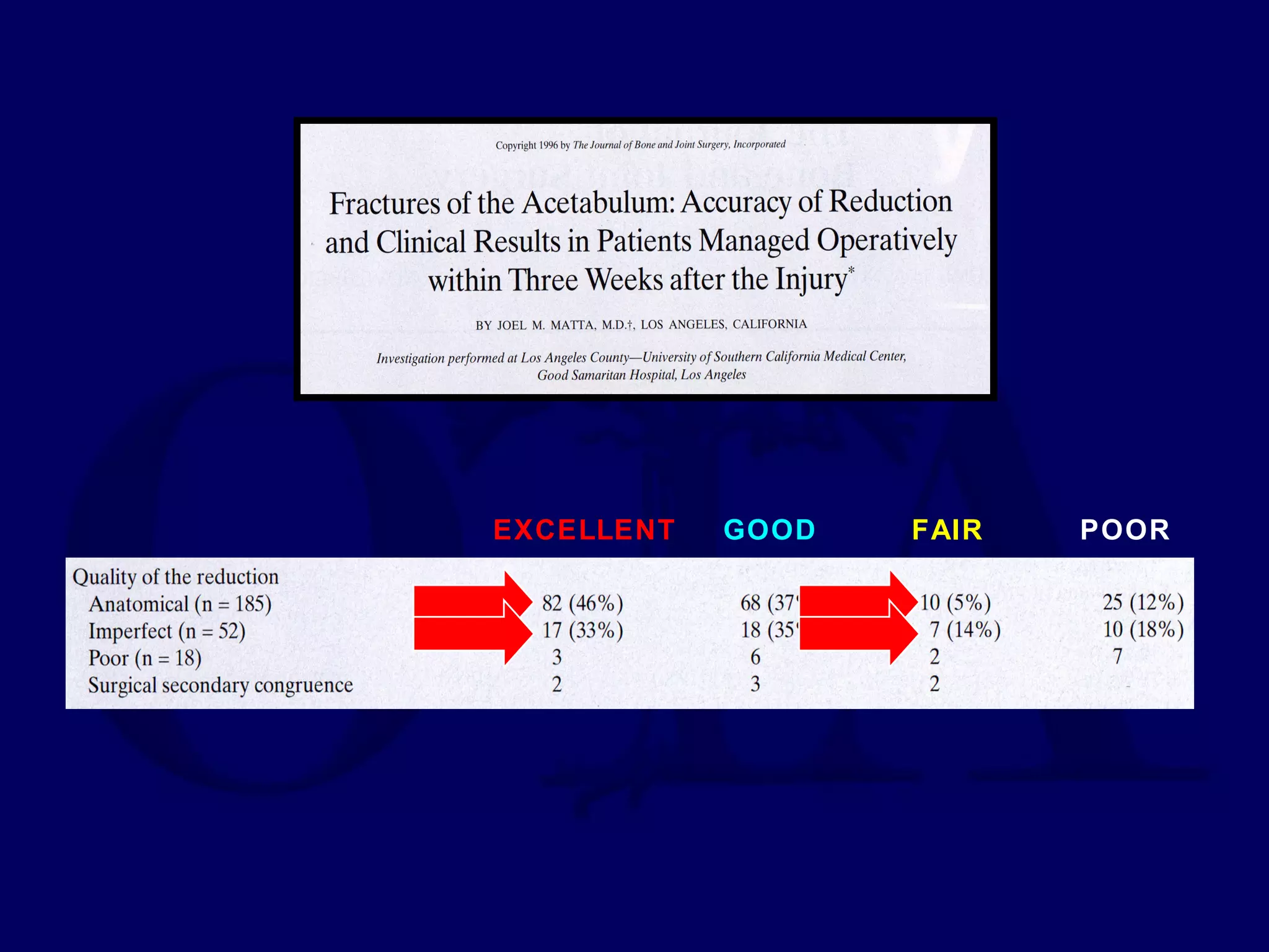
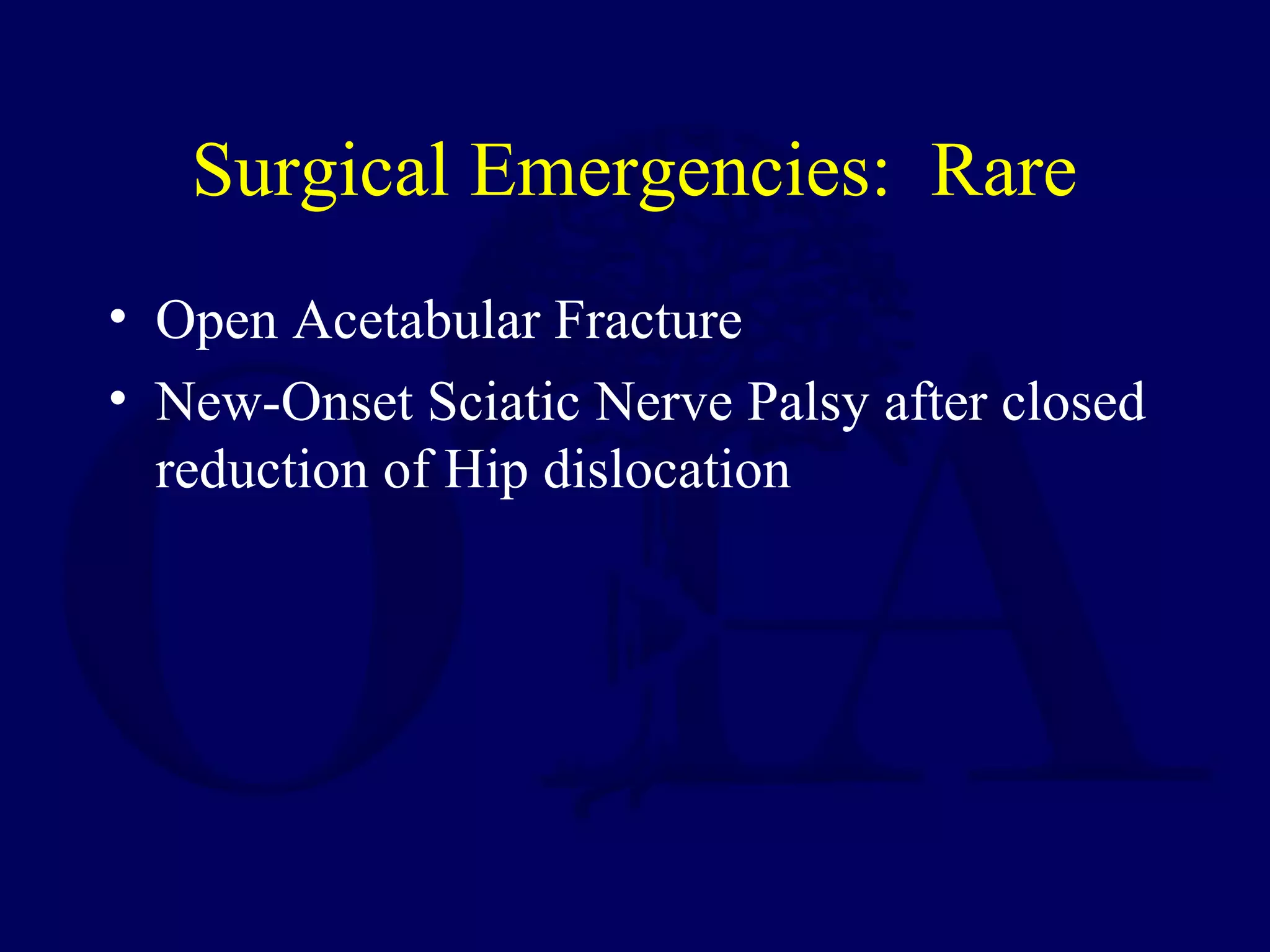
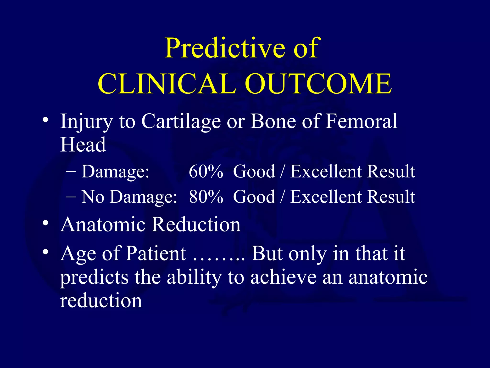
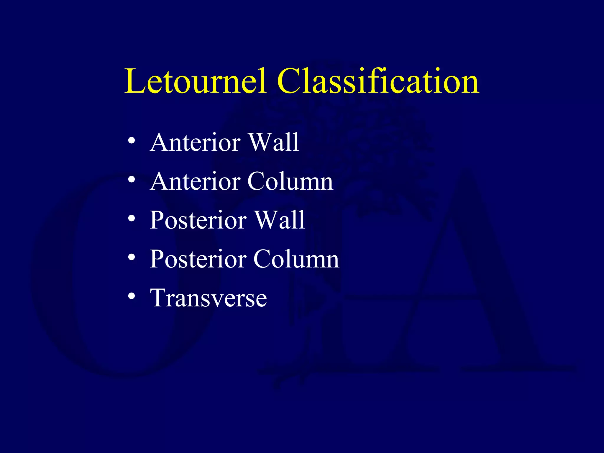
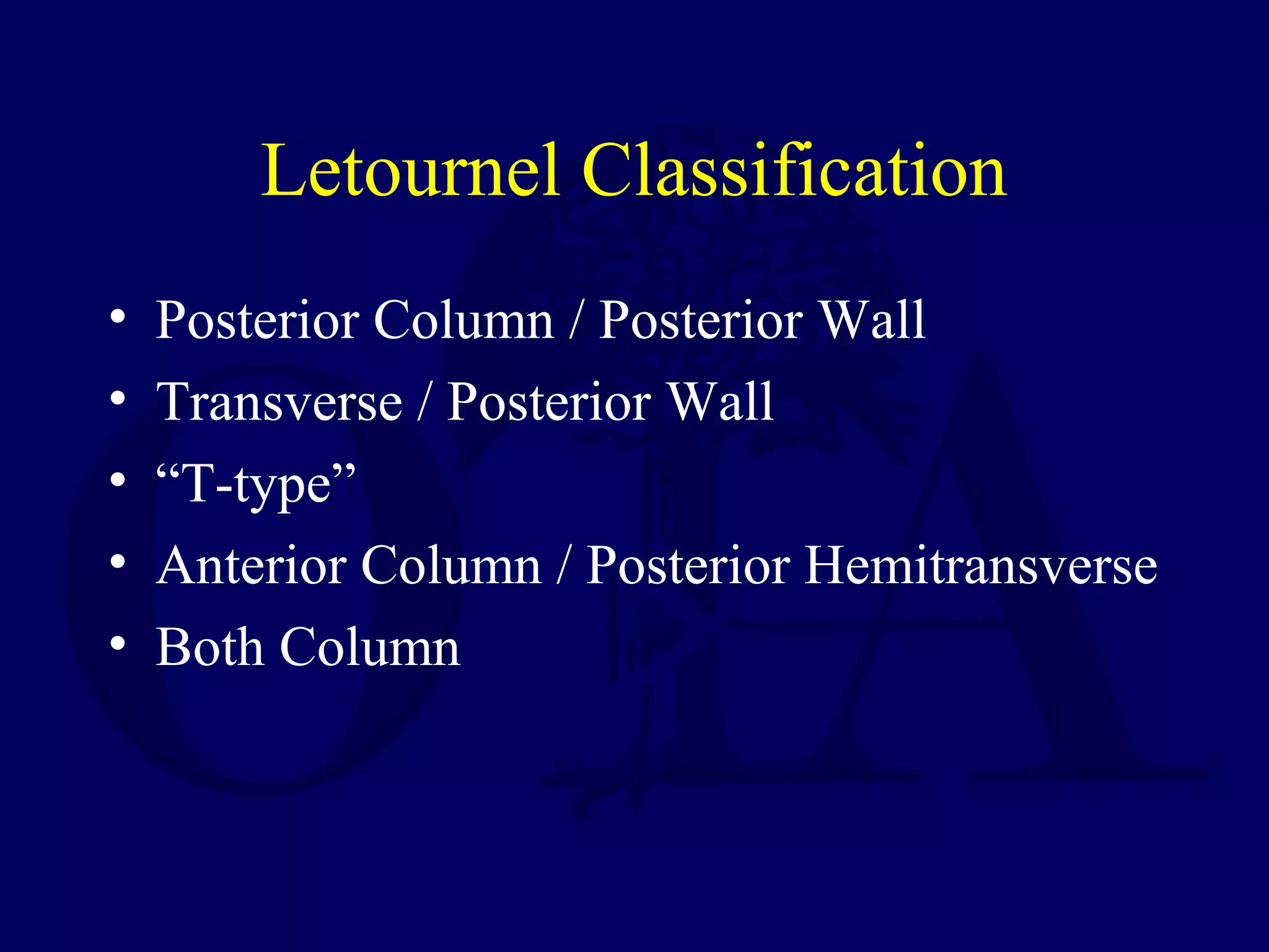
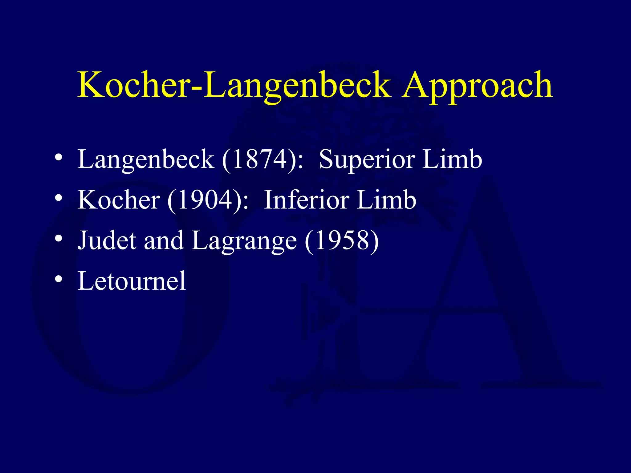
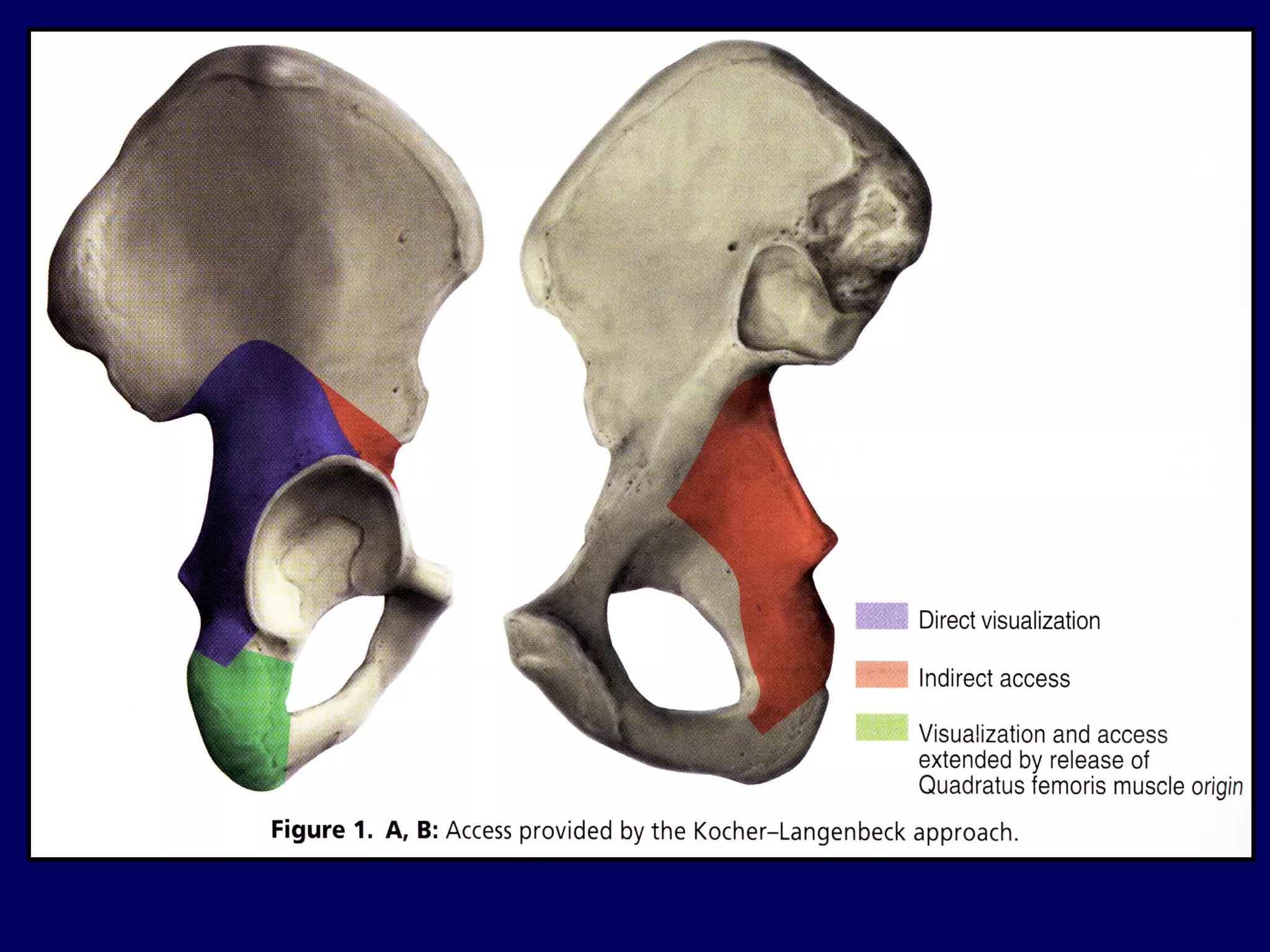
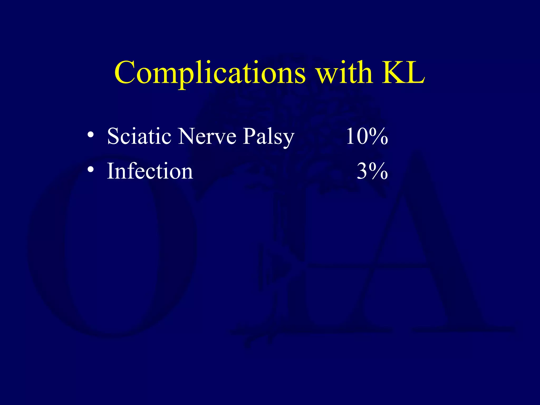
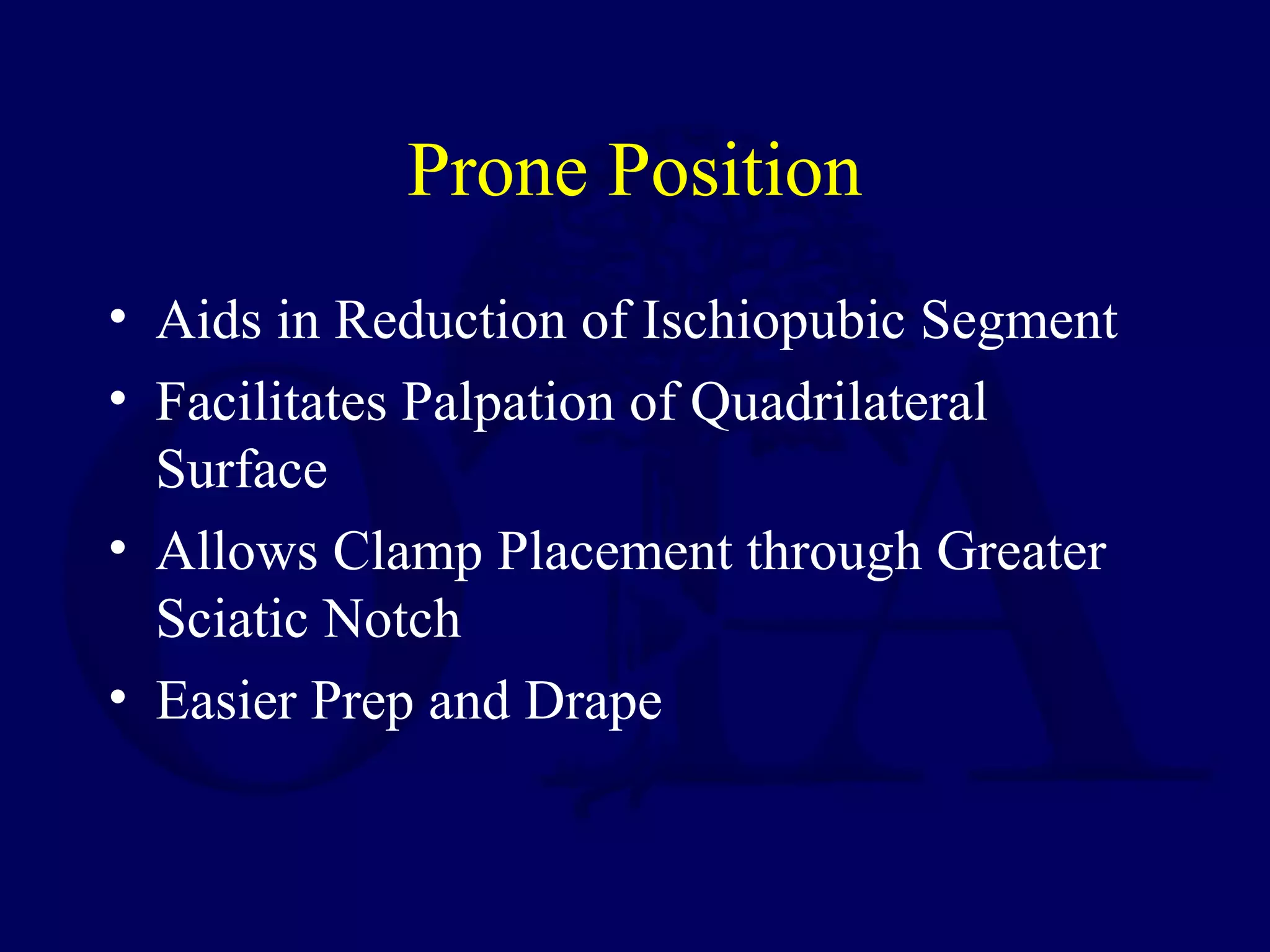
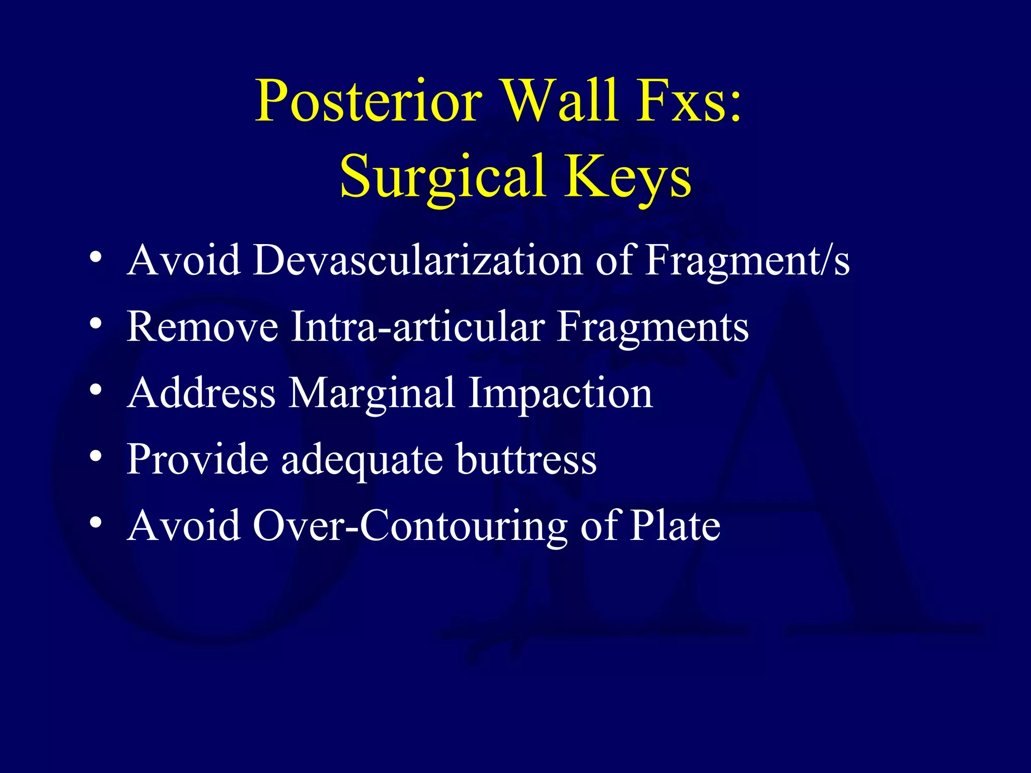
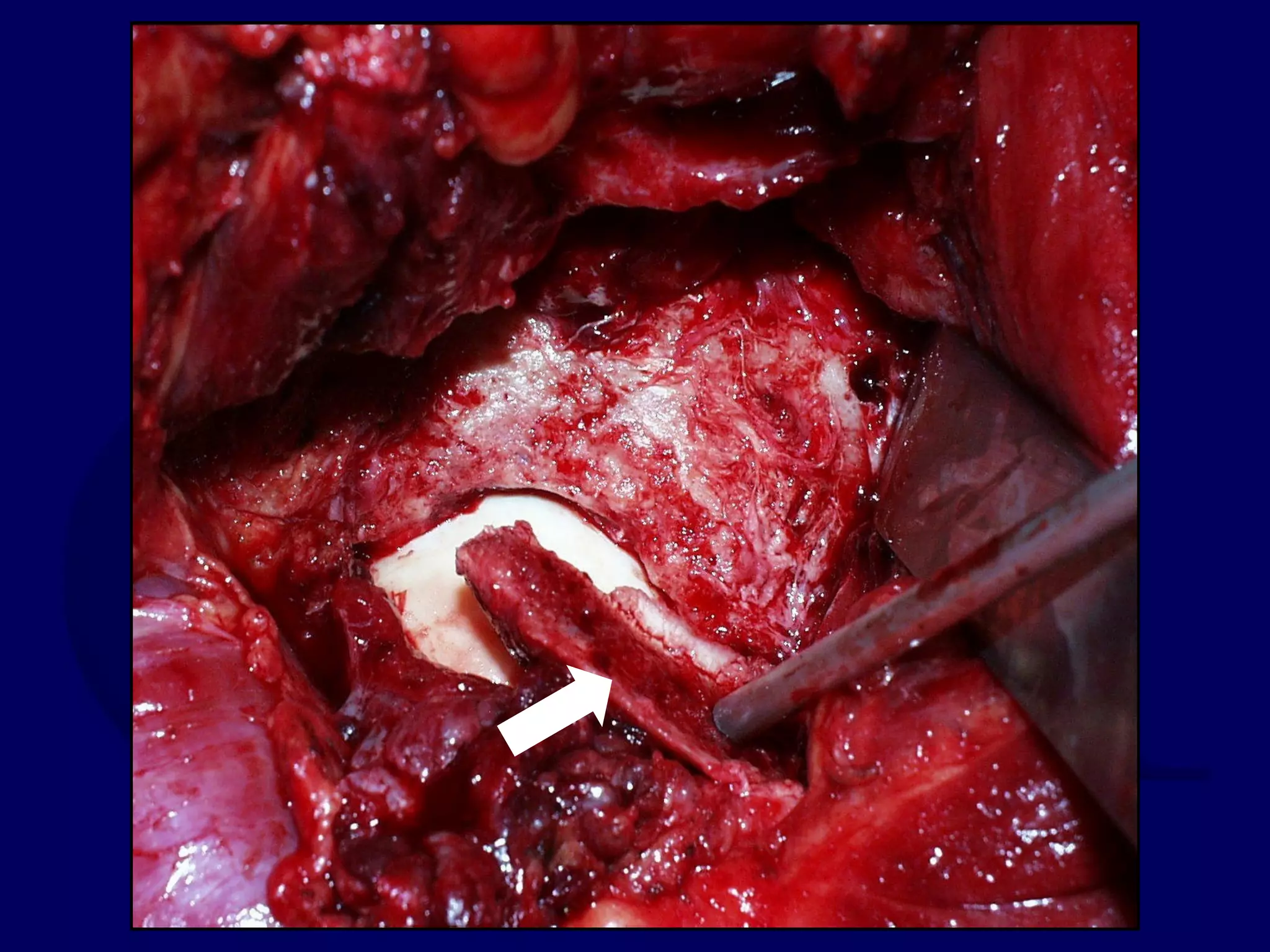
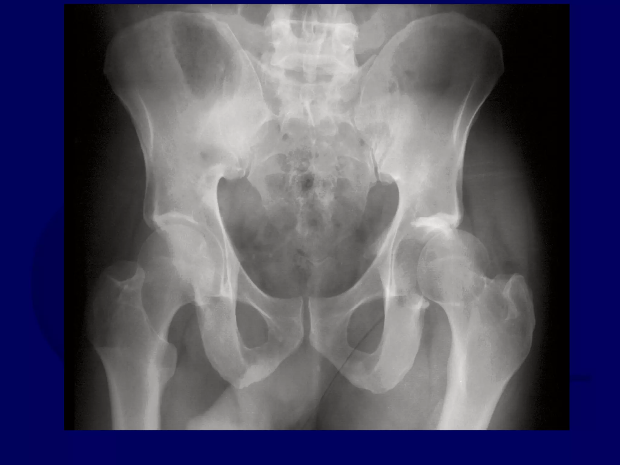
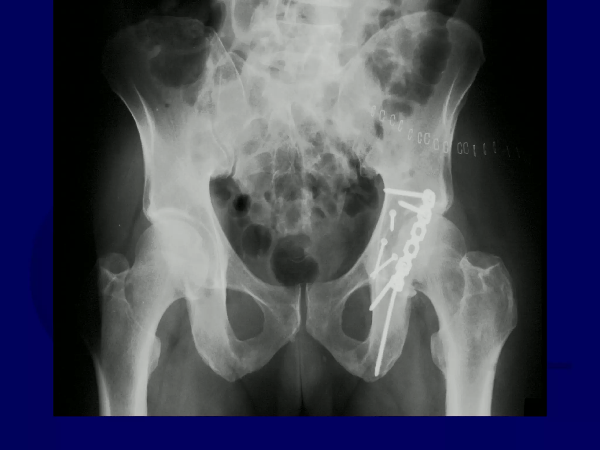
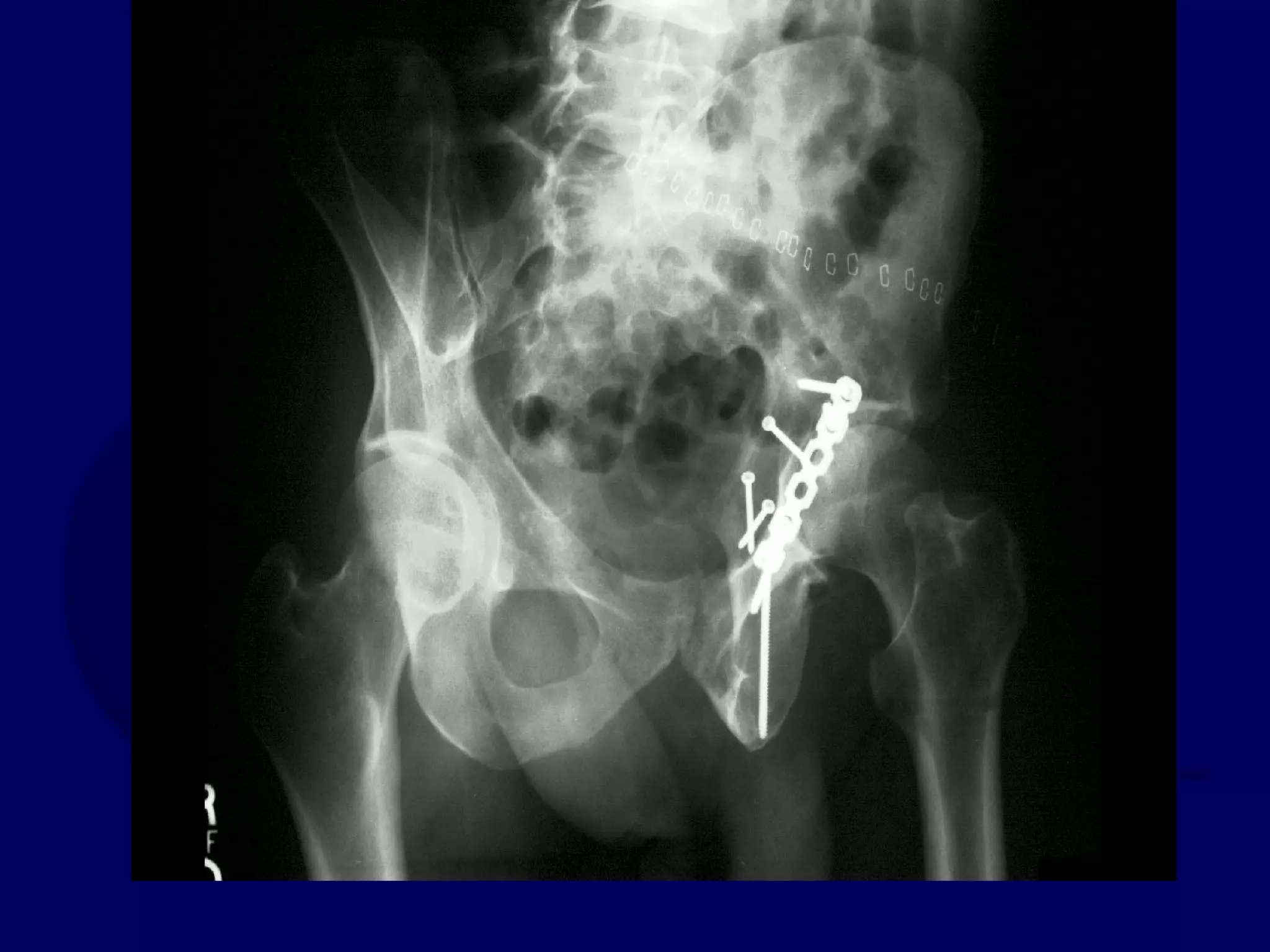
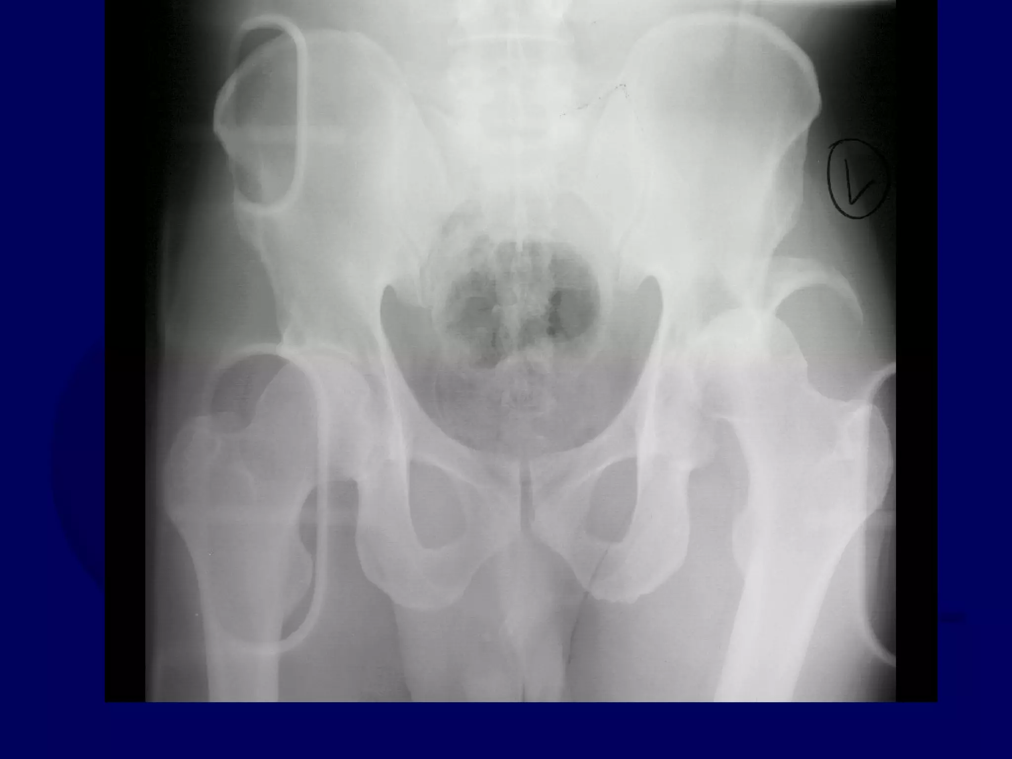
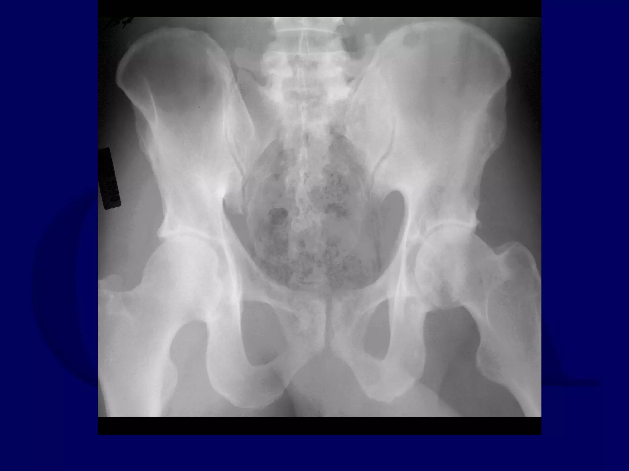
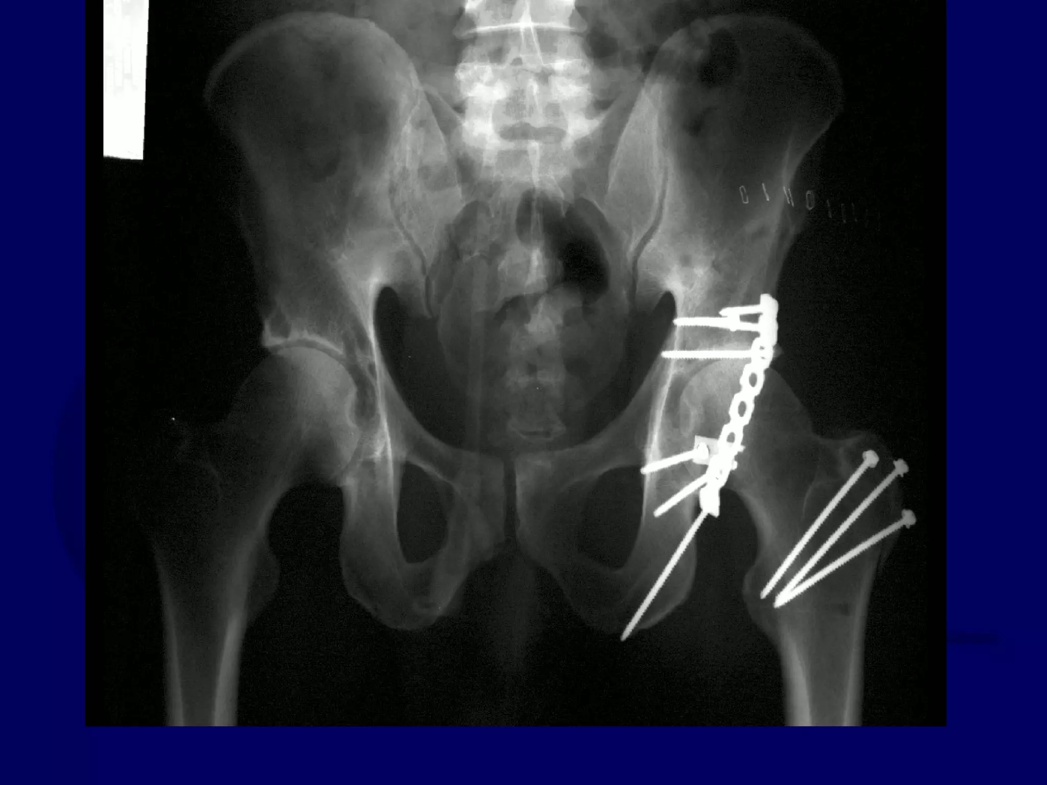
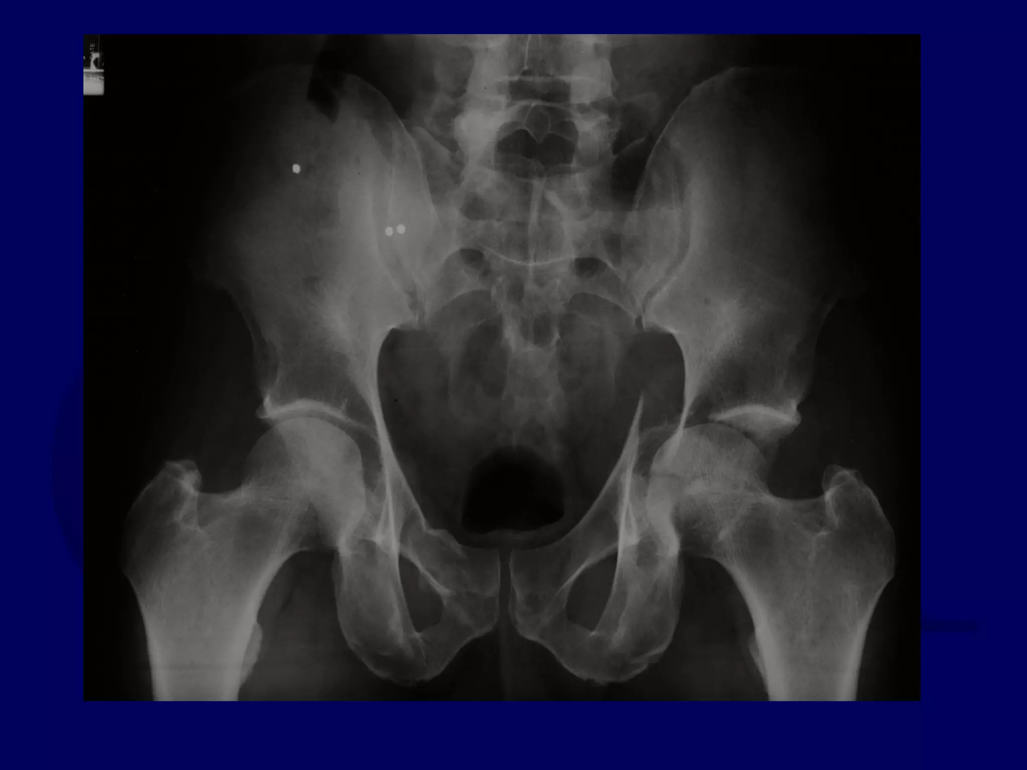
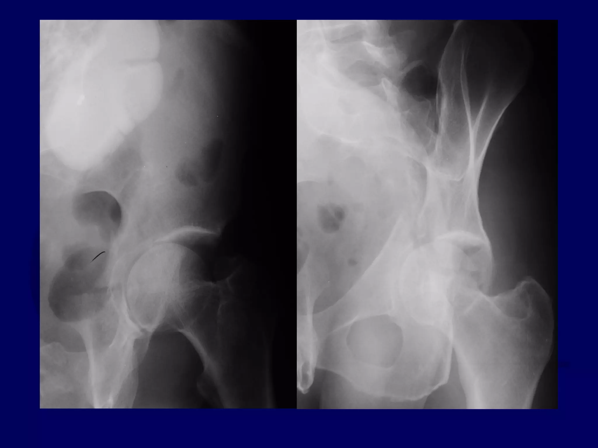
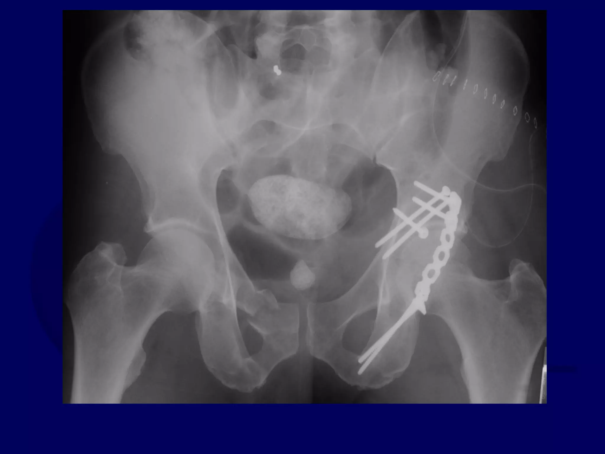
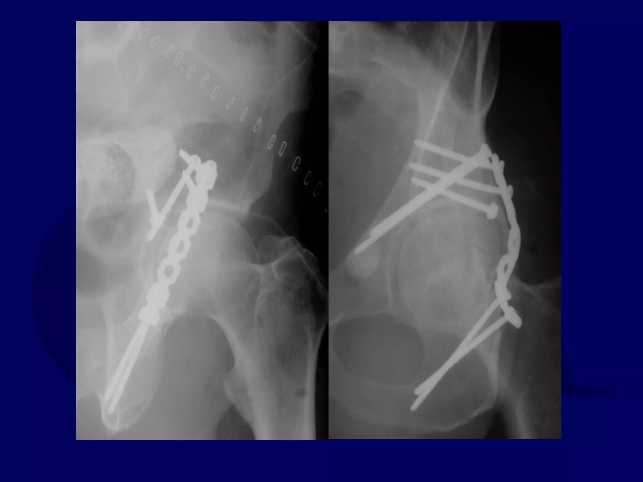
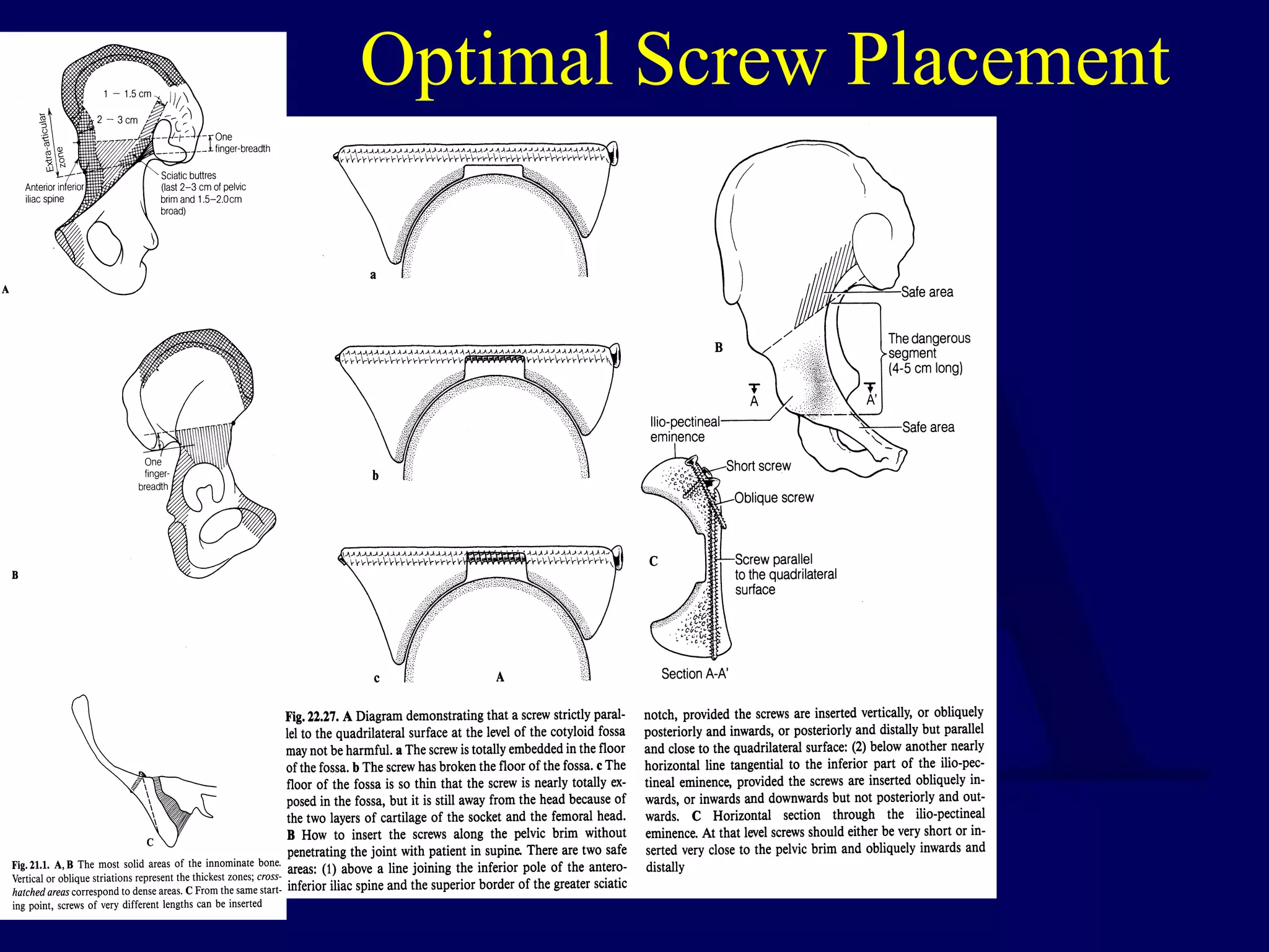
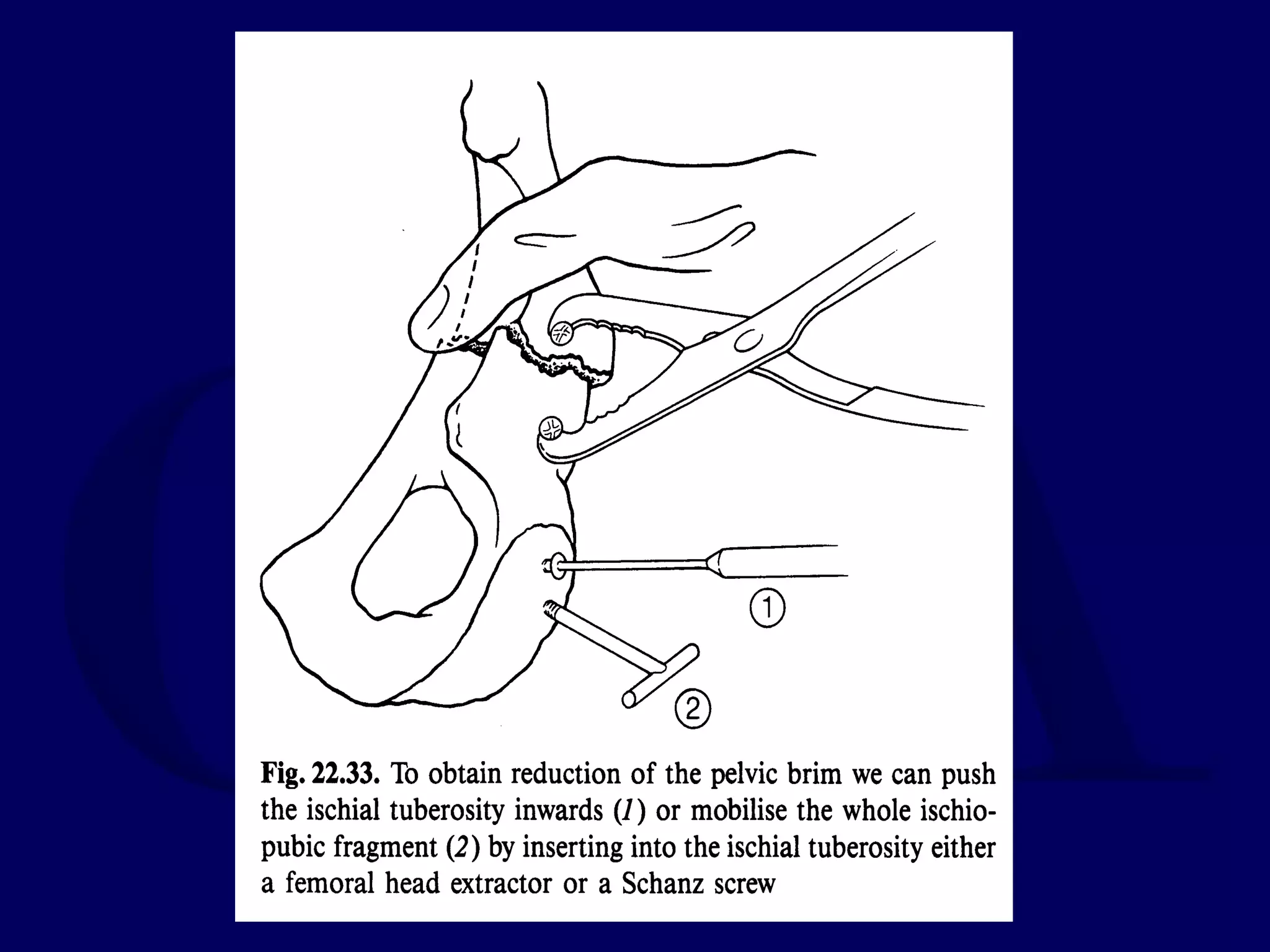
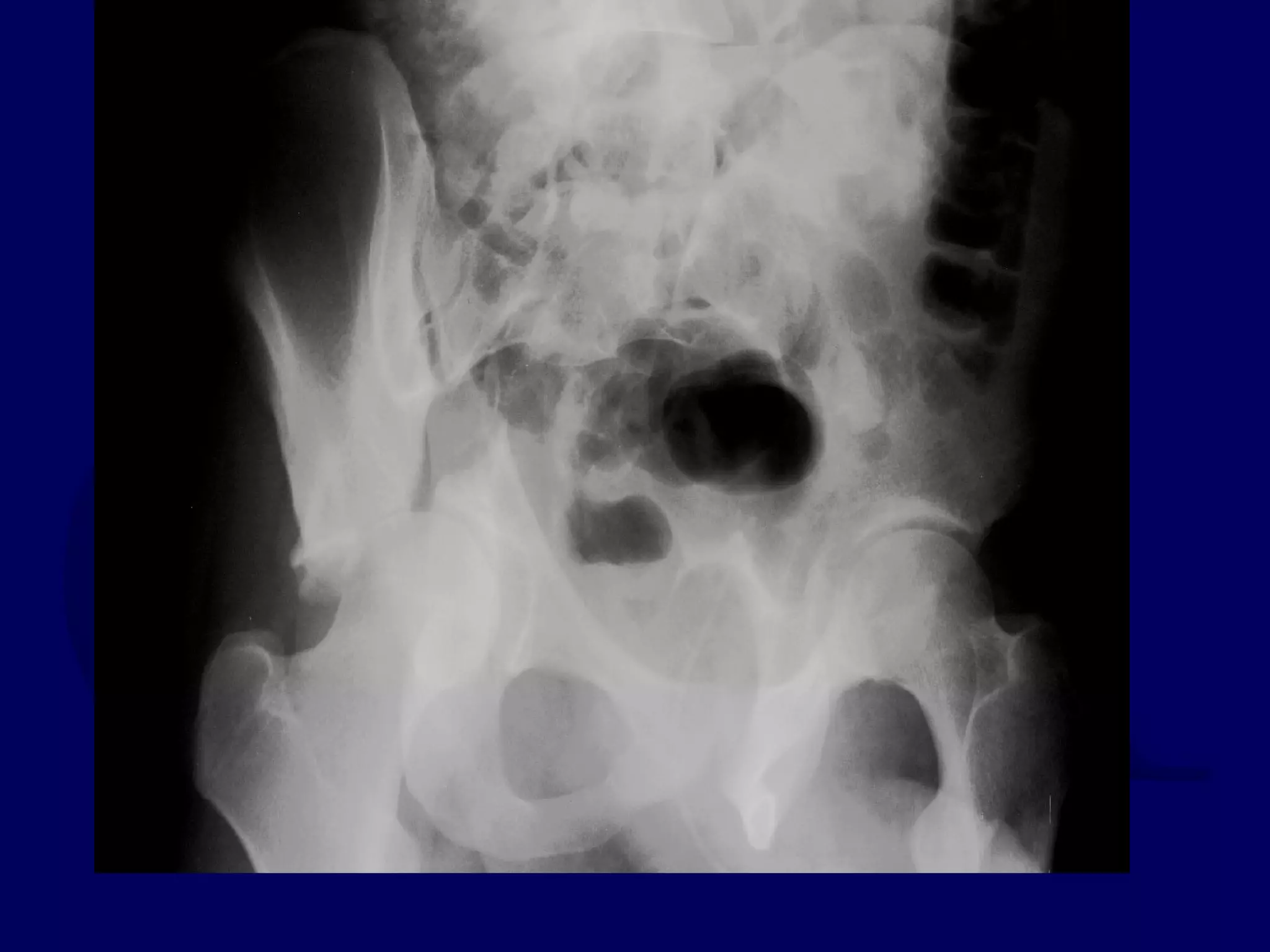
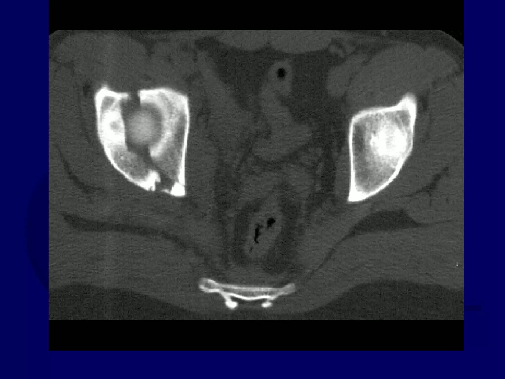
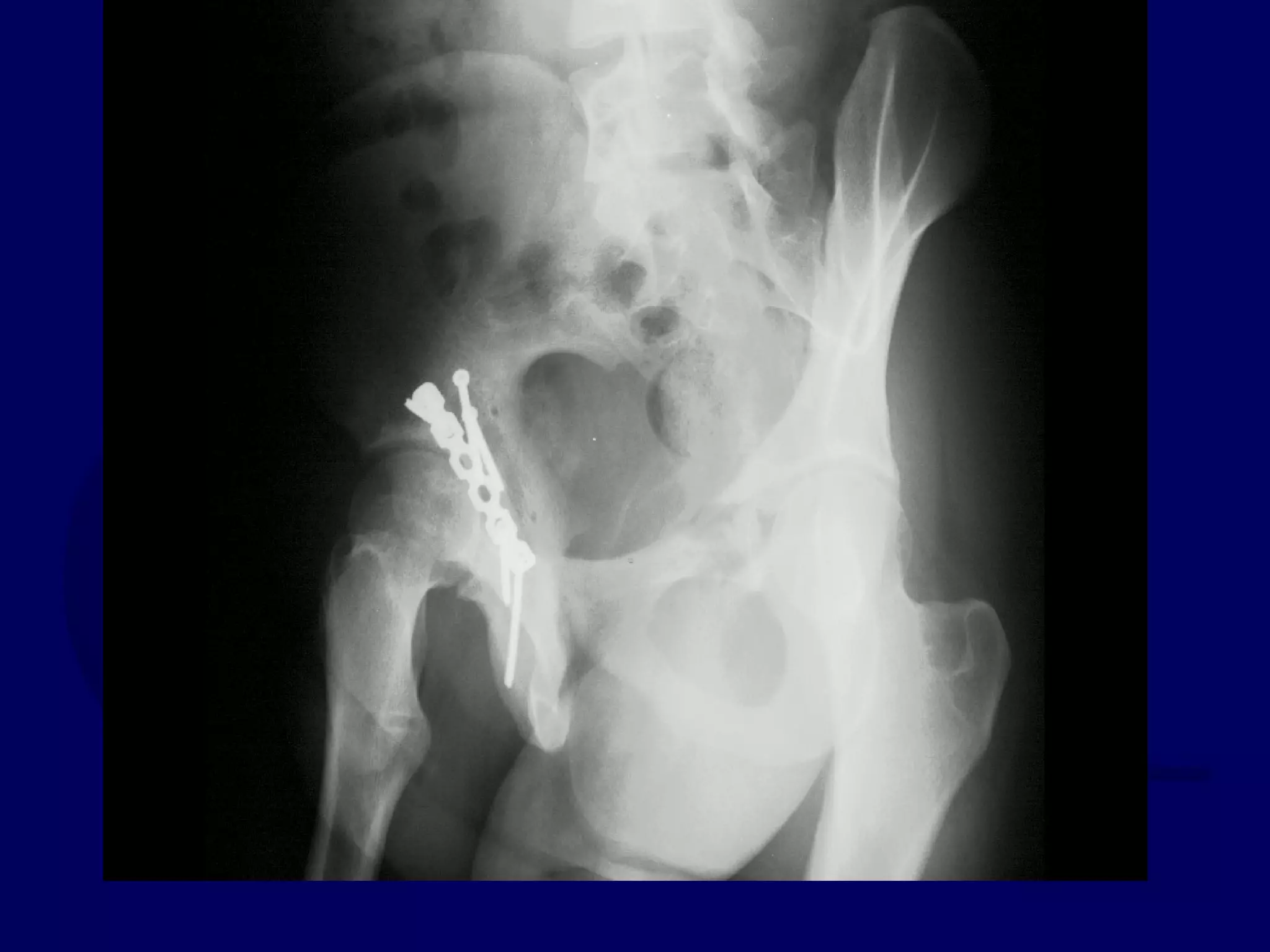
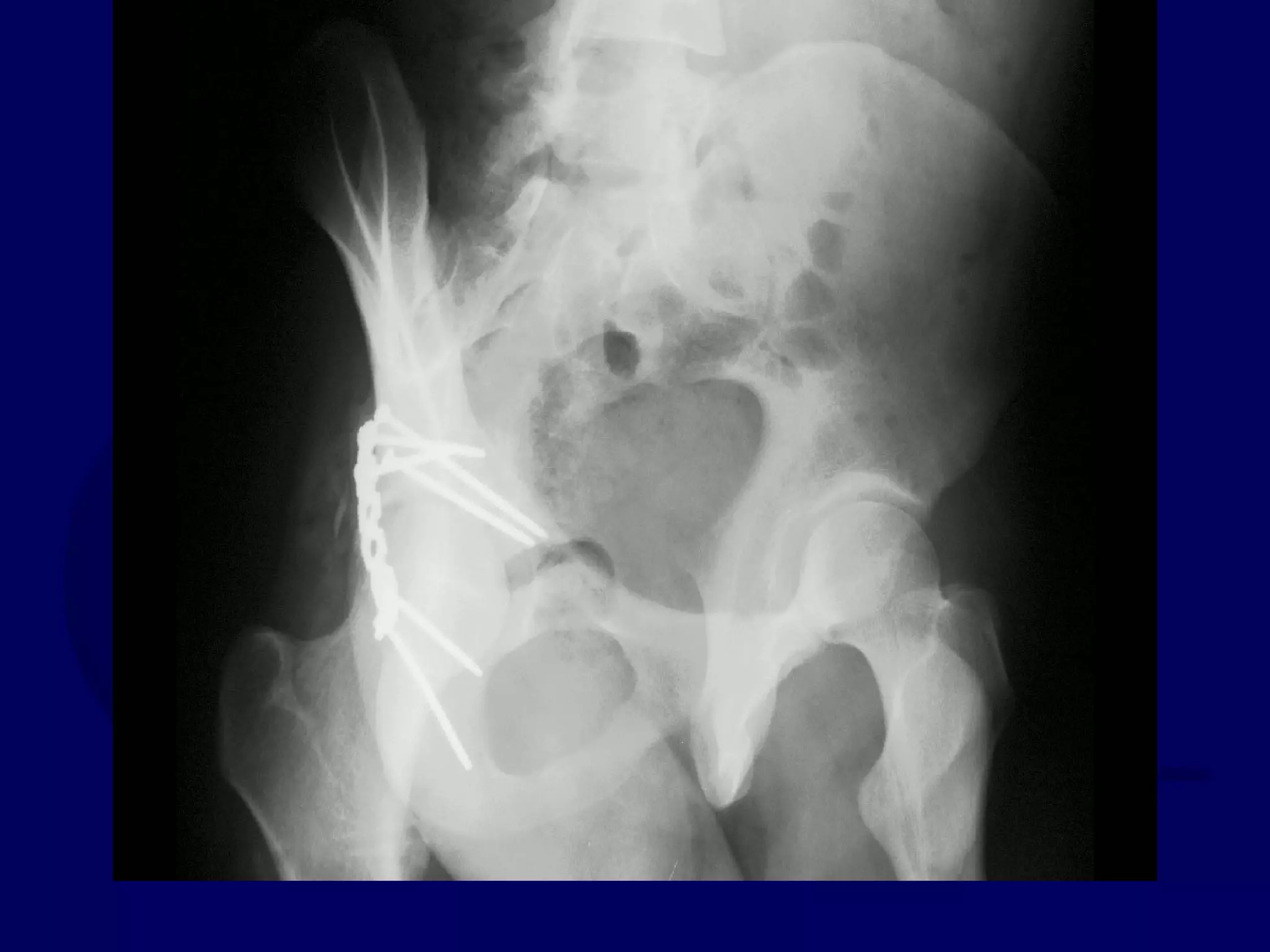
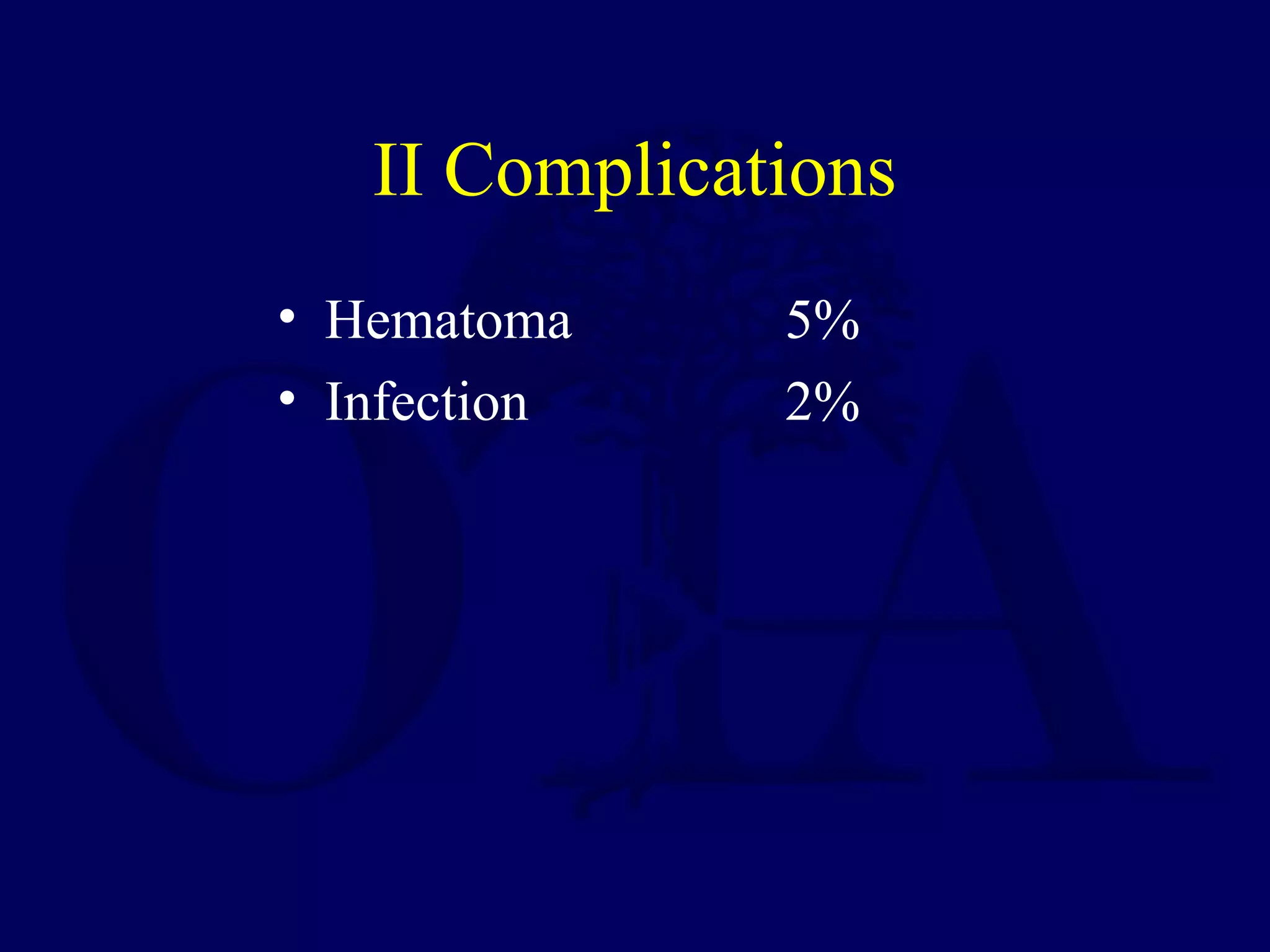
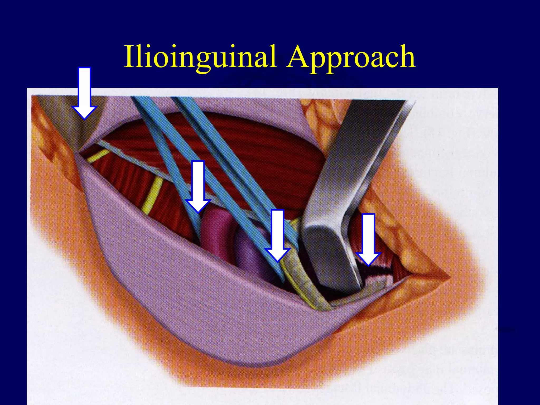
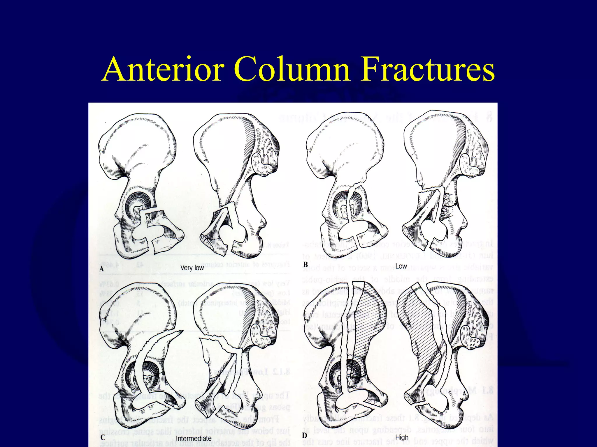
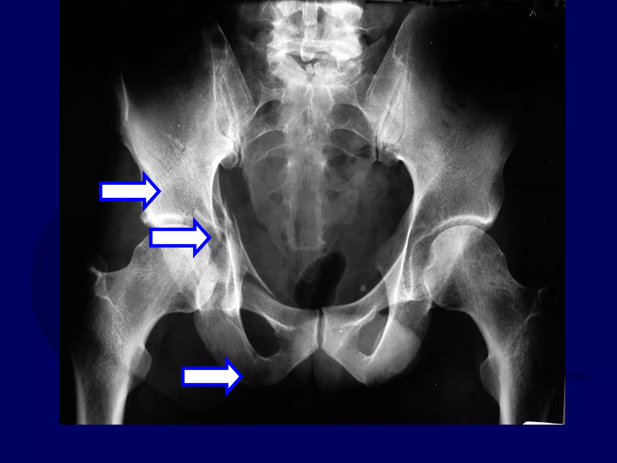
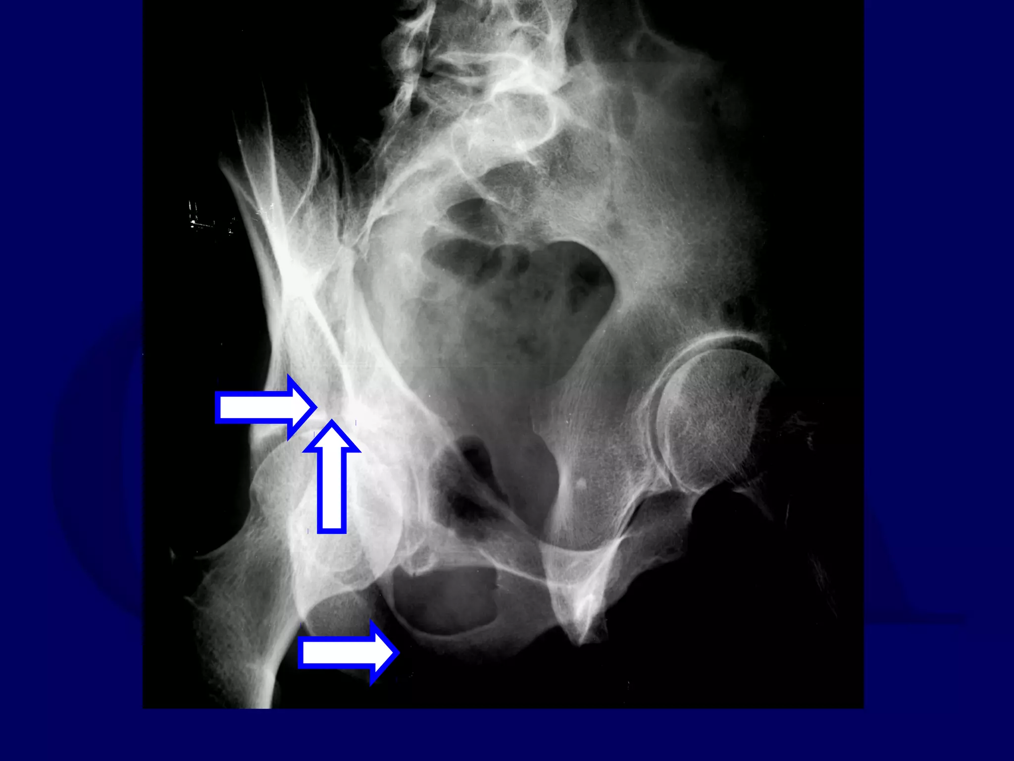
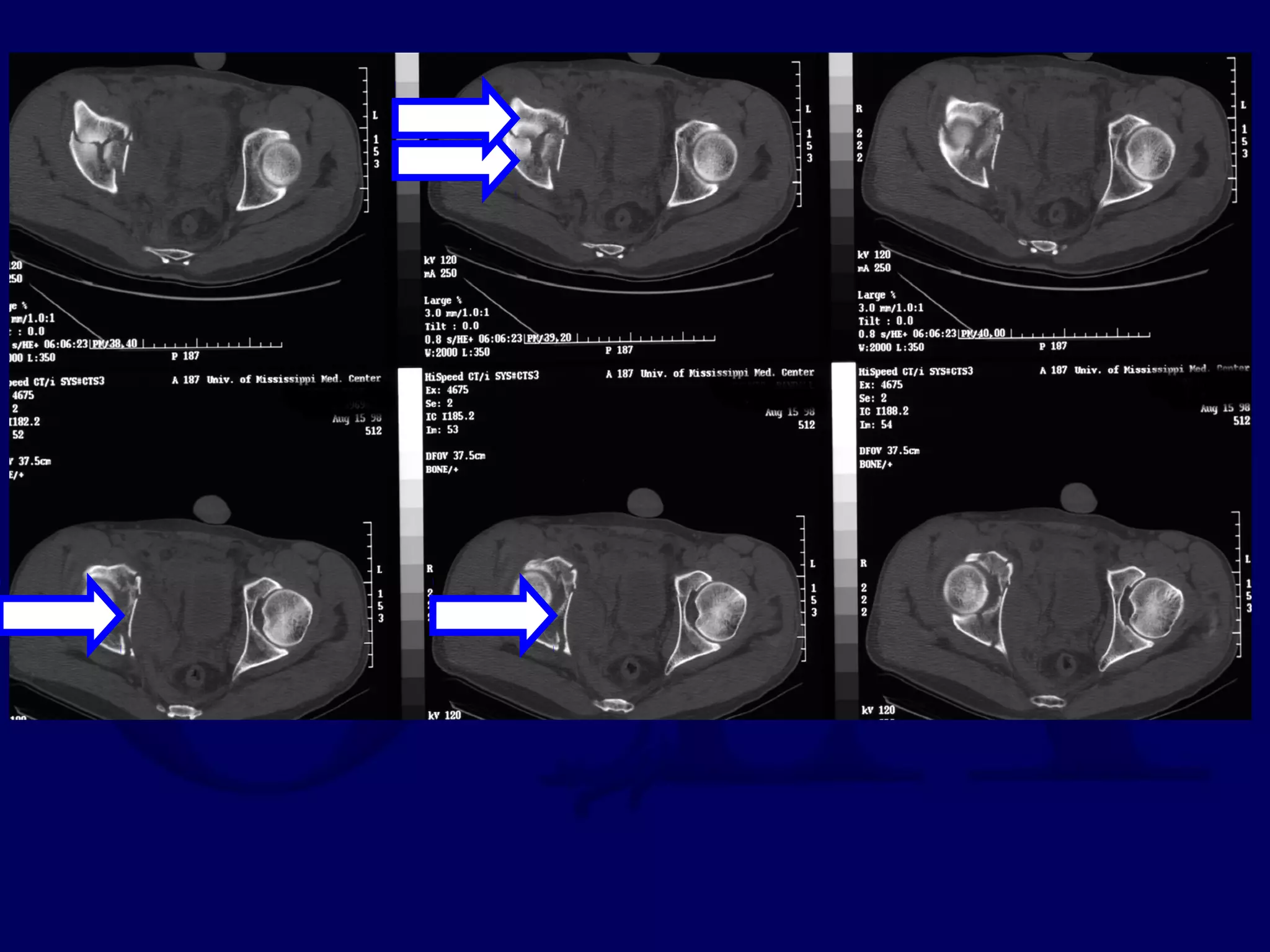
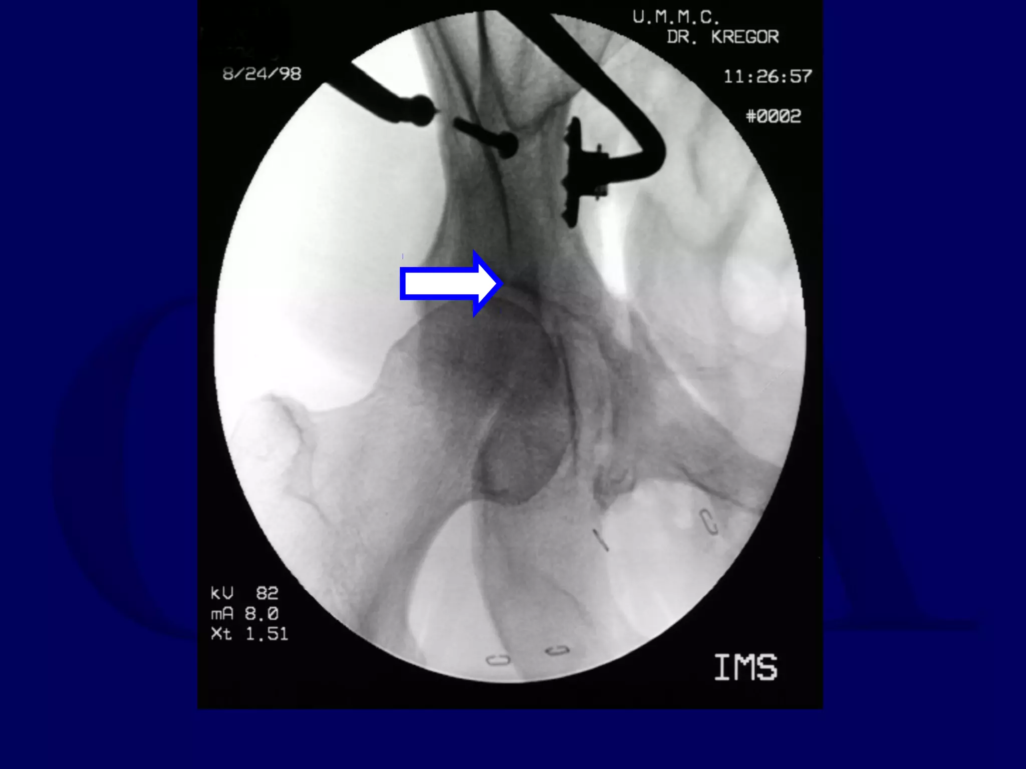
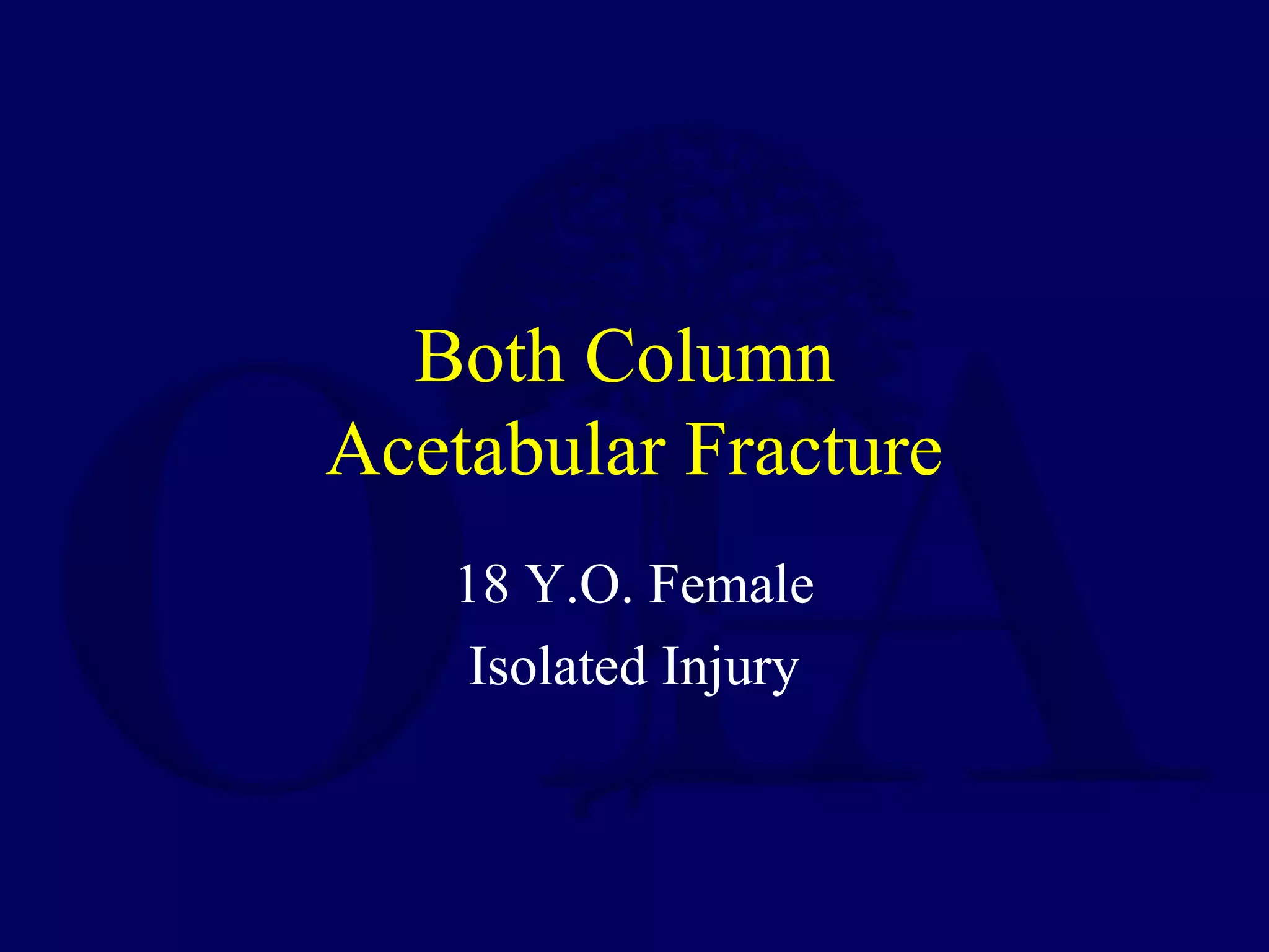
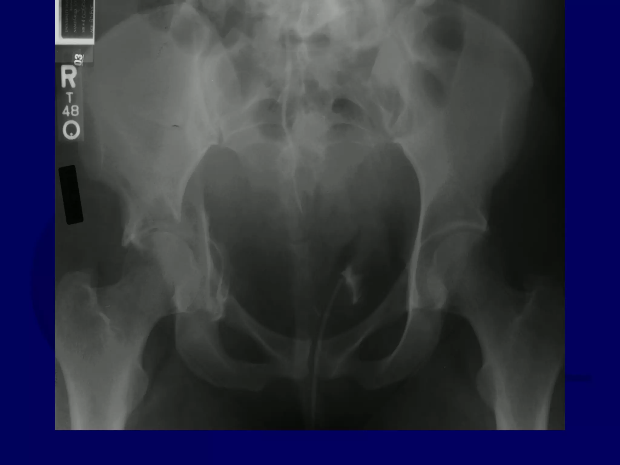
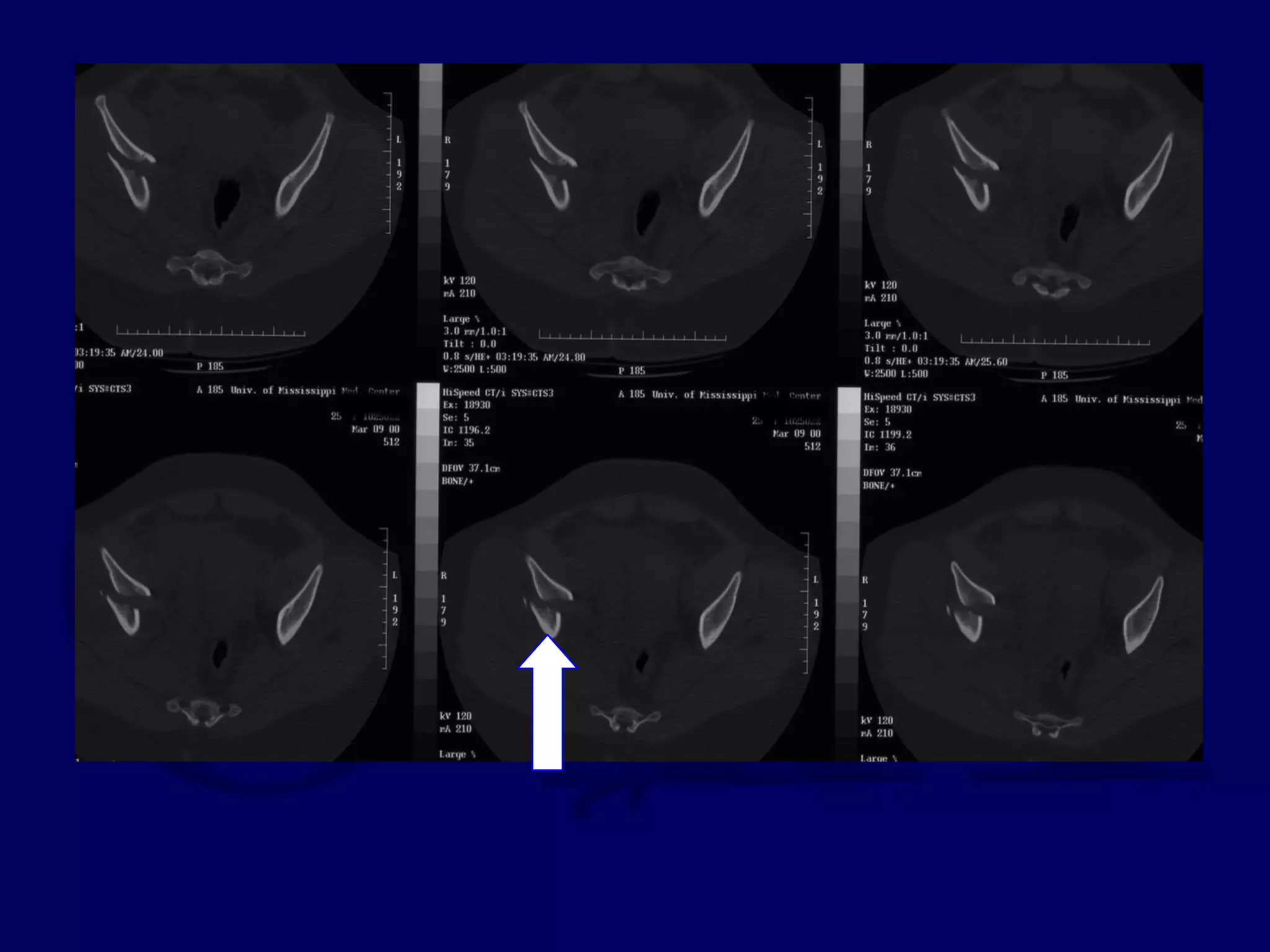
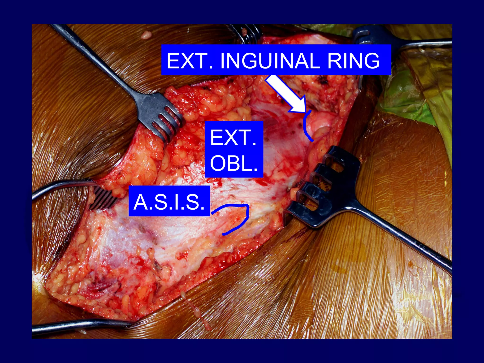
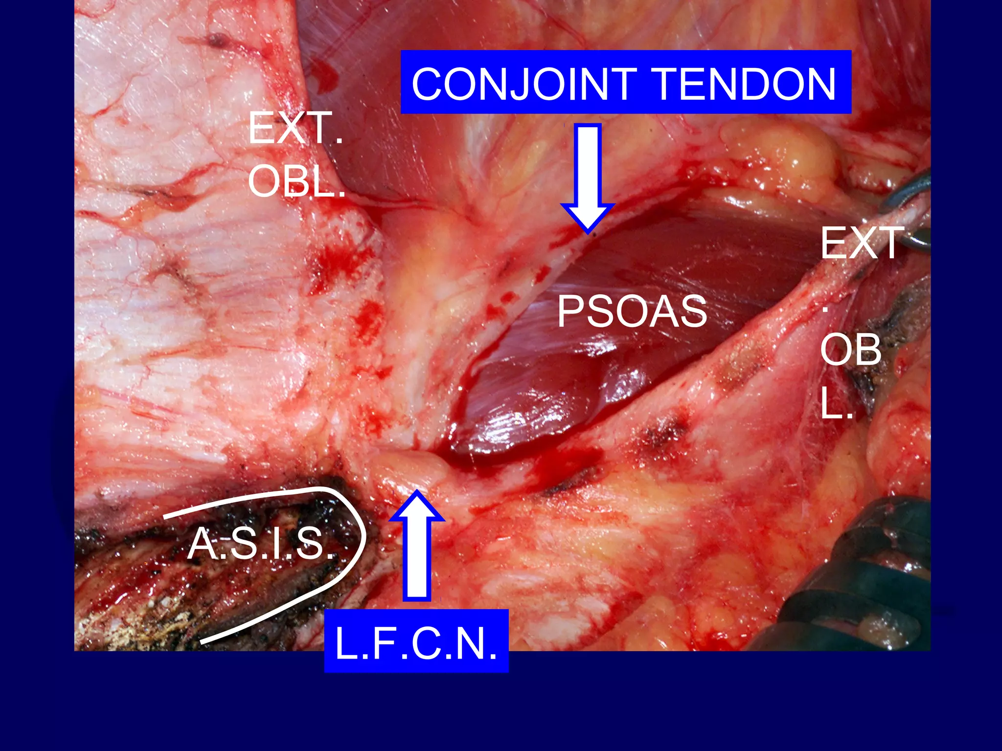
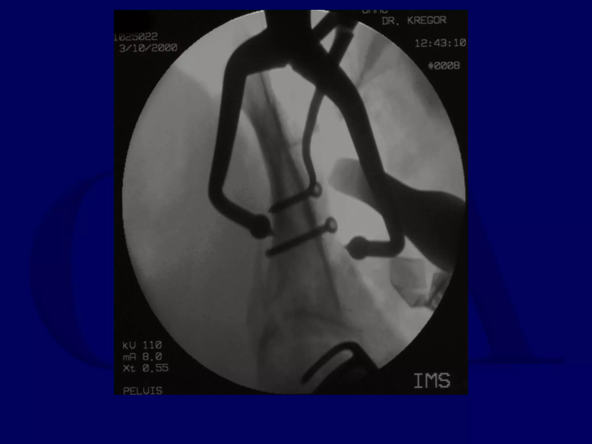
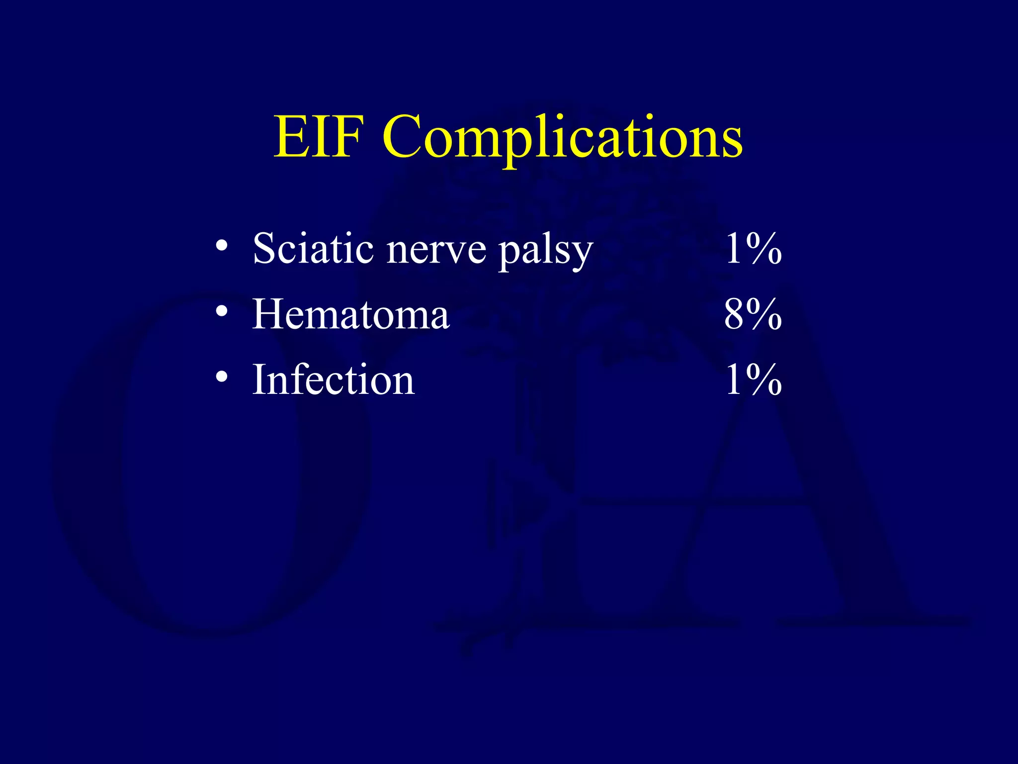
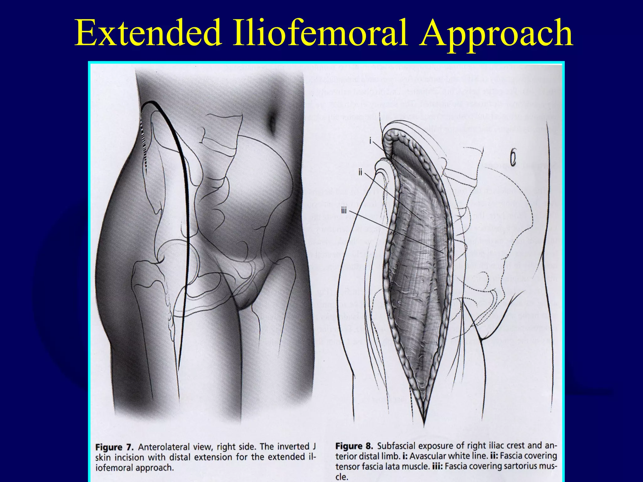
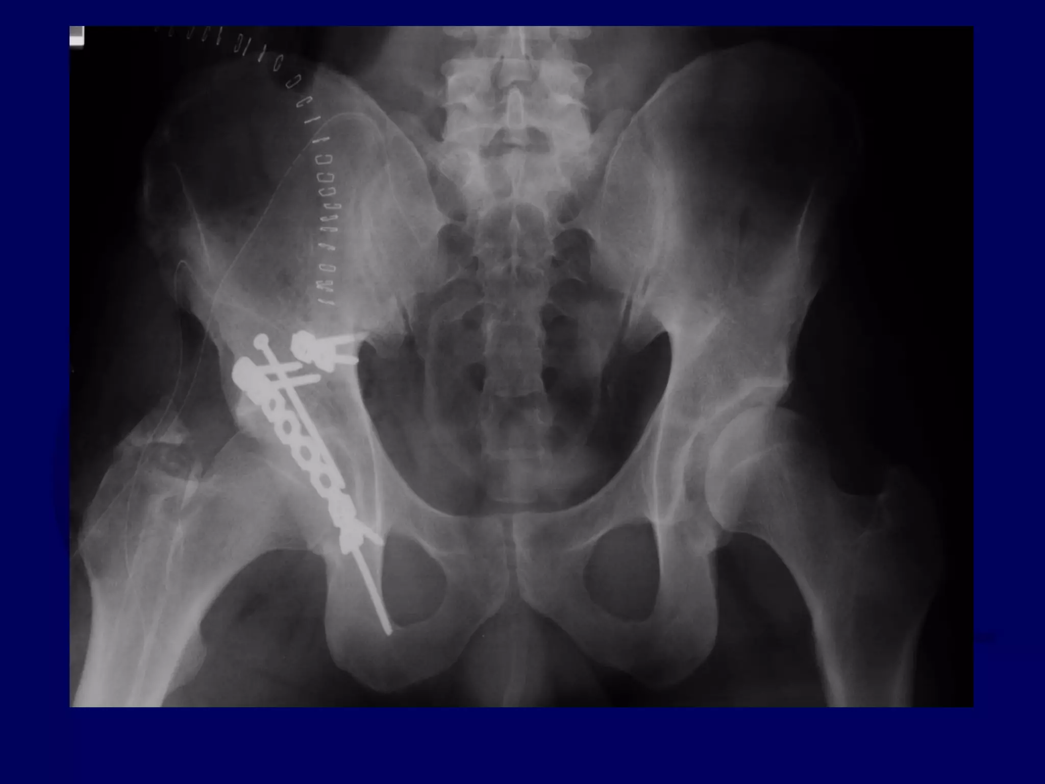
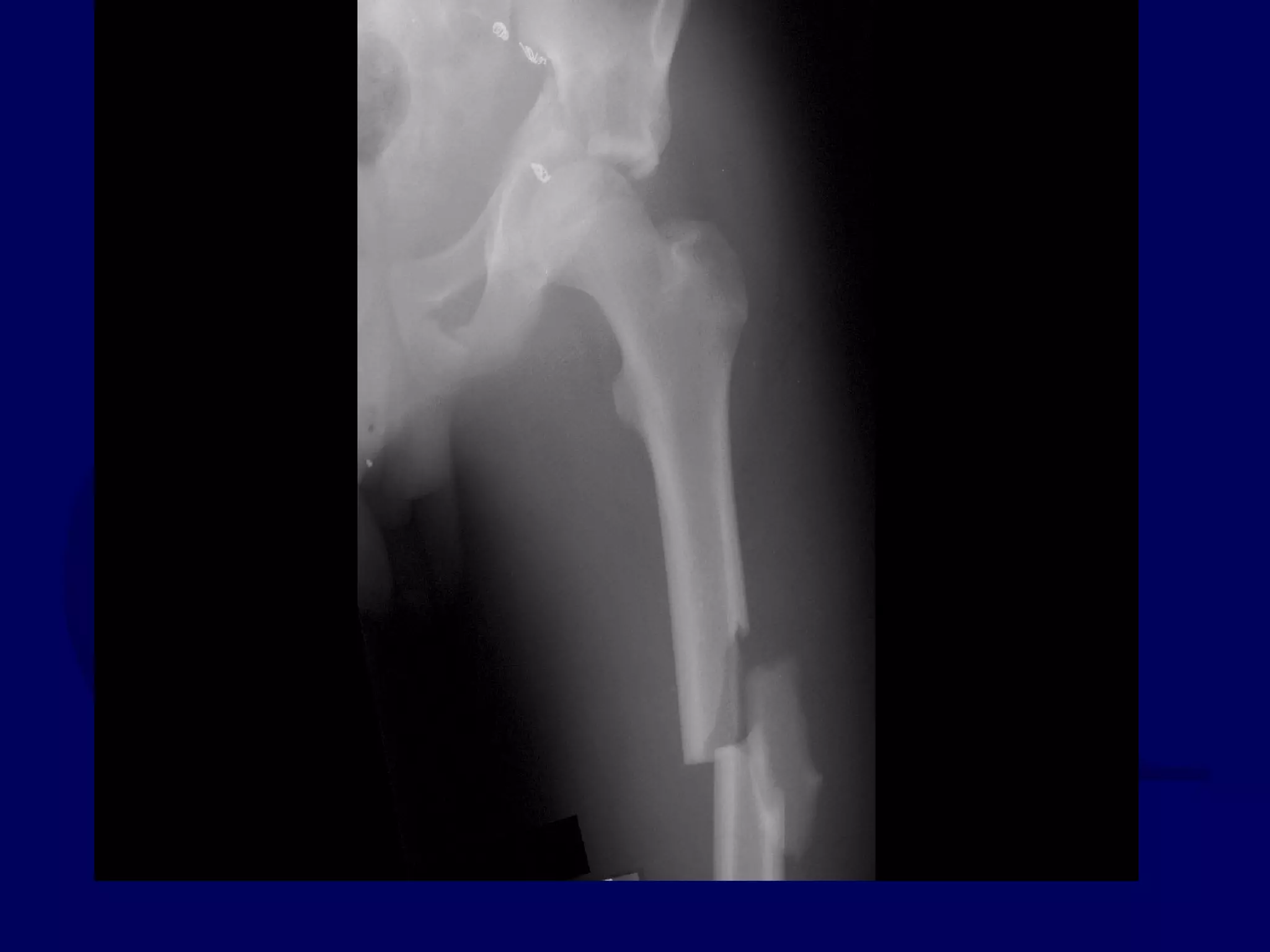
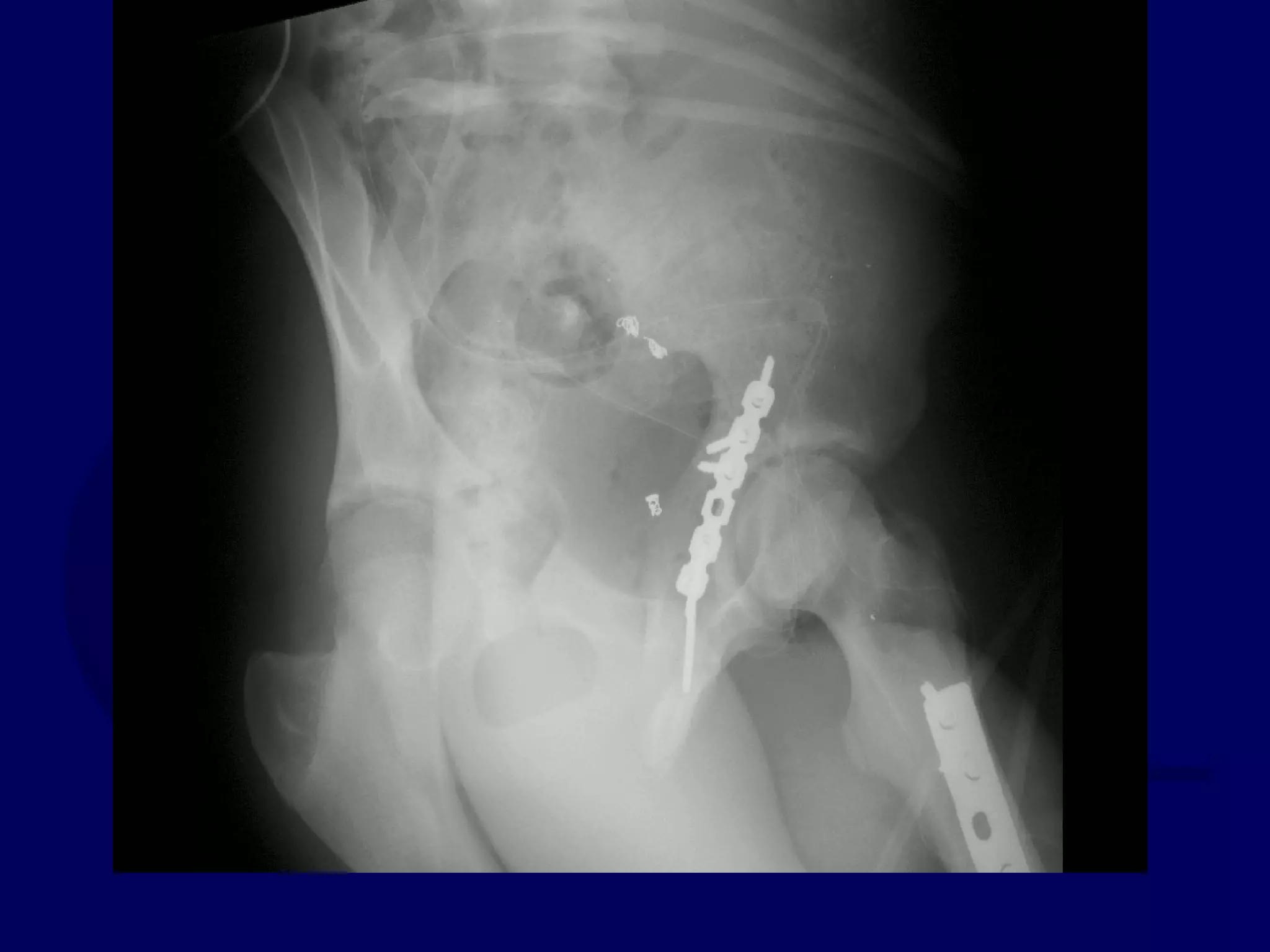
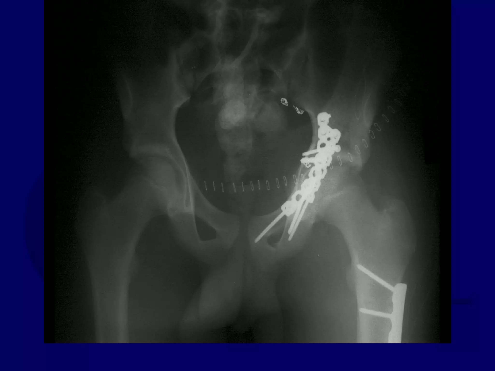
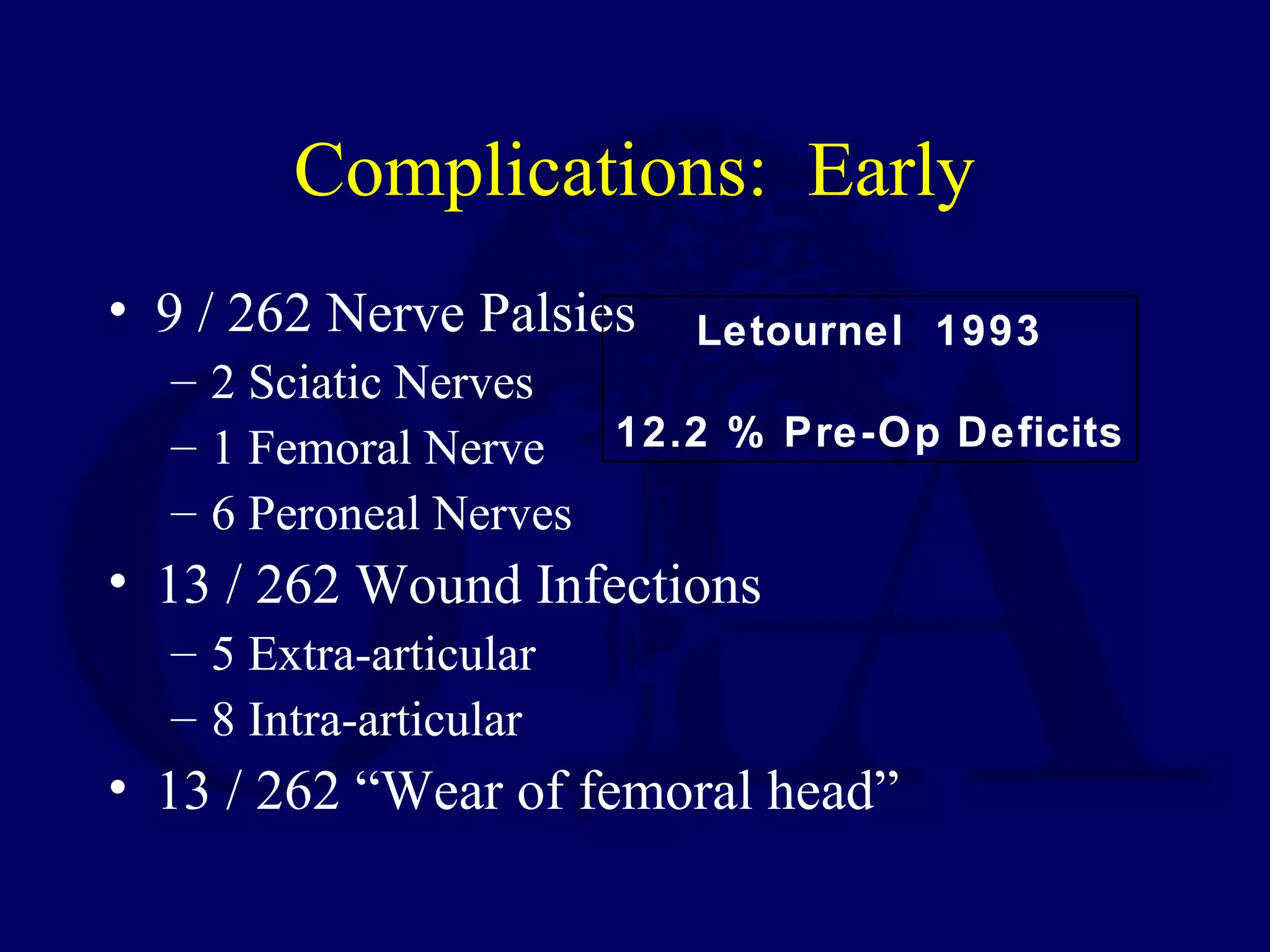
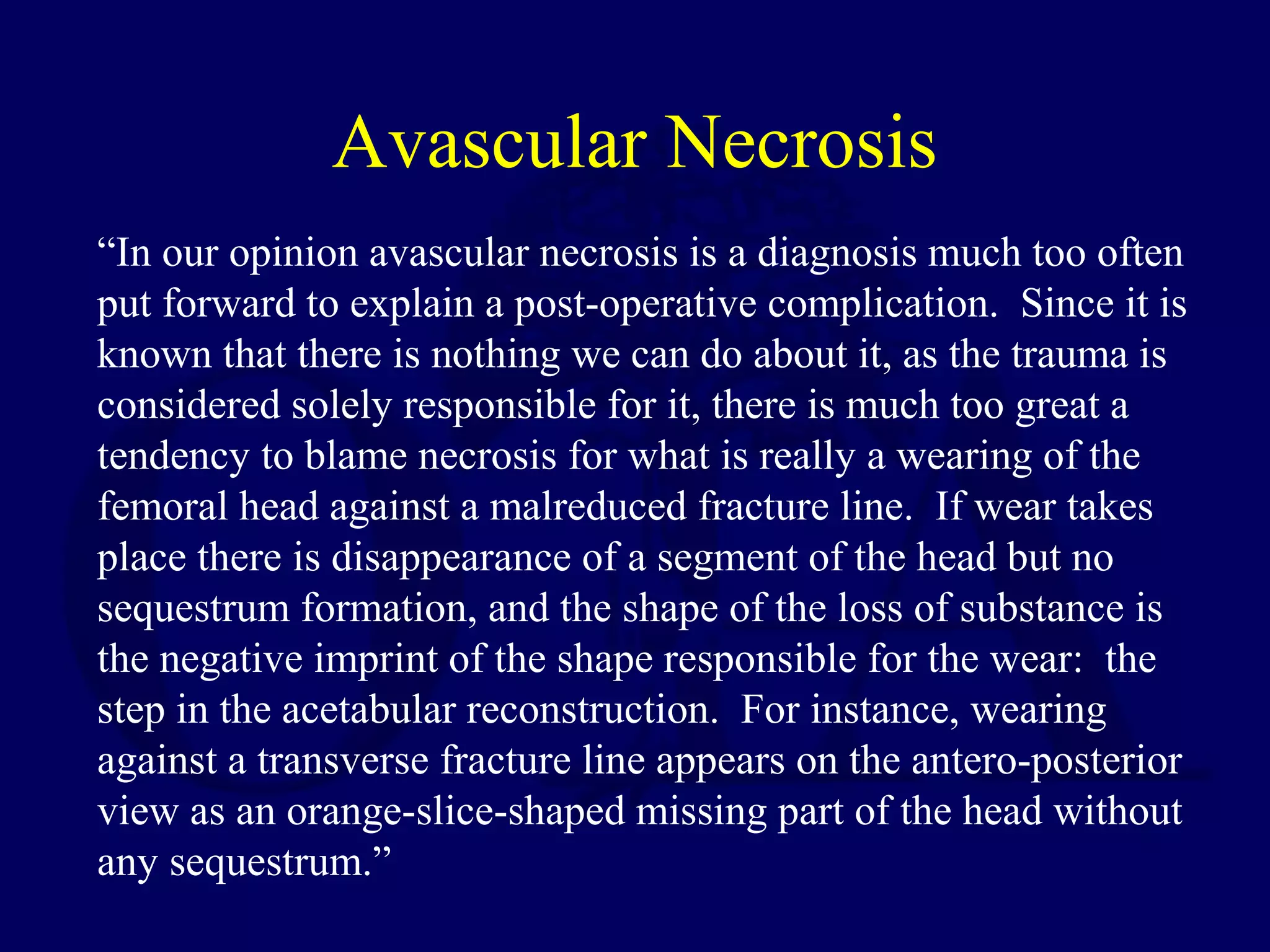
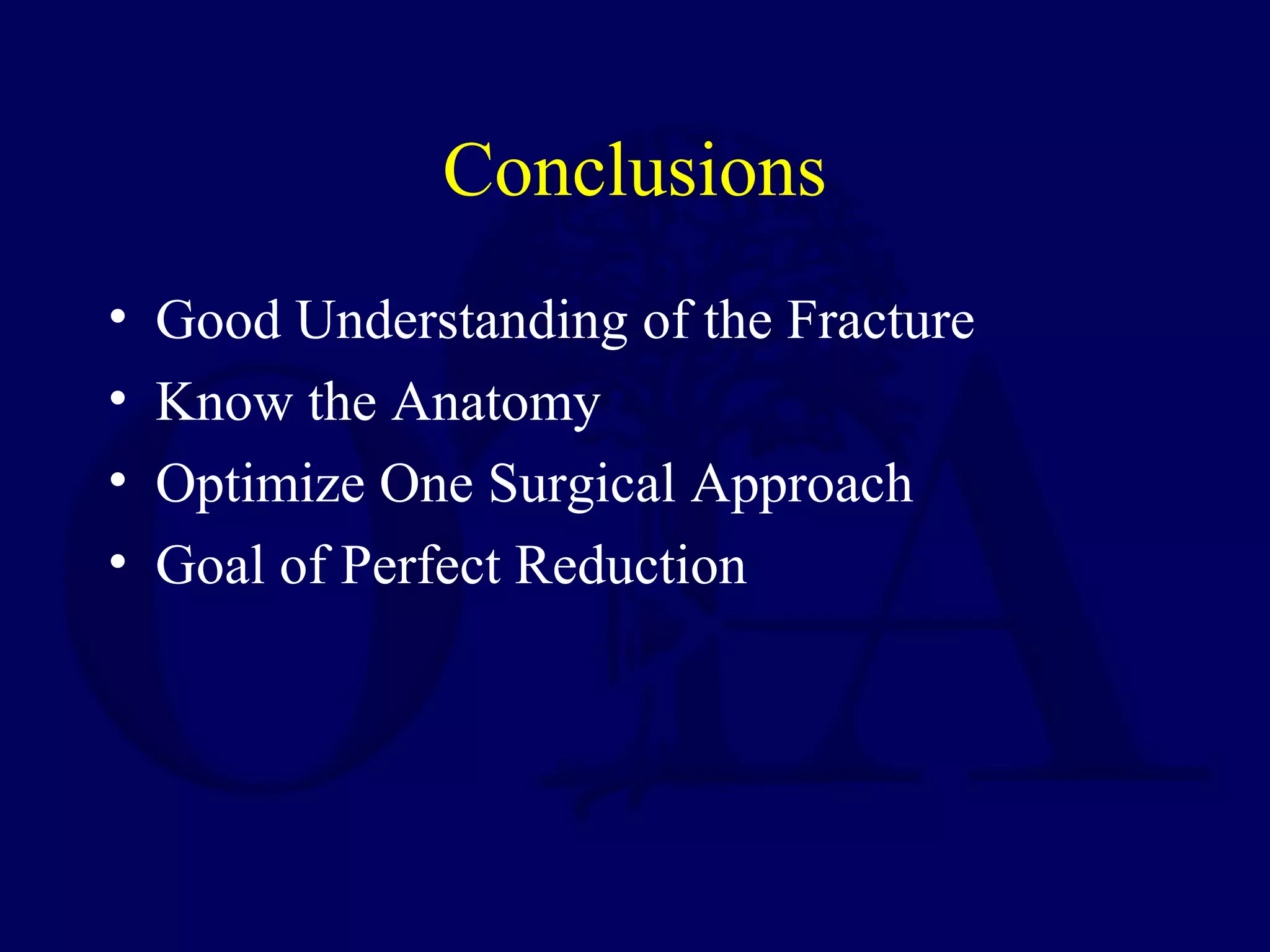
Acetabular Fractures:
Surgical Management
Philip J. Kregor, MD
Orthopedic Traumatology
University of Mississippi Med Center
Jackson, Mississippi
Created March 2004; Reviewed January 2007
Objectives
• Goal of Operative Management
• Specific Approaches for Specific Fractures
• Indications for Kocher-Langenbeck Approach
• Indications for Ilioinguinal Approach
• Reduction Strategies
Letournel School
• Thorough Understanding of Plain Films
• Optimize One Surgical Approach
• Goal of Perfect Concentric Reduction
GOAL: Anatomic Reduction
EXCELLENT GOOD FAIR POOR
Timing of Surgery: Criteria
• Well - resuscitated patient
• Appropriate radiological work-up
• Appropriate understanding of fracture
• Appropriate operative team
Matta 1996
Timing of Surgery and
Anatomical Reductions
• 0-7 Days 74%
• 8-14 Days 71%
• 15-21 Days 57%
Surgical Emergencies: Rare
• Open Acetabular Fracture
• New-Onset Sciatic Nerve Palsy after closed
reduction of Hip dislocation
Surgical Urgencies: Infrequent
• Irreducible Posterior Hip Dislocation
• Medial Dislocation of Femoral Head
against cancellous bone surface of intact
Ilium
Matta 1996
NOT Predictive of
CLINICAL OUTCOME
• Type of fracture pattern
• Posterior dislocation
• Initial displacement
• Presence of intra-articular fragments
• Presence of acetabular impaction
Matta 1996
Predictive of
CLINICAL OUTCOME
• Injury to Cartilage or Bone of Femoral
Head
– Damage: 60% Good / Excellent Result
– No Damage: 80% Good / Excellent Result
• Anatomic Reduction
• Age of Patient …….. But only in that it
predicts the ability to achieve an anatomic
reduction
Approaches to the Acetabulum
• Posterior: Kocher - Langenbeck
• Anterior: Ilioinguinal
• Extensile: Extended Iliofemoral
Letournel Classification
• Anterior Wall
• Anterior Column
• Posterior Wall
• Posterior Column
• Transverse
Letournel Classification
• Posterior Column / Posterior Wall
• Transverse / Posterior Wall
• “T-type”
• Anterior Column / Posterior Hemitransverse
• Both Column
Kocher-Langenbeck Approach
• Langenbeck (1874): Superior Limb
• Kocher (1904): Inferior Limb
• Judet and Lagrange (1958)
• Letournel
Indications in Acute
Acetabular Fxs
• Posterior Wall Fractures
• Posterior Column Fractures
• Posterior Column / Posterior Wall Fractures
• Juxta-tectal / Infra-tectal Transverse or
Transverse with Posterior Wall Fractures
• Some “T-type” Fractures
Access: Kocher-Langenbeck
• Entire Posterior Column
• Greater and Lesser Sciatic Notches
• Ischial Spine
• Retro-Acetabular Surface
• Ischial Tuberosity
• Ischio-Pubic Ramus
Complications with KL
• Sciatic Nerve Palsy 10%
• Infection 3%
Limitations: Kocher-Langenbeck
• Superior Acetabular Region
• Anterior Column
• Fractures High in Greater Sciatic Notch
Prone Position
• Aids in Reduction of Ischiopubic Segment
• Facilitates Palpation of Quadrilateral
Surface
• Allows Clamp Placement through Greater
Sciatic Notch
• Easier Prep and Drape
Judet Table
Posterior Wall Fractures
Posterior Wall Fxs:
Surgical Keys
• Avoid Devascularization of Fragment/s
• Remove Intra-articular Fragments
• Address Marginal Impaction
• Provide adequate buttress
• Avoid Over-Contouring of Plate
Controlled Distraction
of Hip Joint
• Femoral Distractor
• Traction Table
Posterior Wall Fx
63 Y.O. Male
L.W. 00.09.23
L.W. 00.09.23
L.W. 00.09.23
L.W. 00.09.23
L.W. 00.10.25
L.W. 00.10.25
L.W. 00.10.25
Special Case:
Extended Posterior Wall
??? Ganz Trochanteric Flip Osteotomy
to Visualize Fracture
without Devitalizing Abductors
T.D. 00.02.01
T.D. 00.02.01
T.D. 00.02.01
T.D. 00.02.01
T.D. 00.02.17
T.D. 00.02.08
Reduction Aids: Kocher-
Langenbeck Approach
• Distal Femoral Traction
• Distraction of Hip Joint
• Ischial Tuberosity Schantz Pin
• Quadrangular Clamp through Greater
Sciatic Notch
• Farabeuf Clamp
FAERBEUF CLAMPS
M.M. 98.10.27
M.M 98.10.29
M.M. 98.11.04
M.M. 98.11.05
M.M. 98.11.05
Letournel 1993
Optimal Screw Placement
Transtectal Tranverse
Acetabular Fx
18 Y.O. Male
Isolated Injury
Skinny Patient / Treated Early
W.M. 99.11.27
W.M. 99.11.27
W.M. 99.11.27
W.M. 99.11.27
W.M. 00.01.12
W.M. 00.01.12
W.M. 00.01.12
Ilioinguinal Approach:
Indications
• Anterior Wall
• Anterior Column
• Transverse with significant Anterior
Displacement
• Anterior Column / Posterior Hemitransverse
• Both Column
Ilioinguinal Approach: Access
II Complications
• Direct Hernia 1%
• Significant LFC nerve numbness 23%
• External iliac artery thrombosis
1%
II Complications
• Hematoma 5%
• Infection 2%
Ilioinguinal Approach
Anterior Column Fx
Isolated Injury
73 Y.O. Male
J.W. 00.10.14
J.W. 00.10.14
J.W. 00.10.14
Reduction of Anterior Column
to Intact Ilium
Clamp Placement
Lag Screw Placement
J.W. 00.10.19
J.W. 00.11.02
J.W. 00.10.19
Anterior Column /
Posterior Hemitransverse
Anterior Wall or Column
Posterior Half of Transverse Fracture
Anterior Column Fractures
Anterior Wall Fracture
Jeff Mast, M.D.
R.M. 98.08.15
R.M. 98.08.15
R.M. 98.08.15
R.M. 98.08.15
R.M. 98.08.15
R.M. 98.08.15
R.M. 98.08.15
R.M. 98.08.24
R.H. 98.11.22
R.M. 99.02.17
R.M. 99.02.17
R.M. 99.02.17
Both Column
Acetabular Fracture
18 Y.O. Female
Isolated Injury
R.C. 00.03.09
R.C. 00.03.09
R.C. 00.03.09
SPUR
SIGN
R.C. 00.03.09
R.C. 00.03.09
A.S.I.S.
SYMPHYSIS
EXT.
OBL.
A.S.I.S.
EXT. INGUINAL RING
A.S.I.S.
L.F.C.N.
PSOAS
EXT
.
OB
L.
EXT.
OBL.
CONJOINT TENDON
Completion of Iliac Fracture
Reduction of Anterior Column
to Intact Ilium
Reduction of Posterior Column
INTACT ILIUM
R.C. 00.03.10
Extended Iliofemoral Approach
• “T” Type Fractures
• Trans-tectal Transverse Fractures
• Delayed Reconstruction
EIF Complications
• Sciatic nerve palsy 1%
• Hematoma 8%
• Infection 1%
Extended Iliofemoral Approach
R.H. 98.11.21
R.H. 98.11.21
R.H. 98.11.22
Special Case
“T-Type” Acetabular Fracture
Proximal Femur Fracture
14 y.o. Male
Sequential K-L / Ilioinguinal
Approaches
P.J. 00.12.16
P.J. 00.12.16
P.J. 00.12.16
P.J. 00.12.16
Initial Kocher-Langenbeck
Approach
P.J. 00.12.18
P.J. 00.12.18
Subsequent Ilioinguinal
Approach
P.J. 00.12.22
Intra-Operative Assessment of
Reduction
• Visual Assessment of Fracture Reduction
• Palpation of Fracture
– Quadrilateral surface through Greater Sciatic
Notch
– Anterior Column
• C-Arm assessment
• Plain A.P. Radiograph
• Assurance that all Screws are out of Joint
Assessment of Reduction
• Restoration of Pelvic Lines
• Concentric Reduction on all 3 Views
• Goal of Anatomic Reduction
Complications: Early
• 9 / 262 Nerve Palsies
– 2 Sciatic Nerves
– 1 Femoral Nerve
– 6 Peroneal Nerves
• 13 / 262 Wound Infections
– 5 Extra-articular
– 8 Intra-articular
• 13 / 262 “Wear of femoral head”
Letournel 1993
12.2 % Pre-Op Deficits
Letournel 1993
Complications: Long-term
• 0.7 % Nonunion
• 1% Cartilage Necrosis
• 3.1% Avascular Necrosis
• Osteoarthritis
– 10.2 % after perfect reduction
– 35.7 % after imperfect reduction
Letournel 1993
Avascular Necrosis
“In our opinion avascular necrosis is a diagnosis much too often
put forward to explain a post-operative complication. Since it is
known that there is nothing we can do about it, as the trauma is
considered solely responsible for it, there is much too great a
tendency to blame necrosis for what is really a wearing of the
femoral head against a malreduced fracture line. If wear takes
place there is disappearance of a segment of the head but no
sequestrum formation, and the shape of the loss of substance is
the negative imprint of the shape responsible for the wear: the
step in the acetabular reconstruction. For instance, wearing
against a transverse fracture line appears on the antero-posterior
view as an orange-slice-shaped missing part of the head without
any sequestrum.”
Heterotopic Ossification:
Brooker Classification
• I: Islands of bone less than 1 cm in diameter
• II: Larger islands of bone, leaving at least 1 cm
free space between the two bones of the hip
• III: Free space between the ossification and the
pelvis or the femur is less than 1 cm
• IV: Apparent ankylosis of the joint by a bony
bridge uniting the pelvis and the femur
Heterotopic Ossification
• Classification does not predict mobility
• Approach:
– 34% Grade III / IV Extended Iliofemoral
– 11% Grade III / IV Kocher-Langenbeck
– 1 % Grade III / IV Ilioinguinal
• “Ectopic bone formation appears early on
radiography, and maturity is reached 6
months to 1 year after operation.”
Significant HO
(0 , 90° Hip Flexion)
• KL 8%
• II 2%
• EIF 20%
Prophylaxis for HO
• Indomethacin
• 700 cGy radiation
• Combination
DVT Prophylaxis
• Controversial
• Mechanical devices
• Pharmacologic (I.e. LMWH)
Conclusions
• Good Understanding of the Fracture
• Know the Anatomy
• Optimize One Surgical Approach
• Goal of Perfect Reduction
THANK YOU
Return to
Pelvis
Index
E-mail OTA
about
Questions/Comments
If you would like to volunteer as an author for
the Resident Slide Project or recommend updates
to any of the following slides, please send an e-
mail to ota@aaos.org

V06 orif acetabulum