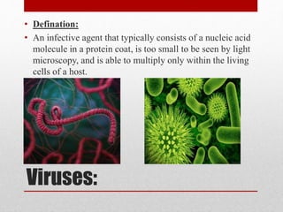
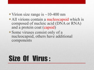
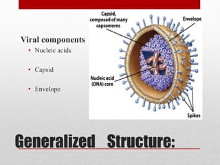
The document discusses viruses, defining them as infectious agents that can only multiply within host cells and consist of nucleic acids and protein coats. It covers virus structure, classification, life cycles, methods of transmission between hosts, discovery, theories of origin, and techniques for studying and cultivating viruses. The document provides an overview of the key aspects of virology, including what viruses are, how they spread and reproduce, approaches to classifying them, and historical developments in the field.
When fresh liquid medium is inoculated with a given number of bacteria and incubated for sufficient period of time, it gives a characteristic growth pattern of bacteria.
If the bacterial population is measured periodically and log of number of viable bacteria is plotted in a graph against time, it gives a characteristic growth curve which is known as growth curve or growth cycle.
Obligate intracellular, unable to self-replicate.
Once inside living cells, viruses induce the host cell to synthesize virus particles.
The genome is either DNA or RNA (single or double stranded).
Viruses do not have a system to produce ATP.
Viruses range in size from 25 to 270 nm.
Viral tropism!!
The classification of viruses is based on nucleic acid type, size and shape of virion, and presence or absence of an envelope.
Viral Structure
I . Virion is the entire viral particle.
2. Capsid is the protein coat that encloses the genetic material.
3. Capsomer is the protein subunit that makes up the capsid.
4. Nucleocapsid is composed of the capsid and genetic material.
5. The envelope is the outer coating composed of a phospholipid bilayer, which is composed of viral-encoded glycoproteins and sometimes viral encoded matrix proteins. The envelope is derived from a host cell's membrane.
Some viruses use the plasma membrane, whereas others use endoplasmic reticulum, Golgi, or nuclear membranes. Naked nucleocapsids are viruses with no envelopes.
Biology I Presentation
FUNGI
We will learn
General characteristics of fungi
Structure of fungi
Economic Importance
Pathogenicity
Brief intro of some fungi
THE SIX KINGDOMS
Fungi are placed in a separate kingdom called the kingdom fungi
OF FUNGI
CHARACTERISTICS
The Characteristics of Fungi
Fungi are NOT plants
Nonphotosynthetic
Eukaryotes
Nonmotile
Most are saprobes (live on dead organisms)
The Characteristics of Fungi
Absorptive heterotrophs (digest food first & then absorb it into their bodies
Release digestive enzymes to break down organic material or their host
Store food energy as glycogen
The Characteristics of Fungi
Important decomposers & recyclers of nutrients in the environment
Most are multicellular, except unicellular yeast
Lack true roots, stems or leaves
fungi as a decomposers
The Characteristics of Fungi
Cell walls are made of chitin (complex polysaccharide)
Body is called the Thallus
Grow as microscopic tubes or filaments called hyphae
The Characteristics of Fungi
Some fungi are internal or external parasites
A few fungi act like predators & capture prey like roundworms
The Characteristics of Fungi
Some are edible, while others are poisonous
The Characteristics of Fungi
Produce both sexual and asexual spores
Classified by their sexual reproductive structures
The Characteristics of Fungi
Grow best in warm, moist environments
Mycology is the study of fungi
Mycologists study fungi
A fungicide is a chemical used to kill fungi
The Characteristics of Fungi
Fungi include puffballs, yeasts, mushrooms, toadstools, rusts, smuts, ringworm, and molds
The antibiotic penicillin is made by the Penicillium mold
FUNGI SIZE
NON-REPRODUCTIVE
Vegetative Structures
Hyphae
Tubular shape
ONE continuous cell
Filled with cytoplasm & nuclei
Multinucleate
Hard cell wall of chitin also in insect exoskeletons
Hyphae
Stolons – horizontal hyphae that connect groups of hyphae to each other
Rhizoids – rootlike parts of hyphae that anchor the fungus
Hyphae
Cross-walls called SEPTA may form compartments
Septa have pores for movement of cytoplasm
Form network called mycelia that run through the thallus (body)
Absorptive Heterotroph
Fungi get carbon from organic sources
Tips of Hyphae release enzymes
Enzymatic breakdown of substrate
Products diffuse back into hyphae
Modifications of hyphae
Fungi may be classified based on cell division (with or without cytokinesis)
Aseptate or coenocytic (without septa)
Septate (with septa)
Modifications of hyphae
Hyphal growth
Hyphae grow from their tips
Mycelium is an extensive, feeding web of hyphae
Mycelia are the ecologically active bodies of fungi
ASEXUAL & SEXUAL SPORES
REPRODUCTIVE STRUCTURES
REPRODUCTION
Most fungi reproduce Asexually and Sexually by spores
ASEXUAL reproduction is most common method & produces genetically identical organisms
Fungi reproduce SEXUALLY when conditions are poor & nutrients
Morphology, Classification, Cultivation and Replication of VirusKrutika Pardeshi
This presentation is Useful for B. Pharmacy SEM III Students to study the Topic Fungi According to PCI Syllabus.
It Consist of Morpholoy of Fungi, Cultivation , Replication and Classification of Virud
General Characters and Classification of Viruses. Includes ICTV classification and Baltimore classification of viruses. A brief explanation of the Viral structure and Lifecycle.
This presentation gives a detail overview on Viruses - Morphology and Classification. The presentation is helpful for students of B. Pharm Second Year and those who wants to gain basic knowledge about Viruses.
Subject - Microbiology
Concept of virology
Viruses
Types of viruses
Viral characteristics
Virion
Size and Shape
Structure
Replication
Viral Variation
Classification
Quiz
BEST OF LUCK
Virus, infectious agent of small size and simple composition that can multiply only in living cells of animals, plants, or bacteria. The name is from a Latin word meaning “slimy liquid” or “poison.”
The increased availability of biomedical data, particularly in the public domain, offers the opportunity to better understand human health and to develop effective therapeutics for a wide range of unmet medical needs. However, data scientists remain stymied by the fact that data remain hard to find and to productively reuse because data and their metadata i) are wholly inaccessible, ii) are in non-standard or incompatible representations, iii) do not conform to community standards, and iv) have unclear or highly restricted terms and conditions that preclude legitimate reuse. These limitations require a rethink on data can be made machine and AI-ready - the key motivation behind the FAIR Guiding Principles. Concurrently, while recent efforts have explored the use of deep learning to fuse disparate data into predictive models for a wide range of biomedical applications, these models often fail even when the correct answer is already known, and fail to explain individual predictions in terms that data scientists can appreciate. These limitations suggest that new methods to produce practical artificial intelligence are still needed.
In this talk, I will discuss our work in (1) building an integrative knowledge infrastructure to prepare FAIR and "AI-ready" data and services along with (2) neurosymbolic AI methods to improve the quality of predictions and to generate plausible explanations. Attention is given to standards, platforms, and methods to wrangle knowledge into simple, but effective semantic and latent representations, and to make these available into standards-compliant and discoverable interfaces that can be used in model building, validation, and explanation. Our work, and those of others in the field, creates a baseline for building trustworthy and easy to deploy AI models in biomedicine.
Bio
Dr. Michel Dumontier is the Distinguished Professor of Data Science at Maastricht University, founder and executive director of the Institute of Data Science, and co-founder of the FAIR (Findable, Accessible, Interoperable and Reusable) data principles. His research explores socio-technological approaches for responsible discovery science, which includes collaborative multi-modal knowledge graphs, privacy-preserving distributed data mining, and AI methods for drug discovery and personalized medicine. His work is supported through the Dutch National Research Agenda, the Netherlands Organisation for Scientific Research, Horizon Europe, the European Open Science Cloud, the US National Institutes of Health, and a Marie-Curie Innovative Training Network. He is the editor-in-chief for the journal Data Science and is internationally recognized for his contributions in bioinformatics, biomedical informatics, and semantic technologies including ontologies and linked data.
Richard's entangled aventures in wonderlandRichard Gill
Since the loophole-free Bell experiments of 2020 and the Nobel prizes in physics of 2022, critics of Bell's work have retreated to the fortress of super-determinism. Now, super-determinism is a derogatory word - it just means "determinism". Palmer, Hance and Hossenfelder argue that quantum mechanics and determinism are not incompatible, using a sophisticated mathematical construction based on a subtle thinning of allowed states and measurements in quantum mechanics, such that what is left appears to make Bell's argument fail, without altering the empirical predictions of quantum mechanics. I think however that it is a smoke screen, and the slogan "lost in math" comes to my mind. I will discuss some other recent disproofs of Bell's theorem using the language of causality based on causal graphs. Causal thinking is also central to law and justice. I will mention surprising connections to my work on serial killer nurse cases, in particular the Dutch case of Lucia de Berk and the current UK case of Lucy Letby.
A brief information about the SCOP protein database used in bioinformatics.
The Structural Classification of Proteins (SCOP) database is a comprehensive and authoritative resource for the structural and evolutionary relationships of proteins. It provides a detailed and curated classification of protein structures, grouping them into families, superfamilies, and folds based on their structural and sequence similarities.
(May 29th, 2024) Advancements in Intravital Microscopy- Insights for Preclini...Scintica Instrumentation
Intravital microscopy (IVM) is a powerful tool utilized to study cellular behavior over time and space in vivo. Much of our understanding of cell biology has been accomplished using various in vitro and ex vivo methods; however, these studies do not necessarily reflect the natural dynamics of biological processes. Unlike traditional cell culture or fixed tissue imaging, IVM allows for the ultra-fast high-resolution imaging of cellular processes over time and space and were studied in its natural environment. Real-time visualization of biological processes in the context of an intact organism helps maintain physiological relevance and provide insights into the progression of disease, response to treatments or developmental processes.
In this webinar we give an overview of advanced applications of the IVM system in preclinical research. IVIM technology is a provider of all-in-one intravital microscopy systems and solutions optimized for in vivo imaging of live animal models at sub-micron resolution. The system’s unique features and user-friendly software enables researchers to probe fast dynamic biological processes such as immune cell tracking, cell-cell interaction as well as vascularization and tumor metastasis with exceptional detail. This webinar will also give an overview of IVM being utilized in drug development, offering a view into the intricate interaction between drugs/nanoparticles and tissues in vivo and allows for the evaluation of therapeutic intervention in a variety of tissues and organs. This interdisciplinary collaboration continues to drive the advancements of novel therapeutic strategies.
THE IMPORTANCE OF MARTIAN ATMOSPHERE SAMPLE RETURN.Sérgio Sacani
The return of a sample of near-surface atmosphere from Mars would facilitate answers to several first-order science questions surrounding the formation and evolution of the planet. One of the important aspects of terrestrial planet formation in general is the role that primary atmospheres played in influencing the chemistry and structure of the planets and their antecedents. Studies of the martian atmosphere can be used to investigate the role of a primary atmosphere in its history. Atmosphere samples would also inform our understanding of the near-surface chemistry of the planet, and ultimately the prospects for life. High-precision isotopic analyses of constituent gases are needed to address these questions, requiring that the analyses are made on returned samples rather than in situ.
Seminar of U.V. Spectroscopy by SAMIR PANDASAMIR PANDA
Spectroscopy is a branch of science dealing the study of interaction of electromagnetic radiation with matter.
Ultraviolet-visible spectroscopy refers to absorption spectroscopy or reflect spectroscopy in the UV-VIS spectral region.
Ultraviolet-visible spectroscopy is an analytical method that can measure the amount of light received by the analyte.
Multi-source connectivity as the driver of solar wind variability in the heli...Sérgio Sacani
The ambient solar wind that flls the heliosphere originates from multiple
sources in the solar corona and is highly structured. It is often described
as high-speed, relatively homogeneous, plasma streams from coronal
holes and slow-speed, highly variable, streams whose source regions are
under debate. A key goal of ESA/NASA’s Solar Orbiter mission is to identify
solar wind sources and understand what drives the complexity seen in the
heliosphere. By combining magnetic feld modelling and spectroscopic
techniques with high-resolution observations and measurements, we show
that the solar wind variability detected in situ by Solar Orbiter in March
2022 is driven by spatio-temporal changes in the magnetic connectivity to
multiple sources in the solar atmosphere. The magnetic feld footpoints
connected to the spacecraft moved from the boundaries of a coronal hole
to one active region (12961) and then across to another region (12957). This
is refected in the in situ measurements, which show the transition from fast
to highly Alfvénic then to slow solar wind that is disrupted by the arrival of
a coronal mass ejection. Our results describe solar wind variability at 0.5 au
but are applicable to near-Earth observatories.
2. Viruses:
• Defination:
• An infective agent that typically consists of a nucleic acid
molecule in a protein coat, is too small to be seen by light
microscopy, and is able to multiply only within the living
cells of a host.
3. Introduction to viruses
Viruses do not have cells that divide; new
viruses are assembled in the infected host cell
But unlike still simpler infectious agents,
viruses contain genes, which gives them the
ability to mutate and evolve.
Evolved from plasmids : pieces of DNA that can
move between cells
while others may have evolved from bacteria.
Over 5,000 species of viruses have been
discovered.
4. Introduction to viruses
A virus consists of two or three parts:
genes, made from either DNA or RNA,
long molecules that carry genetic information
protein coat that protects the genes; and in some viruses,
an envelope of fat
Viruses vary in shape from the
simple helical and icosahedral to
more complex structures.
Viruses range in size from 20 to 300 nanometres; it
would take 30,000 to 750,000 of them, side by side, to
stretch to 1 centimeter.
5. Spreading , Vectors:
Viruses spread in many ways. Just as many viruses
are very specific as to which host species or tissue
they attack,
Plant viruses are often spread from plant to plant by
insects and other organisms, known as vectors.
Some viruses of animals, including humans, are
spread by exposure to infected bodily fluids
Viruses such as influenza are spread through the air
by droplets of moisture when people cough or sneeze.
Viruses such as norovirus are transmitted by
the faecal–oral route, which involves the
contamination of hands, food and water.
6. The human immunodeficiency virus, HIV, is
transmitted by bodily fluids transferred
during sex.
Dengue virus, are spread by blood-sucking
insects.
Rotavirus is often spread by direct contact
with infected children.
Antibiotics have no effect on viruses,
but antiviral drugs have been developed to
treat life-threatening
infections. Vaccines that produce lifelong
immunity can prevent some viral infections.
7. Discovery:
In 1884 the French microbiologist Charles
Chamberland invented a filter, known today as
the Chamberland filter or Chamberland–Pasteur filter, that
has pores smaller than bacteria. Thus he could pass a
solution containing bacteria through the filter and
completely remove them from the solution.
In the early 1890s the Russian biologist Dmitri
Ivanovsky used this filter to study what became known as
the tobacco mosaic virus. His experiments showed that
extracts from the crushed leaves of infected tobacco-plants
remain infectious after filtration.
8. Discovery:
In 1899 the Dutch microbiologist Martinus
Beijerinck observed that the agent multiplied only in dividing
cells. Having failed to demonstrate its particulate nature he
called it a "contagium vivum fluidum", a "soluble living
germ".
In the early 20th century the English bacteriologist Frederick
Twort discovered viruses that infect bacteria
With the invention of the electron microscope in 1931 by the
German engineers Ernst Ruska and Max Knoll came the first
images of viruses.
9. Discovery:
In 1935
American biochemist and virologist Wendell
Meredith Stanley examined the tobacco mosaic
virus and found it to be mostly made from protein.
A short time later, this virus was separated into
protein and RNA parts.
The breakthrough came in 1931, when the
American pathologist Ernest William
Goodpasture grew influenza and several other
viruses in fertilised chickens' eggs.
10. Origins Theories:
Viruses co-exist with life wherever it occurs. They have
probably existed since living cells first evolved.
Viruses do not form fossils so molecular techniques have
been the most useful means of hypothesising how they
arose.
Three main theories speculate on the origins of viruses:
Regressive theory
Viruses may have once been small cells
that parasitised larger cells. Over time, genes not required by
their parasitism were lost. The
bacteria rickettsia and chlamydia are living cells that, like
viruses, can reproduce only inside host cells.
11. Origins Theories:
They lend credence to this theory, as their dependence on
parasitism is likely to have caused the loss of genes that
enabled them to survive outside a cell
Cellular origin theory
Some viruses may have evolved from bits of DNA or
RNA that "escaped" from the genes of a larger
organism. The escaped DNA could have come
from plasmids—pieces of DNA that can move between
cells—while others may have evolved from bacteria.
12. Origins Theories:
Coevolution theory
Viruses may have evolved from complex molecules of
protein and DNA at the same time as cells first appeared on
earth and would have depended on cellular life for many
millions of years
Problems with these theories:
the regressive hypothesis does not explain why even the
smallest of cellular parasites do not resemble viruses in any
way.
The escape hypothesis does not explain the structures of
virus particles.
The coevolution, or virus-first hypothesis, contravenes the
definition viruses, in that they are dependent on host cells.
13. Characteristics
Obligate intracellular parasites of bacteria, protozoa, fungi,
algae, plants, and animals.
Ultramicroscopic size, ranging from 20 nm up to 450 nm
(diameter).
Not cellular in nature; structure is very compact and
economical.
Do not independently fulfill the characteristics of life.
Inactive macromolecules outside the host cell and active only
inside host cells.
Basic structure consists of protein shell (capsid) surrounding
nucleic acid core.
• Nucleic acid can be either DNA or RNA but not both
14. • Nucleic acid can be double-stranded DNA, single-
stranded DNA single-stranded RNA, or double-stranded
RNA.
• Molecules on virus surface impart high specificity for
attachment to host cell.
• Multiply by taking control of host cell’s genetic
material and regulating the synthesis and assembly of new
viruses.
• Lack enzymes for most metabolic processes.
• Lack machinery for synthesizing proteins.
• Most RNA viruses multiply in & are released from the
cytoplasm.
• l Viral infections range from very mild to life
threatening.
16. • Viruses have no nucleus, no organelles, no cytoplasm or
cell membrane—Non-cellular
17. Size of virus ?
• Smallest infectious agents
• Most are so small, they can only be seen with an electron
microscope
• Animal viruses
• Proviruses- around 20 nm in diameter
• Mimi viruses- up to 450 nm in length
• Viewing viruses
• Special stains and an electron microscope
• Negative staining outlines the shape
• Positive staining shows internal details
• Shadow casting technique
18.
19. Structure Of virus
• Questions Relating to Structure
• Is it rigid?
• How big is it?
• Is it flexible?
• Structure Must Serve Virus
• It should provide protection for genome
• It should allow virus to move from one host to next
• It should allow for attachment of virus on to new host
20. Size Of Virus :
Virion size range is ~10-400 nm
All virions contain a nucleocapsid which is
composed of nucleic acid (DNA or RNA)
and a protein coat (capsid)
Some viruses consist only of a
nucleocapsid, others have additional
components
21.
22. Capsids:
nucleocapsid
The capsid and the nucleic acid together are called the
nucleocapsid
virion
Fully formed virus that is able to establish an infection
in a host cell
25. Nucleic Acids:
• Genome:
the sum total of the genetic
information carried by an organism
• Number of viral genes compared with a cell-
quite small
• They only have the genes necessary to invade
host cells and redirect their activity
28. RNA Viruses:
• Mostly single-stranded
• Positive-sense RNA: genomes that are ready for immediate
translation into proteins
• Negative-sense RNA: genomes have to be converted into
the proper form to be made into protein
32. Tools For Studying
Structure :
• Electron Microscopy
• Excellent tool with some limitations
• High resolution
• Image can be a distortion due to specimen processing
• X-ray Diffraction
• Good for naked virions (no envelope)
• Cryoelectron Microscopy
33. Structural Symmetries:
• Icosahedral Symmetry
• 20 triangular faces
• It is a common capsid structure
• Examples of viruses with icosahedral symmetry
• Parvoviruses
• These are simple viruses
• ssDNA genome
• Capsid is formed with 60 copies of single protein
• Polio virus
• Uses 180 copies of 3 subunit proteins
• Much bigger virus
34. Capsid:
• Constructed from identical subunits called capsomers
• Made up of protein molecules
Two different types
• Helical
• Rod-shaped capsomers
• Assemble in to helical nucleocapsid
40. Functions of the Viral
Capsid
• Protects nucleic acids
• Help introduce the viral DNA or RNA into a suitable host
cell
• Stimulate the immune system to produce antibodies that
can protect the host cells against future infections
41.
42.
43. Ebola Virus:
• Morphology under electron microscope
• filamentous, enveloped RNA virus
• approx. 19 kb in length (1 kb = 1000 RNA
bases/nucleotides) or 60-80 nm in diameter
• single-stranded, linear, non-segmented
• negative-sense RNA (encoded in a 3’ to 5’ direction)
• appears to have “spikes” due to glycoprotein on
outside membrane
45. Viral reproduction
• Viruses can reproduce only when they enter cells and
utilize the cellular machinery of their hosts. Viruses’ code
their genes on a single type of nucleic acid, either DNA
or RNA, but viruses lack ribosomes and the enzymes
necessary for protein synthesis. Viruses are able to
reproduce because their genes are translated into proteins
by the cell’s genetic machinery. These proteins lead to the
production of more viruses.
46. Viral multiplication proceeds as following manner.
• Adsorption,
• Penetration,
• Uncoating,
• Synthesis,
• Assembly and Release
• Adsorption.
47. Adsorption/attachment
• Virus encounters susceptible host cells
• Adsorbs specifically to receptor sites on the cell
membrane
• Because of the exact fit required, viruses have a limited
host range
48. Penetration
• Flexible cell membrane of the host is penetrated by the
whole virus or its nucleic acid
• Endocytosis: entire virus engulfed by the cell and
enclosed in a vacuole or vesicle
• The viral envelope can also directly fuse with the host
cell membrane
49. Uncoating
• Enzymes in the vacuole dissolve the envelope and capsid
• The virus is now uncoated
50.
51. synthesis
• Free viral nucleic acid exerts control over the host’s
synthetic and metabolic machinery
• DNA viruses- enter host cell’s nucleus where they are
replicated and assembled
• DNA enters the nucleus and is transcribed into RNA
• The RNA becomes a message for synthesizing viral
proteins (translation)
• New DNA is synthesized using host nucleotides
• RNA viruses- replicated and assembled in the cytoplasm
53. Release
• Nonenveloped and complex viruses are released when the
cell lyses or ruptures
• Enveloped viruses are liberated by budding or exocytosis
• Anywhere from 3,000 to 100,000 virions may be
released, depending on the virus
• Entire length of cycle- anywhere from 8 to 36 hours
55. Cultivation of viruses
• Primary purposes of viral cultivation
• To isolate and identify viruses in clinical specimens
• To prepare viruses for vaccines
• To do detailed research on viral structure, multiplication
cycles, genetics, and effects on host cells
• Using Live Animal Inoculation
• Specially bred strains of white mice, rats, hamsters, guinea
pigs, and rabbits
• Occasionally invertebrates or nonhuman primates are used
• Animal is exposed to the virus by injection
56.
57. Tissue culture technique
• Most viruses are propagated in some sort of cell culture
• The cultures must be developed and maintained
• Animal cell cultures are grown in sterile chambers with
special media
• Cultured cells grow in the form of a monolayer
• Primary or continuous
58. Classification
• Viruses are classified on the basis of habitat (host).which is
trivial system beside this Viruses are classified on following
criteria.
• Structure
• Chemical composition
• Similarities in genetic makeup
• International Committee on the Taxonomy of Viruses, which
includes
• 3 orders
• 63 families “-viridae”
• 263 genera “-virus”
59.
60. Types of Classification:
• 3 Types of systems were proposed to classify the viruses:
• Baltimore Classification.
• Classical System Classification.
• Genetic Classification.
61. Baltimore Classification:
7 groups were made.
Its principles are fundamental to an understanding of virus
classification and genome replication.
The Baltimore classification has + RNA as its central point.
I: dsDNA viruses (e.g. Adenoviruses, Herpesviruses, Poxviruses)
II: ssDNA viruses (+ strand or "sense") DNA (e.g. Parvoviruses)
III: dsRNA viruses (e.g. Reoviruses)
IV: (+)ssRNA viruses (+ strand or sense) RNA
(e.g. Picornaviruses, Togaviruses)
V: (−)ssRNA viruses (− strand or antisense) RNA
(e.g. Orthomyxoviruses, Rhabdoviruses)
VI: ssRNA viruses (+ strand or sense) RNA with DNA
intermediate in life-cycle (e.g. Retroviruses)
VII: dsDNA viruses (e.g. Heptadnaviruses)
62.
63. Classification on basis of
host
• Animal viruses:
• Viruses of animal host
• Rabies, Polio, Mumps, Chicken pox, Small pox, and
Influenza.
• Plant Viruses:
• viruses which show their live characteristics when
attached to plants.
• Tobacco mosaic virus (TMV), Banana streak virus,
Carrot thin leaf virus
• Bacterial Virus: Bacteriophages ( T1, T2, T3, and T4.)
64.
65. Classification on
Genetics basis
• According to genetic consequences viruses are classified as.
DNA Viruses and RNA Viruses
• Genes may be linear or circular
• The smallest have only 4 genes and largest have several
hundred.
• DNA Viruses
• DNA Viruses are the viruses which consist of DNA genome .
They complete their activities by transcription and most of
them attack on organisms of similar genome.
• RNA Viruses
• RNA Viruses are the viruses which consist of RNA genome.
They complete their activities by reverse transcription.
66. Classification on
structural basis
• With relevant to morphology of viral structure viruses are
organized as Enveloped and Nonenveloped viruses.
• However they are also arranges subclasses of DNA and
RNA viruses
68. Benefits
In Genetic Engineering harmless virus are
used as genetic vectors which carry good
genes into cells.
Viral envelop Stimulate the immune system
to produce antibodies that can protect the
host cells against future infections
Viral genome contain enzymes for specific
operations within the host cell
Antiviral drugs block virus replication by
targeting one of the steps in the viral life
cycle
69. Interferon shows potential for treating
and preventing viral infections
Some recently-developed drugs do
combat some viruses, mostly by
interfering with viral nucleic acid
synthesis.
AZT interferes with reverse
transcriptase of HIV.
Acyclovir inhibits herpes virus DNA
synthesis
70. Uses of viruses
• The first vaccine was developed in the late 1700s by
Edward Jenner to fight smallpox.
• Vaccines can help prevent viral infections, but they can
do little to cure most viral infection once they occur.
• Both plasmids and transposons are mobile genetic
elements.
71. • Human Diseases: Warts, common cold, Influenza (flu),
Smallpox, Ebola, Herpes, AIDS, Chicken pox, Rabies are due
to virus actions.
• Viruses can be prevented with vaccines, but NOT treated with
antibiotics.
• Cytopathic effects- virus-induced damage to the cell that alters
its microscopic appearance
• Inclusion bodies- compacted masses of viruses or damaged
cell organelles
• Oncoviruses- mammalian viruses capable of initiating tumors