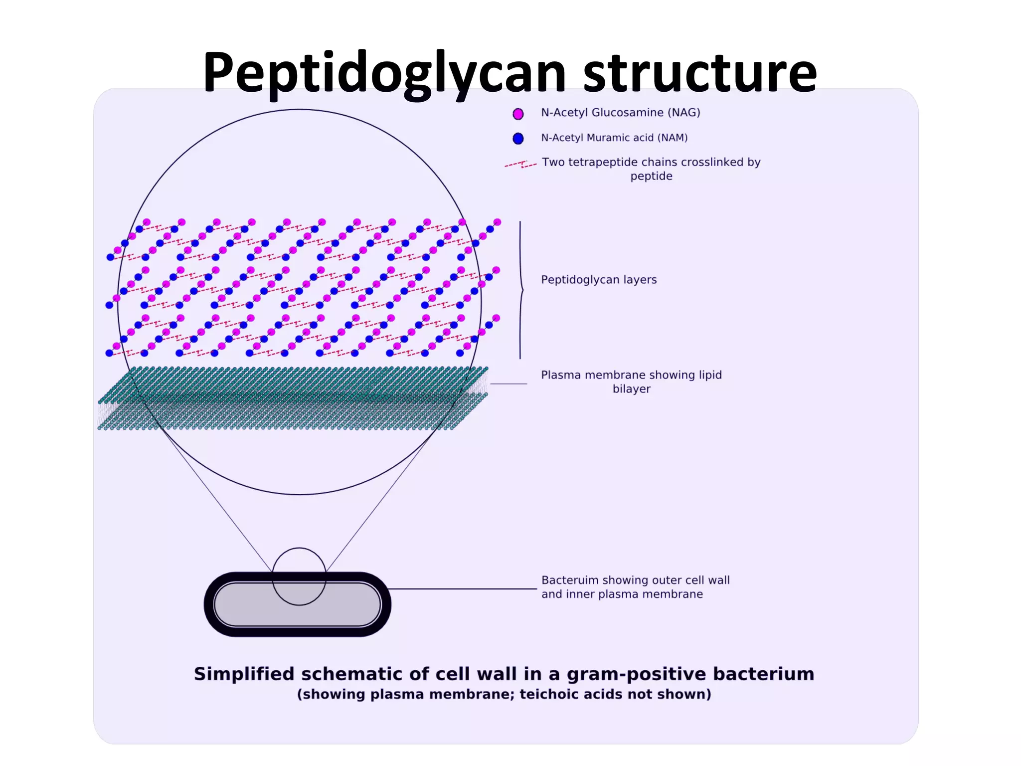
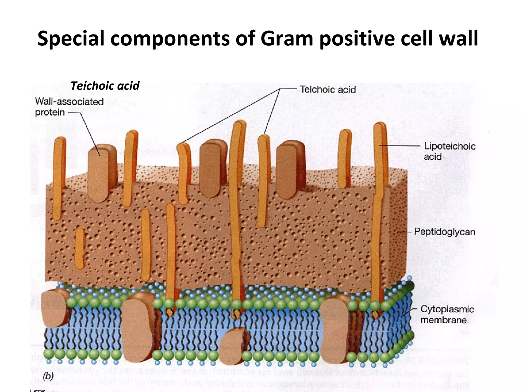
Bacterial Cell Wall
CELL WALL
The cell wall is the outer most layer of the cell. In many
cases the cell wall comes in direct contact with the
environment.
Function
• Protection of the cell.
• Maintains the shapes of the cell.
• Maintains the osmotic integrity of the cell.
• Assist some cells in attaching to other cells or in eluding
antimicrobial drugs.
• Not present in animal cells, so can target cell wall of
bacteria with antibiotics.
• Providing attachment sites for bacteriophages.
• Play an essential role in cell division.
• Providing a rigid platform for surface appendages-
flagella, fimbriae and pili.
Bacterial classification
Peptidoglycan
• Peptidoglycan, also known as murein, is
a polymer consisting of sugars and amino acids that
forms a mesh-like layer outside the cell membrane of
most bacteria forming the cell wall.
• The sugar component consists of alternating residues
of β-(1,4) linked N-acetylglucosamine and N-
acetylmuramic acid.
• These subunits which are related to glucose in their
structure are covalently joined to one another to
form glycan chains.
• Attached to the N-acetylmuramic acid is a peptide
chain of three to five amino acids. The peptide chain
can be cross-linked to the peptide chain of another
strand forming the peptidoglycan.
Peptidoglycan structure
Gram Positive Cell wall
• Usually thick, homogenous, composed mainly
of peptidoglycan.
• It accounts for 50-90% of the dry weight of the
cell wall.
• Contain large amount of teichoic acids
(polymers of glycerol or ribitol joined by
phosphate group).
Special components of Gram positive cell wall
Teichoic acid
Teichoic acid
• Teichoic acids are connected to either peptidoglycan or to
plasma membrane lipids.
• Absent in gram negative bacteria.
Function of Teichoic Acid:
. Antigenic determinant (receptor molecule for bacteriophages).
. Participate in the supply of Mg to the cell by binding Mg++
. Regulate normal cell division.
For most part, protein is not found as a constituent of the G+ cell
wall except M protein on group streptococci.
Gram Negative Cell Wall
• Multi layered and more complex than Gram
positive cell walls.
• Peptidoglycan of gram negative bacteria is
thin and comprises only 10% or less of cell
wall.
• Outer membrane lies outside the thin
peptidoglycan layer.
• Most abundant protein is Braun’s lipoprotein.
Special components of Gram negative cell wall
Periplasm:
• The region between the cytoplasmic membrane and
the outer membrane is filled with a gel-like fluid
called periplasm.
• In gram negative bacteria, all secreted proteins are
contained within the periplasm, unless they are
specifically translocated across the outer membrane.
• Periplasm is filled with the proteins that are involved
in various cellular activities, including nutrient
degradation and transport.
Outer membrane
• Peptidoglycan layer is surrounded by outer
membrane in the gram negative bacteria.
• Its outside leaflet is made up of lipopolysaccharides,
rather than phospholipids.
• For this reason, the outer membrane is also called
the lipopolysaccharide layer or LPS.
• The outer membrane functions as a protective
barrier and excludes many toxic compounds.
• Lipopolysaccharide molecule is extremely important
from a medical stand point.
• It consists of three parts, two of them are medically
significant.
1. Lipid A…..embedded in membrane.
2. Core polysaccharide…..located on the surface of
membrane.
3. O antigens….which are short polysaccharides
extended out from core.
• Lipid A: The chemical makeup of lipid A molecule
plays significant role in our body’s ability to recognize
the presence of invading bacteria.
• It is toxic in nature, as a result the LPS can act as an
endotoxin, causing symptoms like fever, diarrhea
and shock.
• O-antigen: It is composed of carbohydrates,
including glucose, galactose, mannose and some
other sugars in varying combinations.
• The O-antigens can react with their specific
antibodies.

Bacterial cell wall

  • 1.
  • 2.
    CELL WALL The cellwall is the outer most layer of the cell. In many cases the cell wall comes in direct contact with the environment. Function • Protection of the cell. • Maintains the shapes of the cell. • Maintains the osmotic integrity of the cell.
  • 3.
    • Assist somecells in attaching to other cells or in eluding antimicrobial drugs. • Not present in animal cells, so can target cell wall of bacteria with antibiotics. • Providing attachment sites for bacteriophages. • Play an essential role in cell division. • Providing a rigid platform for surface appendages- flagella, fimbriae and pili.
  • 4.
  • 5.
    Peptidoglycan • Peptidoglycan, alsoknown as murein, is a polymer consisting of sugars and amino acids that forms a mesh-like layer outside the cell membrane of most bacteria forming the cell wall. • The sugar component consists of alternating residues of β-(1,4) linked N-acetylglucosamine and N- acetylmuramic acid. • These subunits which are related to glucose in their structure are covalently joined to one another to form glycan chains.
  • 6.
    • Attached tothe N-acetylmuramic acid is a peptide chain of three to five amino acids. The peptide chain can be cross-linked to the peptide chain of another strand forming the peptidoglycan.
  • 7.
  • 8.
  • 9.
    Gram Positive Cellwall • Usually thick, homogenous, composed mainly of peptidoglycan. • It accounts for 50-90% of the dry weight of the cell wall. • Contain large amount of teichoic acids (polymers of glycerol or ribitol joined by phosphate group).
  • 11.
    Special components ofGram positive cell wall Teichoic acid
  • 12.
    Teichoic acid • Teichoicacids are connected to either peptidoglycan or to plasma membrane lipids. • Absent in gram negative bacteria. Function of Teichoic Acid: . Antigenic determinant (receptor molecule for bacteriophages). . Participate in the supply of Mg to the cell by binding Mg++ . Regulate normal cell division. For most part, protein is not found as a constituent of the G+ cell wall except M protein on group streptococci.
  • 13.
    Gram Negative CellWall • Multi layered and more complex than Gram positive cell walls. • Peptidoglycan of gram negative bacteria is thin and comprises only 10% or less of cell wall. • Outer membrane lies outside the thin peptidoglycan layer. • Most abundant protein is Braun’s lipoprotein.
  • 15.
    Special components ofGram negative cell wall
  • 16.
    Periplasm: • The regionbetween the cytoplasmic membrane and the outer membrane is filled with a gel-like fluid called periplasm. • In gram negative bacteria, all secreted proteins are contained within the periplasm, unless they are specifically translocated across the outer membrane. • Periplasm is filled with the proteins that are involved in various cellular activities, including nutrient degradation and transport.
  • 17.
    Outer membrane • Peptidoglycanlayer is surrounded by outer membrane in the gram negative bacteria. • Its outside leaflet is made up of lipopolysaccharides, rather than phospholipids. • For this reason, the outer membrane is also called the lipopolysaccharide layer or LPS. • The outer membrane functions as a protective barrier and excludes many toxic compounds.
  • 18.
    • Lipopolysaccharide moleculeis extremely important from a medical stand point. • It consists of three parts, two of them are medically significant. 1. Lipid A…..embedded in membrane. 2. Core polysaccharide…..located on the surface of membrane. 3. O antigens….which are short polysaccharides extended out from core.
  • 19.
    • Lipid A:The chemical makeup of lipid A molecule plays significant role in our body’s ability to recognize the presence of invading bacteria. • It is toxic in nature, as a result the LPS can act as an endotoxin, causing symptoms like fever, diarrhea and shock. • O-antigen: It is composed of carbohydrates, including glucose, galactose, mannose and some other sugars in varying combinations. • The O-antigens can react with their specific antibodies.