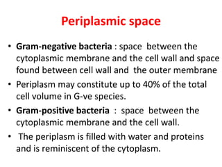
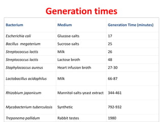
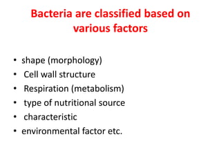
Bacteria have a simple structure compared to eukaryotic cells, lacking organelles. Their small size allows rapid growth and inhabitation of diverse environments. Bacterial cells contain a cytoplasm surrounded by a cell membrane and cell wall. The cytoplasm holds the circular chromosome, ribosomes for protein production, and storage structures. Some bacteria have flagella for mobility or pili for attachment. Gram-positive bacteria have a thick peptidoglycan cell wall, while Gram-negatives have a thin wall and an outer membrane. This membrane structure contributes to differences in antibiotic susceptibility between Gram-positive and Gram-negative bacteria.
When fresh liquid medium is inoculated with a given number of bacteria and incubated for sufficient period of time, it gives a characteristic growth pattern of bacteria.
If the bacterial population is measured periodically and log of number of viable bacteria is plotted in a graph against time, it gives a characteristic growth curve which is known as growth curve or growth cycle.
Bacteria are microscopic, single-celled organisms that thrive in diverse environments. These organisms can live in soil, the ocean and inside the human gut. Humans' relationship with bacteria is complex. Sometimes bacteria lend us a helping hand, such as by curdling milk into yogurt or helping with our digestion.
The physical factors affects the growth of microorganism.
1) Temperature
Temperature is the most important factor that influences the rate of enzyme catalysed reactions and rate of growth.
For every organisms there is an optimum temperature for growth and minimum temperature for inhibiting the growth.
Most extreme the microbes need liquid water to grow.(330C).
some algae and fungi grow at 55-60 degreeC.
Prokaryotes are grow at 100 degreeC.
Based on temperature the microorganisms are classified into two 4.
Classifications of Fungi
Characteristics of all Fungi
Structure of Fungi
Reproduction
Classification of Fungi
Basidiomycota
sexual reproduction occur by basidium , will be present spore is called basidiospore .
Asexual by budding ,fragementation, conidiospores.
Ascomycota
microscopic sexual structure in which nonmotile spores, called ascospores.
Mostly the ascomycota is sexual but some asexual it lacks the ascospore.
Zygomycota
Two spore
mitospores ( or) sporangiospore
chlamitospore (or) zygospore
Deuteromycota
Imperfect Fungi referring to our "imperfect" knowledge of their complete life cycles.
sexual life cycle that is either unknown or absent.
Asexual reproduction is by means of conidia or may be lacking.
culture media
SDA medium – sabouraud dextrose agar
A Power point presentation on General Features of Prokaryotes.
This ppt covers brief information of "General Features of Prokaryotes" useful for introduction lecture as well as for seminar purpose.
Bacteria are unicellular, procaryotic microorganisms which have diverse shape size and structures. Bacteria are found almost everywhere on Earth. Even the human body is full of bacteria, and in fact is estimated to contain more bacterial cells than human cells. Most bacteria in the body are harmless, and some are even helpful. A relatively small number of species cause disease.
Cell Anatomy and physiology ( structure and function for NEET asparients, Biology, MBBS, BPT, Allied, nursing , medical and paramedical students. This is the easiest form of slide share to understand the context better.
Biology Class 11 Chapter 8
FOR FURTHER DETAILS YOU CAN WATCH THE RELATED VIDEO AT THE GIVEN LINK
https://www.youtube.com/channel/UCxo06Nj-QWo_7SNvMyDnJCQ?view_as=subscriber
Chromatography: Principle, types, application.
A complete description of Chromatography along with all the types including HPLC, GAS, COLUMN, ION EXCHANGE, AFFINITY, COLUMN, PAPER, THIN LAYER CHROMATOGRAPHY - Techniques, Steps, principles, application.
Meat : Structure, Composition and Characteristics.Umesh Maskare
Meat - General introduction about meat, production and consumption in all over the World, its Complete structure and Composition with data and Characteristic Properties.
Observation of Io’s Resurfacing via Plume Deposition Using Ground-based Adapt...Sérgio Sacani
Since volcanic activity was first discovered on Io from Voyager images in 1979, changes
on Io’s surface have been monitored from both spacecraft and ground-based telescopes.
Here, we present the highest spatial resolution images of Io ever obtained from a groundbased telescope. These images, acquired by the SHARK-VIS instrument on the Large
Binocular Telescope, show evidence of a major resurfacing event on Io’s trailing hemisphere. When compared to the most recent spacecraft images, the SHARK-VIS images
show that a plume deposit from a powerful eruption at Pillan Patera has covered part
of the long-lived Pele plume deposit. Although this type of resurfacing event may be common on Io, few have been detected due to the rarity of spacecraft visits and the previously low spatial resolution available from Earth-based telescopes. The SHARK-VIS instrument ushers in a new era of high resolution imaging of Io’s surface using adaptive
optics at visible wavelengths.
Cancer cell metabolism: special Reference to Lactate PathwayAADYARAJPANDEY1
Normal Cell Metabolism:
Cellular respiration describes the series of steps that cells use to break down sugar and other chemicals to get the energy we need to function.
Energy is stored in the bonds of glucose and when glucose is broken down, much of that energy is released.
Cell utilize energy in the form of ATP.
The first step of respiration is called glycolysis. In a series of steps, glycolysis breaks glucose into two smaller molecules - a chemical called pyruvate. A small amount of ATP is formed during this process.
Most healthy cells continue the breakdown in a second process, called the Kreb's cycle. The Kreb's cycle allows cells to “burn” the pyruvates made in glycolysis to get more ATP.
The last step in the breakdown of glucose is called oxidative phosphorylation (Ox-Phos).
It takes place in specialized cell structures called mitochondria. This process produces a large amount of ATP. Importantly, cells need oxygen to complete oxidative phosphorylation.
If a cell completes only glycolysis, only 2 molecules of ATP are made per glucose. However, if the cell completes the entire respiration process (glycolysis - Kreb's - oxidative phosphorylation), about 36 molecules of ATP are created, giving it much more energy to use.
IN CANCER CELL:
Unlike healthy cells that "burn" the entire molecule of sugar to capture a large amount of energy as ATP, cancer cells are wasteful.
Cancer cells only partially break down sugar molecules. They overuse the first step of respiration, glycolysis. They frequently do not complete the second step, oxidative phosphorylation.
This results in only 2 molecules of ATP per each glucose molecule instead of the 36 or so ATPs healthy cells gain. As a result, cancer cells need to use a lot more sugar molecules to get enough energy to survive.
introduction to WARBERG PHENOMENA:
WARBURG EFFECT Usually, cancer cells are highly glycolytic (glucose addiction) and take up more glucose than do normal cells from outside.
Otto Heinrich Warburg (; 8 October 1883 – 1 August 1970) In 1931 was awarded the Nobel Prize in Physiology for his "discovery of the nature and mode of action of the respiratory enzyme.
WARNBURG EFFECT : cancer cells under aerobic (well-oxygenated) conditions to metabolize glucose to lactate (aerobic glycolysis) is known as the Warburg effect. Warburg made the observation that tumor slices consume glucose and secrete lactate at a higher rate than normal tissues.
Earliest Galaxies in the JADES Origins Field: Luminosity Function and Cosmic ...Sérgio Sacani
We characterize the earliest galaxy population in the JADES Origins Field (JOF), the deepest
imaging field observed with JWST. We make use of the ancillary Hubble optical images (5 filters
spanning 0.4−0.9µm) and novel JWST images with 14 filters spanning 0.8−5µm, including 7 mediumband filters, and reaching total exposure times of up to 46 hours per filter. We combine all our data
at > 2.3µm to construct an ultradeep image, reaching as deep as ≈ 31.4 AB mag in the stack and
30.3-31.0 AB mag (5σ, r = 0.1” circular aperture) in individual filters. We measure photometric
redshifts and use robust selection criteria to identify a sample of eight galaxy candidates at redshifts
z = 11.5 − 15. These objects show compact half-light radii of R1/2 ∼ 50 − 200pc, stellar masses of
M⋆ ∼ 107−108M⊙, and star-formation rates of SFR ∼ 0.1−1 M⊙ yr−1
. Our search finds no candidates
at 15 < z < 20, placing upper limits at these redshifts. We develop a forward modeling approach to
infer the properties of the evolving luminosity function without binning in redshift or luminosity that
marginalizes over the photometric redshift uncertainty of our candidate galaxies and incorporates the
impact of non-detections. We find a z = 12 luminosity function in good agreement with prior results,
and that the luminosity function normalization and UV luminosity density decline by a factor of ∼ 2.5
from z = 12 to z = 14. We discuss the possible implications of our results in the context of theoretical
models for evolution of the dark matter halo mass function.
The increased availability of biomedical data, particularly in the public domain, offers the opportunity to better understand human health and to develop effective therapeutics for a wide range of unmet medical needs. However, data scientists remain stymied by the fact that data remain hard to find and to productively reuse because data and their metadata i) are wholly inaccessible, ii) are in non-standard or incompatible representations, iii) do not conform to community standards, and iv) have unclear or highly restricted terms and conditions that preclude legitimate reuse. These limitations require a rethink on data can be made machine and AI-ready - the key motivation behind the FAIR Guiding Principles. Concurrently, while recent efforts have explored the use of deep learning to fuse disparate data into predictive models for a wide range of biomedical applications, these models often fail even when the correct answer is already known, and fail to explain individual predictions in terms that data scientists can appreciate. These limitations suggest that new methods to produce practical artificial intelligence are still needed.
In this talk, I will discuss our work in (1) building an integrative knowledge infrastructure to prepare FAIR and "AI-ready" data and services along with (2) neurosymbolic AI methods to improve the quality of predictions and to generate plausible explanations. Attention is given to standards, platforms, and methods to wrangle knowledge into simple, but effective semantic and latent representations, and to make these available into standards-compliant and discoverable interfaces that can be used in model building, validation, and explanation. Our work, and those of others in the field, creates a baseline for building trustworthy and easy to deploy AI models in biomedicine.
Bio
Dr. Michel Dumontier is the Distinguished Professor of Data Science at Maastricht University, founder and executive director of the Institute of Data Science, and co-founder of the FAIR (Findable, Accessible, Interoperable and Reusable) data principles. His research explores socio-technological approaches for responsible discovery science, which includes collaborative multi-modal knowledge graphs, privacy-preserving distributed data mining, and AI methods for drug discovery and personalized medicine. His work is supported through the Dutch National Research Agenda, the Netherlands Organisation for Scientific Research, Horizon Europe, the European Open Science Cloud, the US National Institutes of Health, and a Marie-Curie Innovative Training Network. He is the editor-in-chief for the journal Data Science and is internationally recognized for his contributions in bioinformatics, biomedical informatics, and semantic technologies including ontologies and linked data.
Nutraceutical market, scope and growth: Herbal drug technologyLokesh Patil
As consumer awareness of health and wellness rises, the nutraceutical market—which includes goods like functional meals, drinks, and dietary supplements that provide health advantages beyond basic nutrition—is growing significantly. As healthcare expenses rise, the population ages, and people want natural and preventative health solutions more and more, this industry is increasing quickly. Further driving market expansion are product formulation innovations and the use of cutting-edge technology for customized nutrition. With its worldwide reach, the nutraceutical industry is expected to keep growing and provide significant chances for research and investment in a number of categories, including vitamins, minerals, probiotics, and herbal supplements.
2. STRUCTURE OF BACTERIA
• Smaller and simpler in structure than
eukaryotic cells, with no recognizable
organelles.
• All of the activities performed by organelles
also take place in bacteria, but they are not
carried out by specialized structures.
• The small size, simple design, and broad
metabolic capabilities of bacteria allow them
to grow and divide very rapidly and to inhabit
and flourish in almost any environment.
3. STRUCTURE OF BACTERIA
• They were first seen under a microscope by Anton
van Leeuwenhoek in 1676.
• As microscopes have improved, scientists have
come to understand bacterial cell structure better.
4.
5.
6. Bacterial cell structure
• organized into 3 categories :
• Internal Structures: Cytoplasm, nucleoid,
bacterial chromosome, plasmid, ribosomes, and
storage granules
• Cell envelope: cell membrane, peptidoglycan cell
wall or an outer lipid membrane (only found in
Gram-negative cells)
• External structures (appendages & coverings):
flagella, fimbriae, sex pilus and glycocalyx
8. Cytoplasm
• Portion of the cell that lies within the PM
• substances within the plasma membrane, excluding
the genetic material.
• Gel-like matrix composed of mostly water(4/5 th ),
enzymes, nutrients, wastes, and gases
• Contains cell structures - ribosomes, chromosome,
and plasmids , as well as the components necessary
for bacterial metabolism.
• It is relatively featureless by electron microscope -
although small granules can be seen.
• carries out very important functions for the cell -
growth, metabolism, and replication .
9. Constituents
– Proteins including enzymes
– Vitamins
– Ions
– Nucleic acids and their precursors
– Amino acids and their precursors
– Sugars, carbohydrates and their derivatives
– Fatty acids and their derivatives
10. Nucleoid
• Unlike the eukaryotic (true) cells, bacteria do not
have a membrane enclosed nucleus.
• The nucleoid is a region of cytoplasm where the
chromosomal DNA is located.
• It is not a membrane bound nucleus, but simply an
area of the cytoplasm where the strands of DNA
are found.
11. Plasmids
• small extra-chromosomal DNA
• contain genes for antibiotic resistance or virulence.
• Structure Similar to most bacterial chromosomes, but
considerably smaller.
• plasmids are covalently closed circular DNA
• In a few species linear plasmids have been found.
• Size : Chromosomal DNA is typically about 4000 kb,
• plasmid DNA ranges from 1-200 kb.
• Number of plasmids: 1-700 copies of plasmid in a
cell.
12. Plasmid Function
• The function of plasmids is not always known, but they
are not normally essential for survival of host, although
their presence generally gives the host some
advantage.
• Antibiotic resistance - Some plasmids code for proteins
that degrade antibiotics-a big advantage for pathogens.
• Some encode for proteins which confer virulence
factors on the host. For example- E. coli plasmid Ent
P307 codes for an enterotoxin which makes E. coli
pathogenic.
• Conjugative plasmids - These allow exchange of DNA
between bacterial cells.
13. Plasmids
• Plasmids and the associated traits can be transferred
between bacteria, even from one bacterial species to
another.
• Plasmids are not involved in reproduction.
• Plasmids replicate independently of the chromosome.
• Plasmids are passed to other bacteria by two means.
• For most plasmid types, copies in the cytoplasm are
passed on to daughter cells during binary fission.
14. Plasmids
• Other types of plasmids ,form tube like structure at the
surface called a pilus that passes copies of the plasmid
to other bacteria during conjugation, a process by
which bacteria exchange genetic information.
• Plasmids have been shown to be instrumental in the
transmission of special properties, such as antibiotic
drug resistance, resistance to heavy metals, and
virulence factors necessary for infection of animal or
plant hosts.
• The ability to insert specific genes into plasmids have
made them extremely useful tools in the area of
genetic engineering/RDNA Technology .
15. Ribosomes- protein synthesis machinery
• Consists of RNA and protein
• Abundant in cytoplasm
• Often grouped in long chains called polyribosomes.
• give the cytoplasm of bacteria a granular appearance
in EM.
• smaller than the ribosomes in eukaryotic cells-but
have a similar function
• Bacterial ribosomes have sedimentation rate of 70S;
their subunits have rates of 30S and 50S.
• The unit used to measure sedimentation velocity is
Svedberg
16.
17. Ribosomes
• They translate the genetic code from the molecular language of nucleic
acid to that of amino acids—the building blocks of proteins.
• Bacterial ribosomes are similar to those of eukaryotes, but are smaller and
have a slightly different composition and molecular structure.
• Bacterial ribosomes are never bound to other organelles as they
sometimes are bound to the endoplasmic reticulum in eukaryotes, but are
free-standing structures distributed throughout the cytoplasm.
• There are sufficient differences between bacterial ribosomes and
eukaryotic ribosomes that some antibiotics will inhibit the functioning of
bacterial ribosomes, but not a eukaryote's, thus killing bacteria but not the
eukaryotic organisms they are infecting.
• Streptomycin binds 70S ribosome and stops protein synthesis but it can
not bind 80S ribosome of eukaryotes and thereby eukaryotic cell remains
unaffected.
18. Bacterial Chromosome - Genophore
• The bacterial chromosome consists of a single,
circle of deoxyribonucleic acid.
• DNA is double stranded- two strands line up
antiparrallel to each other and the bases are
linked together with hydrogen bonds.
• It includes most of the genetic material of the
organism .
19.
20. Bacterial Chromosome
• Unlike the DNA in eukaryotic cells, which resides
in the nucleus, DNA in bacterial cells is not
sequestered in a membrane-bound organelle but
appears as a long coil distributed through the
cytoplasm.
• In many bacteria the DNA is present as a single,
circular chromosome and in some cases the DNA
is linear rather than circular.
• some bacteria may contain two chromosomes
21. Bacterial Chromosome
• As in all organisms, bacterial DNA contains the
four nitrogenous bases adenine (A), cytosine
(C), guanine (G), and t
• The amount of DNA in bacterial chromosomes
ranges from 580,000 base pairs in
Mycoplasma gallinarum to 4,700,000 base
pairs in E. coli to 9,140,000 base pairs in
Myxococcus xanthus.
22. Inclusion bodies
• Inclusion bodies: Bacteria can have within their
cytoplasm a variety of small bodies collectively referred
to as inclusion bodies.
• Some are called granules and other are called vesicles.
• Inclusions are considered to be nonliving components
of the cell that do not possess metabolic activity and
are not bounded by membranes.
• The most common inclusions are glycogen, lipid
droplets, crystals, and pigments.
23. Inclusion bodies - Granules
• Granules: Densely compacted substances without a
membrane covering.
• Nutrients and reserves may be stored in the cytoplasm
in the form of glycogen, lipids, polyphosphate, or in
some cases, sulfur or nitrogen for later use.
• Each granule contains specific substances, such as
glycogen (glucose polymer) and polyphosphate
(phosphate polymer, supplies energy to metabolic
processes).
• Sulfur bacteria contains reserve granules of sulfur.
• These granules are depleted in starvation.
24. Inclusion bodies-vesicles
• Some aquatic photosynthetic bacteria and cyano
bacteria have rigid gas-filled vacuoles and it helps in
floating at a certain level - allowing them to move
up or down into water layers with different light
intensities and nutrient levels.
• Some magnetotactic bacterium, eg. Aquaspirillium
magnetotacticum , stores Magnetitite (Ferric oxide).
The presence of such magnetic inclusions enables
these bacteria to responds to magnetic fields.
25. Microcompartments
• Microcompartments are widespread, membrane-
bound organelles that are made of a protein shell
that surrounds and encloses various enzymes.
• Carboxysomes are protein-enclosed bacterial
microcompartments that contain enzymes involved
in carbon fixation.
• Magnetosomes are bacterial microcompartments,
present in magnetotactic bacteria, that contain
magnetic crystals.
27. Plasma Membrane
• Phospholipid bilayer surrounding the cytoplasm and
regulates the flow of substances in and out of the cell.
• Consists of both lipids and proteins.
• Protects the cell from its surroundings.
• Selectively permeable to ions and organic molecules
and controls the movement of substances in and out.
• numerous proteins moving within or upon this layer are
primarily responsible for transport of ions, nutrients
and waste across the membrane.
28.
29.
30. Periplasmic space
• Gram-negative bacteria : space between the
cytoplasmic membrane and the cell wall and space
found between cell wall and the outer membrane
• Periplasm may constitute up to 40% of the total
cell volume in G-ve species.
• Gram-positive bacteria : space between the
cytoplasmic membrane and the cell wall.
• The periplasm is filled with water and proteins
and is reminiscent of the cytoplasm.
31. Periplasmic Space
• However periplasm contains proteins and other
molecules distinct from those in the cytoplasm
because the membrane prevents the free exchange
between these two compartments.
• Periplasmic proteins have various functions in
cellular processes including: transport, degradation,
and motility.
• Periplasm controls molecular traffic entering and
leaving the cell.
32.
33.
34. Cell wall
• Outer covering of most cells that protects the
bacterial cell and gives it shape (spherical, rod and
spiral).
• Composed of peptidoglycan (polysaccharides +
protein)
• Mycoplasma are bacteria that have no cell wall
and therefore have no definite shape.
35. Cell wall
• Peptidoglycan - molecule found only in bacterial cell
walls.
• The rigid structure of peptidoglycan gives the bacterial
cell shape, surrounds the plasma membrane and
provides prokaryotes with protection from the
environment
• From the peptidoglycan inwards all bacterial cells are
similar.
• Going further out, the bacterial world divides into two
major classes: Gram-positive and Gram-negative .
• Amount and location of peptidoglycan in the cell wall
determines whether a bacterium is G+ve or G-ve
36. Peptidoglycan = (polysaccharides + protein),
• Peptidoglycan (murein) - huge polymer of interlocking
chains composed of similar monomers.
• peptidoglycan is made from polysaccharide chains
cross-linked by peptides containing D-amino acids
• The backbone of the peptidoglycan molecule is
composed of two derivatives of glucose:
• N-acetylglucosamine (NAG)
• N-acetlymuramic acid (NAM).
• The NAG and NAM strands are connected by inter
peptide bridges.
37.
38.
39. Gram-positive Cells
• G+ve bacteria possess thick cell wall containing
many layers of peptidoglycan and teichoic acids.
• In G+ ve cells, peptidoglycan is the outermost
structure and makes up as much as 90% of the
thick compact cell wall.
40. Gram-negative
• G-ve bacteria have relatively thin cell wall
consisting of few layers of peptidoglycan
surrounded by a second lipid membrane containing
lipopolysaccharides and lipoproteins
• Peptidoglycan makes up only 5 – 20% of the cell
wall and is not the outermost layer, but lies
between the plasma membrane and an outer
membrane.
41. Gram Staining
• Developed in 1884 by Danish scientist Christian
Gram.
• It is a differential stain.
• In this, bacteria are first stained with crystal violet,
then treated with a mordant - a solution that fixes
the stain inside the cell.
• Bacteria are then washed with a decolorizing agent,
such as alcohol, and counterstained with safranin, a
light red dye.
42. Gram Staining
• Gram-positive bacteria are those that are stained
dark blue or violet by Gram staining.
• Gram-negative bacteria cannot retain the crystal
violet stain, instead take up the counterstain and
appearred or pink.
• The walls of gram-positive bacteria have more
peptidoglycans than do gram-negative bacteria.
Thus, gram-positive bacteria retain the original
violet dye and cannot be counterstained.
43. Cell wall
• If the bacterial cell wall is entirely removed, it is
called a protoplast while if it's partially removed, it
is called a spheroplast.
• Antibiotics such as penicillin inhibit the formation of
peptidoglycan cross-links in the bacterial cell wall.
• The enzyme lysozyme, found in human tears, also
digests the cell wall of bacteria and is the body's
main defense against eye infections.
44. outer membrane
• Similar to the plasma membrane, but is less permeable .
• This membrane has tiny holes or openings called porins.
• Porins block the entrance of harmful chemicals and antibiotics,
making G-ve bacteria much more difficult to treat than G+ve cells.
• Composed of lipopolysaccharides (LPS).
• LPS is a harmful substance classified as an endotoxin.
• Lipopolysaccharides, which acts as an endotoxin, are composed of
polysaccharides and lipid A (responsible for much of the toxicity of
G-ve bacteria).
• These differences in structure can produce differences in antibiotic
susceptibility
• Ex: vancomycin can kill only Gram +ve bacteria and is ineffective
against Gram -ve pathogens, such as Haemophilus influenzae or
Pseudomonas aeruginosa.
46. Flagella
• Singular: flagellum
• Long, whip-like semi-rigid cylindrical structures that
aids in cellular locomotion
• Function much like the propeller on a ship.
• about 20 nm in diameter and up to 20 micromts in
length.
• Diameter of a prokaryotic flagellum is about 1/10 th
of that of eukaryotic.
• Flagella are driven by the energy released by the
transfer of ions down an electrochemical gradient
across the cell membrane.
47. Flagella
• Made up of protein subunits called flagellin.
• Each flagellum is attached to cell membrane with
the help of proteins other than flagellin.
• The basal region has a hook like structure and a
complex basal body. The basal body consists of a
central rod or shaft surrounded by a set of rings.
48. Flagella
• Bacterial spp differ in the number and arrangement
of flagella on their surface.
• Bacteria may have one, a few, or many flagella in
different positions on the cell.
• Monotrichous - single flagellum
• amphitrichous a flagellum at each end
lophotrichous - clusters of flagella at the poles of
the cell
• peritrichous - flagella distributed over the entire
surface of the cell .
51. Flagella
• Motile bacteria are attracted or repelled by certain
stimuli in behaviors called taxis: these include
chemotaxis, phototaxis, and magnetotaxis.
• The flagella beat in a propeller-like motion to help
the bacterium move toward nutrients; away from
toxic chemicals; towards the light (photosynthetic
cyanobacteria).
• Prokaryotes exhibit a variety of movements:
move , swim ,tumble ,glide, swarm in response to
environmental stimuli.
52. FIMBRIAE AND PILI
• Hollow, hair like structures made of protein
• Involved in attachment to solid surfaces or to other
cells and are essential for the virulence of some
bacterial pathogens.
• Fimbriae fine filaments of protein just 2–10 nm in
diameter and up to several micrometers in length.
• They are distributed over the surface of the cell,
and resemble fine hairs when seen under the
electron microscope.
53. FIMBRIAE AND PILI
• Pili: (sing. pilus) are cellular appendages, slightly
larger than fimbriae
• Involved in attachment to surfaces.
• Specialized pili, the sex pili, allows the transfer of
genetic material from one bacteria to another in a
process called conjugation where they are called
conjugation pili or "sex pili".
• type IV pili - generate movement.
• Helps in colonization and pathogenicity.
54. Glycocalyx
• Glycocalyx : sticky coating produced by many
bacteria covering the surface of cell.
• The glycocalyx is composed of polysaccharides
(sugars) and proteins.
• The bacterial glycocalyx has 2 forms
• a highly structured rigid capsule
• a disorganised loose slime layer -
• Capsules are found on many pathogenic bacteria
55. Glycocalyx
• The glycocalyx has several functions including :
protection, attachment to surfaces and formation
of biofilms.
• The glycocalyx helps protect the bacteria cell by
preventing immune cells from attaching to it and
destroying it through phagocytosis.
56. Bacterial reproduction
• Cell growth and reproduction by cell division are
tightly linked in unicellular organisms.
• Bacteria grow to a fixed size and then reproduce
through binary fission, a form of asexual reproduction
• Under optimal conditions, bacteria can grow and
divide extremely rapidly, and bacterial populations can
double as quickly as every 9.8 minutes.
• In cell division, two identical clone daughter cells are
produced.
• Budding involves a cell forming a protrusion that
breaks away and produces a daughter cell
57. Binary fission
• Most prokaryotes reproduce by a process of binary fission, in
which the cell grows in volume until it divides in half to yield two
identical daughter cells.
• Each daughter cell can continue to grow at the same rate as its
parent.
• For this process to occur, the cell must grow over its entire
surface until the time of cell division, when a new hemispherical
pole forms at the division septum in the middle of the cell.
• The septum grows inward from the plasma membrane along the
midpoint and forms as the side wall which pinches inward,
dividing the cell in two.
• In order for the cell to divide in half, the peptidoglycan structure
must be different in the hemispherical cap than in the straight
portion of the cell wall, and different wall-cross-linking enzymes
must be active at the septum than elsewhere.
58. Binary fission
• Binary fission begins with the single DNA molecule
replicating and both copies attaching to the cell
membrane.
• Next, the cell membrane begins to grow between
the two DNA molecules. Once the bacterium just
about doubles its original size, the cell membrane
begins to pinch inward.
• A cell wall then forms between the two DNA
molecules dividing the original cell into two identical
daughter cells
59.
60. Budding
• A group of environmental bacteria reproduces by budding.
• In this process a small bud forms at one end of the mother
cell
• As growth proceeds, the size of the mother cell remains
about constant, but the bud enlarges.
• When the bud is about the same size as the mother cell, it
separates. This type of reproduction is analogous to that in
budding fungi, such as brewer’s yeast (Saccharomyces
cerevisiae).
• One difference between fission and budding is that, in the
latter, the mother cell often has different properties from the
offspring.
• Ex: In some strains, mother cells have a flagellum and are
motile, whereas the daughter buds lack flagella.
65. Three mechanisms of genetic recombination
• Conjugation
• Transformation
• Transduction
66. CONJUGATION
• Two bacterial cells come together and mate such that a gene transfer occurs
between them.
• Can only occur between cells of opposite mating types.
– The donor (or "male") carries a fertility factor (F+).
– The recipient ("female") does not (F−).
• One cell, the donor cell (F+), gives up DNA; and another cell, the recipient
cell (F−), receives the DNA.
• The transfer is nonreciprocal, and a special pilus called the sex pilus joins
the donor and recipient during the transfer.
• The channel for transfer is usually a special conjugation tube formed during
contact between the two cells.
• The DNA most often transferred is a copy of the F factor plasmid.
• The factor moves to the recipient, and when it enters the recipient, it is
copied to produce a double-stranded DNA for integration.
67.
68. BACTERIAL TRANSFORMATION
• Discovered by Frederick Griffith in 1928.
• Many bacteria can acquire new genes by taking up DNA
molecules (ex: plasmid) from their surroundings.
• When bacteria undergo lysis, they release considerable
amounts of DNA into the environment.
• This DNA may be picked up by a competent cell- one
capable of taking up the DNA and undergoing a
transformation.
• To be competent, bacteria must be in the logarithmic
stage of growth, and a competence factor needed for the
transformation must be present.
69. BACTERIAL TRANSDUCTION
• Bacterial viruses ( bacteriophages) transfer
DNA fragments from one bacterium (the
donor) to another bacterium (the recipient).
• The viruses involved contain a strand of DNA
enclosed in an outer coat of protein.
70.
71. After a bacteriophage enters a bacterium, it may encourage the
bacterium to make copies of the phage.
At the conclusion of the process, the host bacterium undergoes
lysis and releases new phages. This cycle is called the lytic cycle.
Under other circumstances, the virus may attach to the bacterial
chromosome and integrate its DNA into the bacterial DNA. It may
remain here for a period of time before detaching and continuing its
replicative process. This cycle is known as the lysogenic cycle.
Under these conditions, the virus does not destroy the host
bacterium, but remains in a lysogenic condition with it. The virus is
called a temperate phage, also known as a prophage.
At a later time, the virus can detach, and the lytic cycle will
ensue.
It will express not only its genes, but also the genes acquired
from the donor bacterium.
73. Bacterial Growth
• Growth of Bacteria is the orderly increase of
all the chemical constituents of the bacteria.
• Multiplication is the consequence of growth.
• Death of bacteria is the irreversible loss of
ability to reproduce.
74. Generation /doubling time
• Generation time (g) : The time it takes the cells to
double.
• The average generative time is about 20-30 minutes
in majority of medically important bacteria.
• They are some exceptions among pathogenic
bacteria.
• Mycobacterium tuberculosis - 18 hrs.
• Mycobacterium leprae -10-20 days
• Length of generative time is in direct dependence
on the length of incubation period of infections.
75. Growth Kinetics
• Bacterial growth follows four phases.
• lag phase
• log phase
• stationary phase
• death phase
76.
77. Lag phase
• Immediately following the seeding of a culture medium.
• A period of adaptation for the cells to their new environment
• cells are adapting to the high-nutrient environment and
preparing for fast growth.
• The lag phase has high biosynthesis rates, as proteins
and metabolic intermediates are built up in adequate
quantities for rapid growth & multiplication to proceed.
• New enzymes are synthesized.
• A slight increase in cell mass and volume, but no increase in
cell number.
78. Duration of the lag phase varies with
- the species
- size of inoculum - Prolonged by low inoculum volume,
poor inoculum condition (high % of dead cells)
- age of inoculum
- Nature of the culture medium (Prolonged by nutrient-
poor medium)
- And environmental factors like temperature, pH etc
79. Log/Exponential growth phase
• In this phase, the cells have adjusted to their new
environment and multiply rapidly (exponentially)
• The bacteria will grow and divide at a doubling time
characteristic of the strains and determined by the
conditions during the exponential phase.
• During this phase, the number of bacteria will increase to
2n, in which n is the no.of generations.
• Balanced growth –all components of a cell grow at the
same rate.
80. Deceleration growth phase
Very short phase, during which growth decelerates due
to either:
• Depletion of one or more essential nutrients
• The accumulation of toxic by-products of growth (e.g.
Ethanol in yeast fermentations)
• Period of unbalanced growth: Cells undergo internal
restructuring to increase their chances of survival
81. Stationary Phase
With the exhaustion of nutrients or build-up of toxic waste
substances and secondary metabolic products in the
medium , the bacteria stop growing and enter the
stationary phase.
- The growth rate equals the death rate – The number of progeny cells
formed is just enough to replace the number of cells that die.
- There is no net growth in the organism population – The viable count
remains stationary as an equilibrium exists between the dying cells
and newly formed cells.
82. Death Phase
- Phase of decline
- The living organism population decreases with time,
due to a lack of nutrients and accumulation of toxic
metabolic by-products.
- Cell death may also be caused by autolytic enzymes.
86. Nutrients
• Nutrients in growth media must contain all the
elements necessary for the synthesis of new
organisms.
• Hydrogen donors and acceptors
• Carbon source
• Nitrogen source
• Minerals : sulphur and phosphorus
• Growth factors: amino acids, purines, pyrimidines;
vitamins
• Trace elements: Mg, Fe, Mn.
87.
88. • Microorganisms are sensitive to temperature changes
– Usually unicellular
– Enzymes have temperature optima
– If temperature is too high, proteins denature, including
enzymes, carriers and structural components
• Temperature ranges are enormous (-20 to 100oC)
Temperature
89. Temperature
– Organisms exhibit distinct cardinal temperatures
(minimal, maximal, and optimal growth temps)
– If an organism has a limited growth temperature
range = stenothermal (e.g. N. gonorrhoeae)
– If an organism has a wide growth temperature
range = eurythermal (E. faecalis)
90. Temperature
Psychrophiles can grow well at 0oC, have
optimal growth at 15oC or lower, and
usually will not grow above 20oC
• Arctic/Antarctic ocean
• Protein synthesis, enzymatic activity and
transport systems have evolved to function at
low temperatures
• Cell walls contain high levels of unsaturated fatty
acids (semi-fluid when cold)
91. Temperature
– Psychrotrophs can also grow at 0oC, but have growth
optima between 20oC and 30oC, and growth maxima at
about 35oC
• Many are responsible for food spoilage in refrigerators
– Mesophiles have growth minima of 15 to 20oC, optima of
20 to 45oC, and maxima of about 45oC or lower
• Majority of human pathogens
92. Temperature
–Thermophiles have growth minima around
45oC, and optima of 55 to 65oC
• Hot springs, hot water pipes, compost heaps
• Lipids in PM more saturated than mesophiles.
–Hyperthermophiles have growth minima
around 55oC and optima of 80 to 110oC
• Sea floor, sulfur vents
95. pH
– pH is the negative logarithm of the hydrogen
ion concentration
– Acidophiles grow best between pH 0 and 5.5
– Neutrophiles grow best between pH 5.5 and 8.0
– Alkalophiles grow best between pH 8.5 and 11.5
– Extreme alkalophiles grow best at pH 10.0 or higher
96. pH
–Sudden pH changes can inactivate enzymes
and damage plasma membrane
• Reason for buffering culture medium, usually
with a weak acid/conjugate base pair (e.g.
KH2PO4/K2HPO4 – monobasic potassium/dibasic
potassium)
99. Oxygen concentration
– Obligate aerobes are completely dependent
on atmospheric O2 for growth
• Oxygen is used as the terminal electron acceptor
for electron transport in aerobic respiration
– Facultative anaerobes do not require O2 for
growth, but do grow better in its presence
– Aerotolerant anaerobes ignore O2 and grow
equally well whether it is present or not
100. Oxygen concentration
–Obligate (strict) anaerobes do not tolerate
O2 and die in its presence.
–Microaerophiles are damaged by the
normal atmospheric level of O2 (20%) but
require lower levels (2 to 10%) for growth
101. Oxygen and growth
Environment
Group Aerobic Anaerobic O2 Effect
Obligate Aerobe Growth No growth Required (utilized for
aerobic respiration)
Microaerophile
Growth if
level not
too high
No growth
Required but at levels
below 0.2 atm
Obligate Anaerobe No growth Growth Toxic
Facultative
(An)aerobe
Growth Growth
Not required for growth
but utilized when available
Aerotolerant
Anaerobe
Growth Growth Not required and not
utilized
103. Water availability
• Water is solvent for biomolecules, and its availability is
critical for cellular growth
• The availability of water depends upon its presence in
the atmosphere (relative humidity) or its presence in
solution or a substance (water activity, (Aw))
• Aw of pure water (100%) is 1.0; affected by dissolved
solutes such as salts or sugars.
• Microorganisms live over a range of aW from 1.0 to 0.7.
The aW of human blood is 0.99; seawater = 0.98; maple
syrup = 0.90; Great Salt Lake = 0.75. Water activities in
agricultural soils range between 0.9 and 1.0.
105. Pressure
–Barotolerant organisms are adversely
affected by increased pressure, but not as
severely as are nontolerant organisms
–Barophilic organisms require, or grow more
rapidly in the presence of increased
pressure
–Light: Optimum condition for growth is
darkness.
106. Radiation
-Ultraviolet radiation damages cells by causing
the formation of thymine dimers in DNA.
–Ionizing radiation such as X rays or gamma rays
are even more harmful to microorganisms than
ultraviolet radiation
• Low levels produce mutations and may
indirectly result in death
• High levels are directly lethal by direct
damage to cellular macromolecules or
through the production of oxygen free
radicals
107. (B) Sources of Metabolic Energy
• Mainly three mechanisms generate metabolic
energy. These are
• Fermentation
• Respiration and
• Photosynthesis.
An organism to grow, at least one of these
mechanisms must be used.
109. • Used to maintain cells in the exponential
growth phase at a constant biomass
concentration for extended periods of
time
• Conditions are met by continual provision
of nutrients and removal of wastes =
OPEN SYSTEM
• Constant conditions are maintained
111. • Balanced (exponential) growth occurs when
all cellular components are synthesized at
constant rates relative to one another
• Unbalanced growth occurs when the rate of
synthesis of some components change
relative to the rate of synthesis of other
components.
–This usually occurs when the environmental
conditions change
113. MORPHOLOGY
• Bacteria display a wide diversity of shapes and sizes called
morphologies
• Cannot be seen with human eyes (microscopic)
• Their presence was only first recognized in 1677, when the Dutch
naturalist Antonie van Leeuwenhoek saw microscopic organisms
in a variety of substances with the aid of primitive microscopes.
• Now bacteria are usually examined under light microscopes
capable of more than 1,000-fold magnification
• Details of their internal structure can be observed only with the
aid of much more powerful transmission electron microscopes.
• Unless special phase-contrast microscopes are used, bacteria
have to be stained with a coloured dye so that they will stand
out from their background.
114. Size
• Bacteria are the smallest living creatures
• Most bacteria are 0.2 um in diameter and 2-8 um in length.
• Bacterial cells are about one tenth the size of eukaryotic cells
• are typically 0.5 – 5.0 micrometres in length.
• Giant bacteria for example, Thiomargarita namibiensis,
Titanospirillum namibiensis and Epulopiscium fishelsoni — are
up to half a mm long and are visible to the unaided eye
• E. fishelsoni reaches 0.7 mm.
• Among the smallest bacteria are members of the genus
Mycoplasma, which measure about 0.1 to 0.25 μm in
diameter, as small as the largest viruses.
• Some bacteria may be even smaller, but these ultramicro
bacteria are not well-studied.
115. Size
• E. coli, a normal inhabitant of the intestinal tract of humans
and animals, is about 2 μm long and 0.5 μm in diameter
• spherical cells of Staphylococcus aureus - up to 1 μm in
diameter.
• the rod-shaped Bordetella pertussis, causative agent of
whooping cough - 0.2 to 0.5 μm in diameter and 0.5 to 1 μm
in length
• corkscrew-shaped Treponema pallidum, causative agent of
syphilis averaging only 0.15 μm in diameter but 10 to 13 μm
in length.
• Some bacteria are relatively large, such as Azotobacter, which
has diameters of 2 to 5 μm or more
• cyanobacterium Synechococcus averages 6 μm by 12 μm
• Achromatium, which has a minimum width of 5 μm and a
maximum length of 100 μm, depending on the species.
116. Cell Shape
• Bacteria come in a wide variety of shapes.
• Coccus – are spherical or oval cells.
• Bacillus - are round-ended cylinder shaped cells.
• Vibrios comma shaped ,curved rods and derive the
name from their characteristic vibratory motility
• Spirilla – are rigid spiral forms(coil).
• Spirochetes - are long, slender, and flexible spiral
forms(from speira meaning coil and chaite meaning
hair)
• Filamentous – resembles radiating rays of sun
117. Cell Shape
• coccobacilli - Some bacilli are so short and fat that
they look like cocci and are referred to as
coccobacilli.
• A small number of species even have tetrahedral
or cuboidal shapes.
• More recently, bacteria were discovered deep
under the Earth's crust that grow as long rods with
a star-shaped cross-section.
• The large surface area to volume ratio of this
morphology may give these bacteria an advantage
in nutrient-poor environments.
118. Cell Shape
• is generally characteristic of a given bacterial species
• but can vary depending on growth conditions.
• Some bacteria have complex life cycles involving the
production of stalks and appendages (e.g. Caulobacter)
and some produce elaborate structures bearing
reproductive spores (e.g. Myxococcus, Streptomyces).
• Bacteria generally form distinctive cell morphologies
when examined by light microscopy and distinct colony
morphologies when grown on Petri plates.
• These are often the first characteristics observed by a
microbiologist to determine the identity of an unknown
bacterial culture
119. Cell Shape
• This wide variety of shapes is determined by
the bacterial cell wall and cytoskeleton
• Shape of the cell is important because it can
influence the ability of bacteria to acquire
nutrients, attach to surfaces, swim through
liquids and escape predators
120. Arrangement of cells
• Cellular arrangements occur singularly, in pairs,
in chains and in clusters.
128. • Culture techniques are designed to promote the
growth and identify particular bacteria,while
restricting the growth of the other bacteria in the
sample.
• In the laboratory, bacteria are usually grown using
solid growth media such as agar plates or liquid
media such as broth.
• Solid, agar-based media can be used to identify
colonial characteristics (shape, size, elevation, margin
type) and to isolate pure cultures of a bacterial strain
• liquid growth media are used when measurement of
growth or large volumes of cells are required.
129. • Growth in stirred liquid media occurs as an even cell
suspension, making the cultures easy to divide and
transfer
• isolating single bacteria from liquid media is difficult.
• The use of selective media (media with specific
nutrients added or deficient, or with antibiotics added)
can help identify specific org’s.
• Most laboratory techniques for growing bacteria use
high levels of nutrients to produce large amounts of
cells cheaply and quickly.
• However, in natural environments nutrients are limited,
meaning that bacteria cannot continue to reproduce
indefinitely
130. Cultural characteristics
Basic conditions for cultivation
• Optimum environmental moisture. It is possible to
cultivate bacteria in liquid media or solid media with a
gelling agent (agar) binding about 90% of water.
• Optimum temperature for cultivation of bacteria of
medical importance is about 370C. Saprophytic bacteria
are able to grow at lower temperatures.
• Optimum pH of culture media is usually 7.2-7.4
Lactobacillus spp need acid pH and vibrio cholera needs
alkaline pH for the growth.
• Optimum constituents of bacteriological culture media.
• All culture media share a number of common constituents
necessary to enable bacteria to grow in vitro.
131. Optimum Quantity of oxygen in
cultivation environment.
• Bacteria obtain energy either by oxidation or by fermentation i.e.,
oxidation – reduction procedure without oxygen.
• Bacteria are classified into four basic groups according to their
relation to atmospheric oxygen:
• Obligate aerobes: Reproduce only in the presence of oxygen
• Facultative anaerobes : reproduce in both aerobic and anaerobic
environments. Their complete enzymatic equipment allows them
to live and grow in the presence or absence of oxygen.
• Obligate anaerobes: grow only in the absence of free oxygen (i,e
unable to grow and reproduce in the presence of oxygen). Some
species are so sensitive that they die if exposed to oxygen.
• Anaerobic Aerotolerant: microbes do not need oxygen for their
growth and it is not fatal for them
132. Colony morphology
• Form - the basic shape of the colony
ex: circular, filamentous etc.
• Size – The diameter of the colony.
• Elevation - This describes the side view of a colony.
Turn the Petri dish on end.
• Margin/border - magnified shape of the edge of the
colony
• Surface - colony appearance
ex: smooth, glistening, rough, wrinkled or dull.
• Opacity - ex transparent (clear), opaque, translucent
(like looking through frosted glass), etc.
• Colour (pigmentation) ex: white, buff, red, purple, etc.
133.
134. Colony morphology
• Colony morphology is a method that scientists use
to describe the characteristics of an individual
colony of bacteria growing on agar in a Petri dish. It
can be used to help to identify them.
• Each distinct colony represents an individual
bacterial cell or group that has divided repeatedly.
Being kept in one place, the resulting cells have
accumulated to form a visible patch.
• Most bacterial colonies appear white or a creamy
yellow in colour, and are fairly circular in shape.
135. Effect of media
• different types of media, which contain different
nutrients can affect the cultural characteristics of
bacteria.
• Some types of media are much more nutritive and will
encourage hearty growth. Some types of media may
restrict growth.
• Colonial morphology may also be affected by the
temperature at which the bacteria is incubated. Some
bacteria grow better at body temperature and grow
weakly at room temperature, or vice versa.
• Some bacteria express certain characteristics, such as
the formation of pigment, more strongly at some
temperatures than at others.
139. Bacterial Classification Based on Shapes
• Bacilli: Rod shaped bacteria.
• Diplobacilli, tetrad , palisade (two cells arranged parallel) or
sterptobacilli (chain arrangement). e.g. E.Coli and Salmonella
• Coccus: Spherical or oval cells shaped bacteria which is further
classified as monococcus, diplococci, streptococci ,Staphylcocci
e.g. Staphylococcus and Streptococcus
• Spiral: Spiral shaped bacteria are called spirilla
e.g. Treponema and Borellia
sub divided into spirilla (rigid spiral forms) and
spirochetes(flexible spiral forms).
• Comma shaped: Vibrio
• Branching filamentous forms : Actinomycetes
140. Bacterial Classification Based on
Staining Methods
• Gram positive bacteria - take up crystal violet dye
and retain their blue or violet color.
Gram negative bacteria - do not take up crystal
violet dye, and thus appear red or pink.
141. Classification Based on Respiration
• Aerobic Respiration : sugars are broken down in
the presence of oxygen to produce carbon dioxide,
water, and energy.
• Anaerobic Respiration : anaerobic respiration
breaks down sugars and releases energy in the
absence of oxygen.
• anaerobic respiration is typically slower and less
efficient than aerobic respiration.
• anaerobic respiration involves chemicals other
than oxygen and carbondioxide.
142. Classification Based on Respiration
• Facultative Anaerobic Respiration : Facultative
Anaerobes are able to perform either aerobic /
anaerobic respiration depending on the oxygen
content of their environment.
• Ex: Coliform bacteria
• Microaerophiles : sugars are broken down in the
presence of minute amounts of oxygen to
produce energy.
143. Classification Based on Environment
• Mesophiles - which require moderate temp to survive.
Neutrophiles - require moderate conditions to survive.
Extremophiles - can survive in extreme conditions.
Acidophiles - which can tolerate low pH conditions.
Alkaliphiles - which can tolerate high pH conditions.
Thermophiles - which can resist high temperature.
Psychrophiles - can survive extremely cold conditions.
Halophiles - can survive in highly saline conditions.
Osmophiles - can survive in high sugar osmotic
conditions.
144. Classification Based on Flagella
• Atrichous (no flagella),
• monotrichous (uni flagella)
• amphitrichous (bi flagella)
• polytrichous (more flagella)
147. Nutritional Source
• bacteria are also classified based on the type of
energy source utilized by them for survival.
• Autotrophs: obtain the carbon it requires from
carbon dioxide
• Photoautotrophs: directly use sunlight in order to
produce sugar from carbon dioxide.
• Chemoautotrophs : depend on various chemical
reactions.
• use inorganic energy sources, such as hydrogen
sulfide, elemental sulfur, ferrous iron, molecular
hydrogen, and ammonia.
148. Nutritional Source
• Heterotrophs : Heterotrophic bacteria obtain
sugar from the environment they are in (ex: the
living cells or organisms they are in).
• symbiotic
• saprophytes
• parasite
150. Classification of bacteria
• With over millions of bacteria present in the planet, it
is not an easy job to identify, isolate and study a
particular species or particular bacteria as such.
• Microbiologists categorized bacteria based on basic
and important factors making all the bacteria fall under
any one of the categories and thus making the process
of isolation and identification much easier.
• Bacteria are classified based on various factors like
shape (morphology), Cell wall structure, Respiration
(metabolism), type of nutritional source, characteristic
and environmental factor.
151. Bacteria are classified based on
various factors
• shape (morphology)
• Cell wall structure
• Respiration (metabolism)
• type of nutritional source
• characteristic
• environmental factor etc.
152. • Chemostat
– A continuous culture device that maintains a
constant growth rate by:
• supplying a medium containing a limited amount of an
essential nutrient at a fixed rate
• removing medium that contains microorganisms at the
same rate
– As fresh media is added to the chamber, bacteria
are removed
– Limiting nutrients control growth rates
– Cell density depends on nutrient concentration
153. • Turbidostat
A continuous culture device that regulates the flow rate of
media through the vessel in order to maintain a
predetermined turbidity or cell density
• There is no limiting nutrient
• Absorbance is measured by a photocell (optical sensing device)