Download as PDF, PPTX
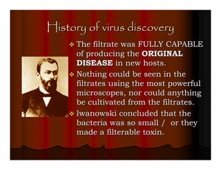
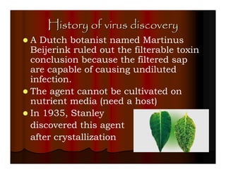
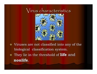

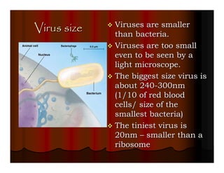
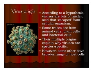
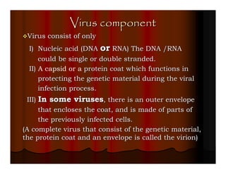
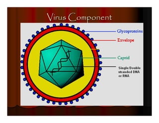
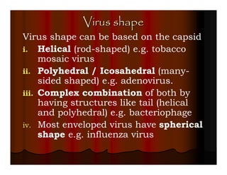
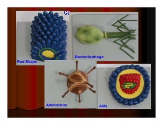

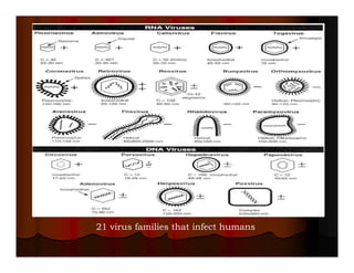
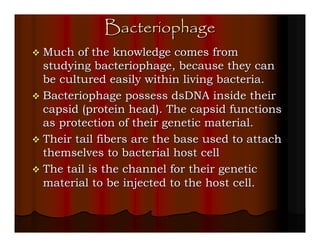
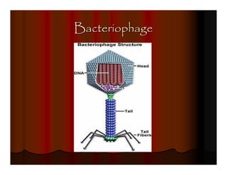
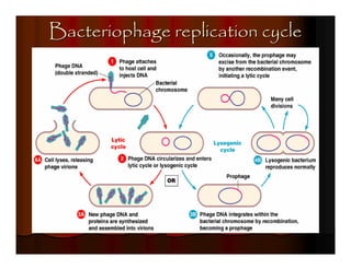
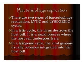
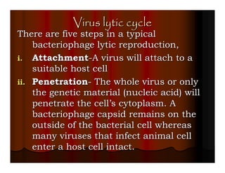
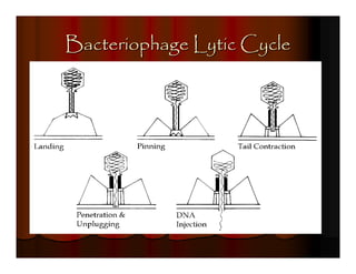
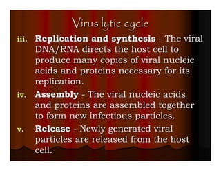
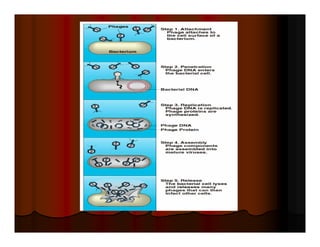
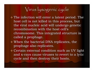
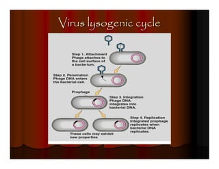
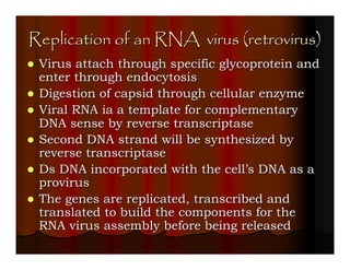
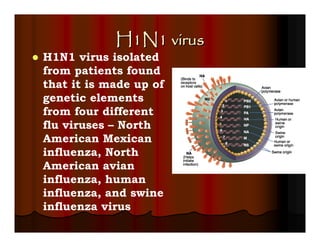
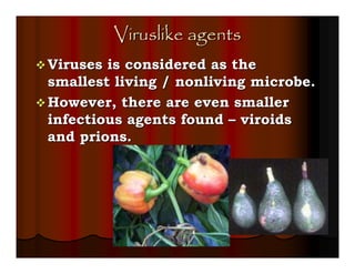
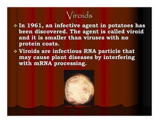
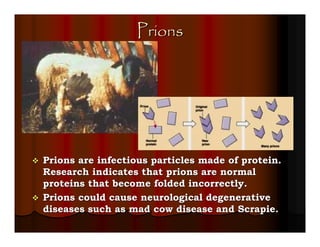

This document provides an overview of viruses, including their history of discovery, characteristics, components, shapes, classification, bacteriophages, replication cycles, enveloped viruses, and other related infectious agents like viroids and prions. It discusses key scientists and experiments that contributed to the understanding of viruses. The replication cycles of lytic and lysogenic bacteriophages as well as enveloped DNA and RNA viruses are described.


