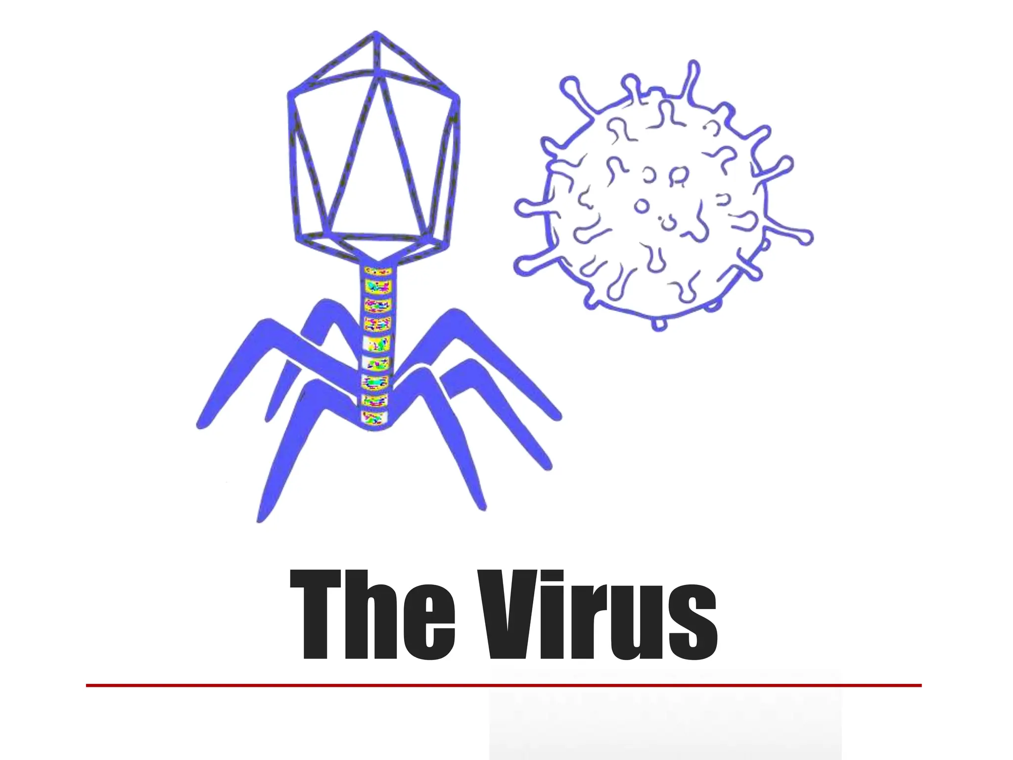
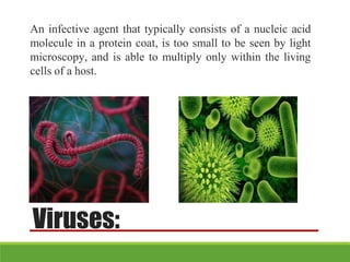
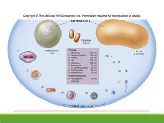
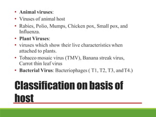
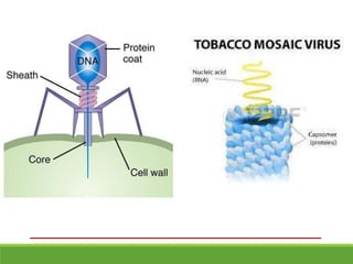
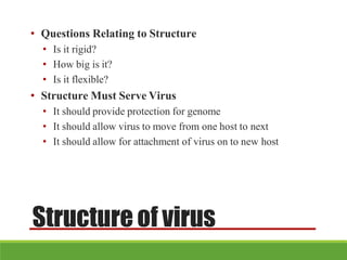
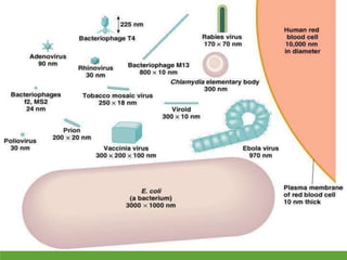
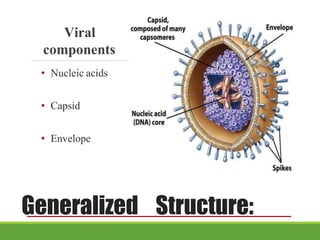
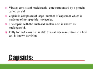
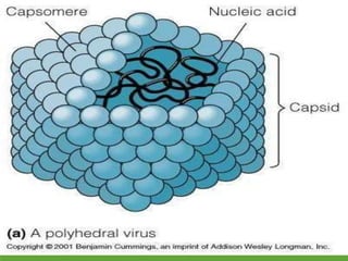
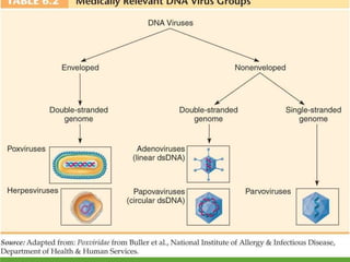
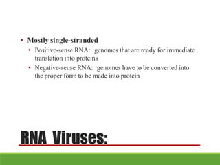
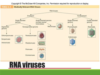
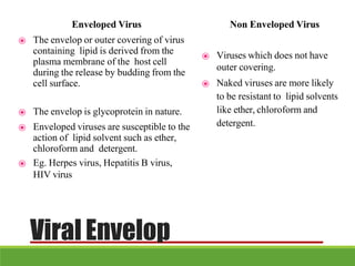
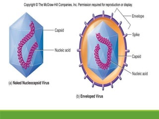
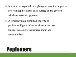
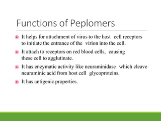
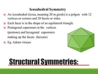
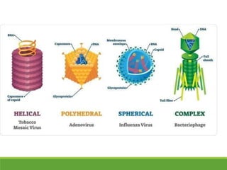
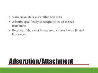
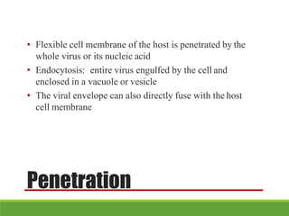
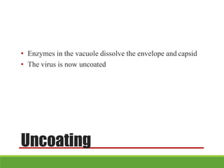
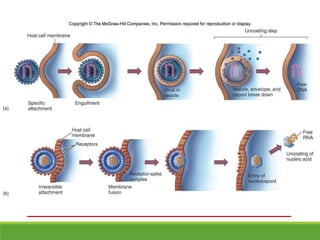
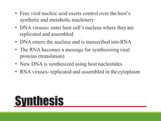
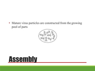
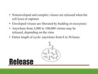
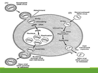
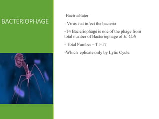
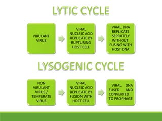
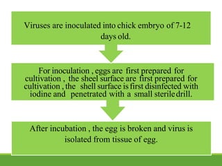
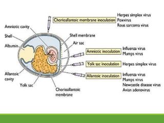
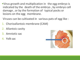
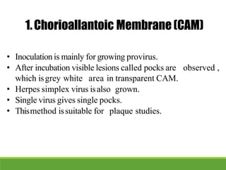
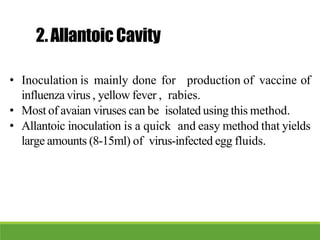
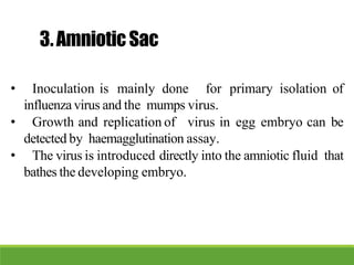
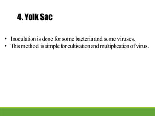
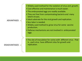
Viruses are obligate intracellular parasites that contain genetic material surrounded by a protein coat. They come in a variety of shapes and sizes but are too small to be seen by light microscopy. Viruses infect all types of living organisms, including animals, plants, bacteria and archaea. They reproduce by taking over the host cell's machinery and forcing it to produce new virus particles. There are thousands of known virus species that are classified based on their structure, genome type and pathogenicity.


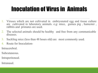
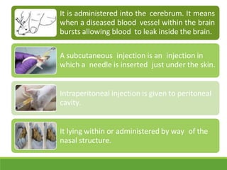
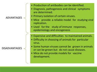
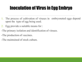


![PERI-PROSTHETIC FRACTURE NAIL-PLATE CONSTRUCT [NPC].pptx](https://cdn.slidesharecdn.com/ss_thumbnails/drarunkumardrmohamedashrafperiprostheticfrasturenail-plateconstructnpc-260209164459-7e9d15a1-thumbnail.jpg?width=640&height=640&fit=bounds)


![CTEV [ clubfoot] DR ARUN LAL ,DR MOHAMED ASHRAF travancore medical college k...](https://cdn.slidesharecdn.com/ss_thumbnails/ctevclubfootdrarunlaldrmohamedashraftravancoremedicalcollegekollamkeralaindia-260208063247-18fc466c-thumbnail.jpg?width=640&height=640&fit=bounds)


