Morphology, Classification, Cultivation and Replication of Virus
•Download as PPTX, PDF•
33 likes•12,207 views

This presentation is Useful for B. Pharmacy SEM III Students to study the Topic Fungi According to PCI Syllabus. It Consist of Morpholoy of Fungi, Cultivation , Replication and Classification of Virud

Recommended
Recommended
More Related Content
What's hot
What's hot (20)
Similar to Morphology, Classification, Cultivation and Replication of Virus
Similar to Morphology, Classification, Cultivation and Replication of Virus (20)
More from Krutika Pardeshi
More from Krutika Pardeshi (7)
Recently uploaded
Recently uploaded (20)
Morphology, Classification, Cultivation and Replication of Virus
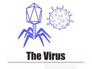
- 1. The Virus
- 3. Viruses: An infective agent that typically consists of a nucleic acid molecule in a protein coat, is too small to be seen by light microscopy, and is able to multiply only within the living cells of a host.
- 4. Introduction to viruses Viruses do not have cells that divide; new viruses are assembled in the infected host cell But unlike still simpler infectious agents, viruses contain genes, which gives them the ability to mutate and evolve. Evolved from plasmids: pieces of DNA that can move between cells while others may have evolved from bacteria. Over 5,000 species of viruses have been discovered.
- 5. Introduction to viruses A virus consists of two or three parts: genes, made from either DNA or RNA, long molecules that carry genetic information protein coat that protects the genes; and in some viruses, an envelope of fat Viruses vary in shape from the simple helical and icosahedral to more complex structures. Viruses range in size from 20 to 300 nanometres; it would take 30,000 to 750,000 of them, side by side, to stretch to 1 centimeter.
- 6. Viruses spread in many ways. Just as many viruses are very specific as to which host species or tissue they attack, Plant viruses are often spread from plant to plant by insects and other organisms, known as vectors. Some viruses of animals, including humans, are spread by exposure to infected bodily fluids Viruses such as influenza are spread through the air by droplets of moisture when people cough or sneeze. Viruses such as norovirus are transmitted by the faecal–oral route, which involves the contamination of hands, food and water. Spreading , Vectors:
- 7. The human immunodeficiency virus, HIV, is transmitted by bodily fluids transferred during sex. Dengue virus, are spread by blood-sucking insects. Rotavirus is often spread by direct contact with infected children. Antibiotics have no effect on viruses, but antiviral drugs have been developed to treat life- threatening infections. Vaccines that produce lifelong immunity can prevent some viral infections.
- 8. Characteristics Obligate intracellular parasites of bacteria, protozoa, fungi, algae, plants, and animals. Ultramicroscopic size, ranging from 20 nm up to 450 nm (diameter). Not cellular in nature; structure is very compact and economical. Do not independently fulfill the characteristics of life. Inactive macromolecules outside the host cell and active only inside host cells. Basic structure consists of protein shell (capsid) surrounding nucleic acid core. • Nucleic acid can be either DNA or RNA but notboth
- 9. • Nucleic acid can be double-stranded DNA, single- stranded DNA single- stranded RNA, ordouble-stranded RNA. • Molecules on virus surface impart high specificity for • attachment to host cell. • Multiply by taking control of host cell’s genetic material and regulating the synthesis and assembly of new viruses. • Lack enzymes for most metabolic processes. • Lack machinery for synthesizing proteins. • Most RNA viruses multiply in & are released fromthe cytoplasm. • Viral infections range from very mild to life threatening.
- 10. A comparison with bacterialcell
- 11. • Viruses have no nucleus, no organelles, no cytoplasm or cell membrane—Non-cellular
- 12. Size of virus ? • Smallest infectious agents • Most are so small, they can only be seen with an electron microscope • Proviruses- around 20 nm in diameter • Mimi viruses- up to 450 nm in length • Special stains and an electron microscope • Negative staining outlines the shape • Positive staining shows internal details
- 14. Classification • Viruses are classified on the basis of habitat (host).which is trivial system beside this Viruses are classified on following criteria. • Structure • Chemical composition • Similarities in genetic makeup • International Committee on the Taxonomy of Viruses, which includes • 3 orders • 63 families “-viridae” • 263 genera “-virus”
- 15. Types of Classification: • 3 Types of systems were proposed to classify the viruses: • Baltimore Classification. • Classical System Classification. • Genetic Classification.
- 16. 7 groups were made. Its principles are fundamental to an understanding of virus classification and genome replication. The Baltimore classification has + RNA as its central point. I: dsDNA viruses (e.g. Adenoviruses, Herpesviruses, Poxviruses) II: ssDNA viruses (+ strand or "sense") DNA (e.g. Parvoviruses) III: dsRNA viruses (e.g. Reoviruses) IV: (+)ssRNA viruses (+ strand or sense) RNA (e.g. Picornaviruses, Togaviruses) V: (−)ssRNA viruses (− strand or antisense) RNA (e.g. Orthomyxoviruses, Rhabdoviruses) VI: ssRNA viruses (+ strand or sense) RNA with DNA intermediate in life-cycle (e.g. Retroviruses) VII: dsDNA viruses (e.g. Heptadnaviruses) Baltimore Classification:
- 17. Classification on basis of host • Animal viruses: • Viruses of animal host • Rabies, Polio, Mumps, Chicken pox, Small pox, and Influenza. • Plant Viruses: • viruses which show their live characteristics when attached to plants. • Tobacco mosaic virus (TMV), Banana streak virus, Carrot thin leaf virus • Bacterial Virus: Bacteriophages ( T1, T2, T3, andT4.)
- 19. Classification on Genetics basis • According to genetic consequences viruses are classified as. DNA Viruses and RNAViruses • Genes may be linear or circular • The smallest have only 4 genes and largest have several hundred. • DNAViruses • DNA Viruses are the viruses which consist of DNA genome . They complete their activities by transcription and most of them attack on organisms of similar genome. • RNAViruses • RNA Viruses are the viruses which consist of RNAgenome. They complete their activities by reverse transcription.
- 20. Classification on structural basis • With relevant to morphology of viral structure viruses are organized as Enveloped and Nonenveloped viruses. • However they are also arranges subclasses of DNAand RNAviruses
- 21. Structure of virus • Questions Relating to Structure • Is it rigid? • How big is it? • Is it flexible? • Structure Must Serve Virus • It should provide protection for genome • It should allow virus to move from one host to next • It should allow for attachment of virus on to new host
- 23. Generalized Structure: Viral components • Nucleic acids • Capsid • Envelope
- 24. Capsids: Viruses consists of nucleic acid core surrounded by a protein called capsid. Capsid is composed of large number of capsomer which is made up of polypeptide molecules. The capsid with the enclosed nucleic acid is known as nucleocapsid. Fully formed virus that is able to establish an infection in a host cell is known as virion.
- 26. Functions of Capsid It protects the viral genome from physical destruction and enzymatic inactivation by nucleases in biological material. It provides the binding site which enable the virus to attach to specific site on the host cell. It facilitates the assembly and packaging of viral genetic information. It serves as a vehicle of transmission from host to another. It is antigenic and specific for each viruses It provides the structural symmetry to the virus particle.
- 27. Nucleic Acids: • Genome: • the sum total of the genetic information carried by an organism • Number of viral genes compared with a cell- quite small • They only have the genes necessary to invade host cells and redirect their activity
- 28. DNA Viruses : • ssDND (single stranded DNA) • dsDNA (double stranded DNA)
- 30. RNA Viruses: • Mostly single-stranded • Positive-sense RNA: genomes that are ready for immediate translation into proteins • Negative-sense RNA: genomes have to be converted into the proper form to be made into protein
- 31. RNA viruses
- 32. Enveloped Virus The envelop or outer covering of virus containing lipid is derived from the plasma membrane of the host cell during the release by budding from the cell surface. The envelop is glycoprotein in nature. Enveloped viruses are susceptible to the action of lipid solvent such as ether, chloroform and detergent. Eg. Herpes virus, Hepatitis B virus, HIV virus Non Enveloped Virus Viruses which does not have outer covering. Naked viruses are more likely to be resistant to lipid solvents like ether, chloroform and detergent. Viral Envelop
- 34. In mature virus particle, the glycoproteins often appear as projecting spikes on the outer surface of the envelop which are known as peplomers. A virus may have more than one type of peplomers. E.g the influenza virus carries two types of peplomers, the hemagglutinin and neuraminidase Peplomers
- 35. Functions of Peplomers It helps for attachment of virus to the host cell receptors to initiate the entrance of the virion into the cell. It attach to receptors on red blood cells, causing these cell to agglutinate. It has enzymatic activity like neuraminidase which cleave neuraminic acid from host cell glycoproteins. It has antigenic properties.
- 37. Structural Symmetries: Icosahedral Symmetry An icosahedral (icosa, meaning 20 in greek) is a polgon with 12 vertices or corners and 20 facets or sides. Each facet is in the shape of an equilateral triangle. Pentagonal capsomers at the vertices (pentons) and hexagonal capsomers making up the facets (hexons) Eg. Adeno viruses
- 38. Helical Symmetry The nucleic acid and capsomers are wound together in the form of helix or spiral. Eg. Influenza virus, parainfluenz virus, rabies virus. Structural Symmetries:
- 39. Complex Symmetry Viruses which don not show either icoshedral or helical symmetry due to complexity of their structure are referred to have complex symmetry. Eg. Pox viruses Structural Symmetries:
- 41. The genomic information necessary for viral replication is contained in the viral nucleic acid but lacking biosynthetic enzymes. The virus depend on the synthetic machinery of the host cell for replication. Viral multiplication proceeds as following manner. • Adsorption, • Penetration, • Uncoating, • Synthesis, • Assembly and Release • Adsorption. Viral Replication
- 42. Adsorption/Attachment • Virus encounters susceptible host cells • Adsorbs specifically to receptor sites on the cell membrane • Because of the exact fit required, viruses have a limited host range
- 43. Penetration • Flexible cell membrane of the host is penetrated by the whole virus or its nucleic acid • Endocytosis: entire virus engulfed by the cell and enclosed in a vacuole or vesicle • The viral envelope can also directly fuse with the host cell membrane
- 44. Uncoating • Enzymes in the vacuole dissolve the envelope and capsid • The virus is now uncoated
- 46. Synthesis • Free viral nucleic acid exerts control over the host’s synthetic and metabolic machinery • DNA viruses- enter host cell’s nucleus where theyare replicated and assembled • DNA enters the nucleus and is transcribed into RNA • The RNA becomes a message for synthesizing viral proteins (translation) • New DNA is synthesized using host nucleotides • RNA viruses- replicated and assembled in thecytoplasm
- 47. Assembly • Mature virus particles are constructed from the growing pool of parts
- 48. Release • Nonenveloped and complex viruses are released when the cell lyses or ruptures • Enveloped viruses are liberated by budding or exocytosis • Anywhere from 3,000 to 100,000 virions may be released, depending on the virus • Entire length of cycle- anywhere from 8 to 36 hours
- 50. Bacteriophage lifecycle LYTIC CYCLE & LYSOGENIC CYCLE
- 51. BACTERIOPHAGE -Bactria Eater - Virus that infect the bacteria -T4 Bacteriophage is one of the phage from total number of Bacteriophage of E. Coli - Total Number – T1-T7 -Which replicate only by Lytic Cycle.
- 52. VIRULANT VIRUS VIRAL NUCLEIC ACID REPLICATE BY RUPTURING HOST CELL VIRAL DNA REPLICATE SEPRATELY WITHOUT FUSING WITH HOST DNA NON VIRULANT VIRUS / TEMPERATE VIRUS VIRAL NUCLEIC ACID REPLICATE BY FUSION WITH HOST CELL VIRAL DNA FUSED AND CONVERTED TO PROPHAGE LYTIC CYCLE LYSOGENIC CYCLE
- 53. CULTIVATION OF VIRUS PURPOSE OF VIRUS CULTIVATION • To isolate and identify viruses in clinical samples. • To prepare viruses for vaccine production. • To do research on viral structure, replication , genetics and effects on host cells.
- 54. METHODS FOR CULTIVATION OF VIRUSES Inoculation of Virus into Animal Inoculation of Virus into Embryonated Eggs. Tissue culture
- 55. Inoculation of Virus in Animals 1. Viruses which are not cultivated in embryonated egg and tissue culture are cultivated in laboratory animals. e.g: mice, guinea pig , hamester , rabbits and primates are used. 2. The selected animals should be healthy and free from any communicable diseases. 3. Suckling mice (less than 48 hours old) are most commonly used. 4. Route for Inoculation- Intracerebral. Subcutaneous. Intraperitoneal. Intranasal.
- 56. It is administered into the cerebrum. It means when a diseased blood vessel within the brain bursts allowing blood to leak inside the brain. A subcutaneous injection is an injection in which a needle is inserted just under the skin. Intraperitoneal injection is given to peritoneal cavity. It lying within or administered by way of the nasal structure.
- 57. ADVANTAGES • Production of antibodies can be identified. • Diagnosis, pathogenesis and clinical symptoms are determined. • Primary isolation of certain viruses. • Mice provide a reliable model for studying viral replication. • Used for the study of immune responses, epidemiology and oncogenesis DISADVANTAGES • Expensive and difficulties to maintained animals. • Difficulty in choosing of animals for particular virus. • Some human viruses cannot be grown in animals or can be grown but do not cause diseases. • Mice do not provide models for vaccine development.
- 58. Inoculation of Virus in Egg Embryo 1. The process of cultivation of viruses in embryonated eggs depend upon the type of egg being used. 2. Egg provide a suitable means for : -The primary isolation and identification of viruses. -The production of vaccines. -The maintained of stock culture.
- 59. After incubation , the egg is broken and virus is isolated from tissue of egg. For inoculation , eggs are first prepared for cultivation , the sheel surface are first prepared for cultivation , the shell surface is first disinfected with iodine and penetrated with a small steriledrill. Viruses are inoculated into chick embryo of 7-12 days old.
- 61. Virus growth and multiplication in the egg embryo is indicated by the death of the embryo , by embryo cell damage , or by the formation of typical pocks or lesions on the egg membrane. Viruses can be cultivated in various pats of egg like : 1. Chorioallantoic membrane (CAM) 2. Allantoic cavity 3. Amniotic sac 4. Yolk sac
- 62. 1.Chorioallantoic Membrane (CAM) • Inoculation is mainly for growing provirus. • After incubation visible lesions called pocks are observed , which is grey white area in transparent CAM. • Herpes simplex virus isalso grown. • Single virus gives single pocks. • Thismethod is suitable for plaque studies.
- 63. 2. Allantoic Cavity • Inoculation is mainly done for production of vaccine of influenza virus , yellow fever , rabies. • Most of avaian viruses can be isolated using this method. • Allantoic inoculation is a quick and easy method that yields large amounts (8-15ml) of virus-infected egg fluids.
- 64. 3.Amniotic Sac • Inoculation is mainly done for primary isolation of influenza virus and the mumps virus. • Growth and replication of virus in egg embryo can be detected by haemagglutination assay. • The virus is introduced directly into the amniotic fluid that bathes the developing embryo.
- 65. 4. Yolk Sac • Inoculation is done for some bacteria and some viruses. • Thismethod is simple for cultivation and multiplication of virus.
- 66. ADVANTAGES • Widely used method for the isolation of virus and growth. • Cost effective and maintenance is much easier. • The embryonated eggs are readily available. • They are free from contaminating bacteria and many latent viruses. • Ideal substrate for the viral growth and replication. • less labor is needed. • Widely used method to grow virus for some vaccine production. • Defense mechanisms are not involved in embryonated eggs. DISADVANTAGES • The site of inoculation for varies with different virus . That is , each virus have different sites for growth and replication.
- 67. Inoculation of Virus Using Tissue Culture There are three types of tissue culture: - Organ culture. - Explant culture. - Cell culture.
- 68. 1. Small bits of organs can be maintained in vitro for days and weeks, preserving their original architecture and function. 2. Organs culture are useful for the isolation of some viruses which appear to be highly specialised parasite of certain organs. 3. For example, the tracheal ring organ culture is employed for the isolation of coronavirus, a respiratory pathogen. Inoculation of Virus Using Organ Culture
- 69. 1. Fragments of minced tissue can be grown as ‘explants’ embedded in plasma clots. 2. They may also be cultivated in suspension. 3. This method is now seldom employed in virology. 4. Adenoid tissue explant culture were used for the isolation of adenoviruses. Inoculation of Virus Using Explant Culture
- 70. This is the type of culture routinely employed for growing viruses Inoculation of Virus Using Cell Culture Cell Culture Primary Cell Culture Diploid Cell Culture Continuous Cell Lines
- 71. - Tissue are dissociated into the component cell by the action of proteolytic enzyme. -The cells are washed , counted and suspended in a growth medium. -Such media will enable most cell types to multiply with a division time of 24-48 hrs. - The cell suspension is dispensed in bottles, tubes or petridishes - The cell adhere to the glass surface and on incubation, divide to form a confluent monolayer sheet of cells covering the surface within about a week.
- 73. 1. These are normal cells obtained from fresh organs of animals or human being and cultured. 2. They are capable of only limited growth in culture and cannot be maintained in serial culture.e.g. monkey kidney cell culture.human embryonic kidney. chick embryo cell culture. 3. They are commonly employed for primary isolation of viruses and in preparation of vaccine. Primary Cell Culture
- 74. 1. These are cells of single type, contain the same number of chromosome as the parent cells and are diploid. 2. The diploid cell strains can be subculture for limited number of times. 3. After about 50 serial passage they undergo senescence. 4. They are also employed for the production of viral vaccine. Eg. human embryonic lung cell strain WI-38 Diploid Cell Culture
- 75. 1. Animal cells capable of indefinite growth are called continuous cell lines or cell lines. 2. These are the cells of a single type , usually derived from cancer cells , that are capable of continuous serial cultivation indefinitely. 3. Standard cell lines derived from human cancers , such as HeLa , HEp – 2 and KB cell lines have been used in laboratories throughout the world for many years. 4. These cell linesmay be maintained by serial subcultivation or stored in the cold ( 5. -70⁰C ) for use when necessary. 6. Some cell lines are now permitted to be used for vaccine manufacture, for example: Vero cells for rabies vaccine. Continuous Cell Line
- 76. Advantages • Relative ease, broad spectrum, cheaper and sensitivity Disadvantages • The process requires trained technicians with experience in working on a full time basis. • Tissue or serum for analysis is sent to central laboratories to identify virus. • State health laboratory and hospital Laboratory do not isolate and identify virus for clinical work.