This document summarizes the structure of bacterial cells. It describes the key components including the cell wall, plasma membrane, and intracellular and extracellular structures. The cell wall differs between gram-positive and gram-negative bacteria. Gram-positive walls are thicker and contain higher amounts of peptidoglycan while gram-negative walls are thinner and contain an outer lipopolysaccharide membrane. Intracellular structures discussed include the nucleoid, ribosomes, plasmids, and mesosomes. Extracellular structures include flagella, pili, and capsules. Bacteria range in size from 0.4 to 1.5 micrometers and have characteristic shapes including cocci, bacilli, spirilla and spirochetes.
Introduction
• All bacteriaare unicellular organisms that
reproduce by binary fission.
• Most bacteria are capable of independent
metabolic existence and growth, but species
of Chlamydia and rickettsia are obligately
intracellular organism.
• Bacterial cells are extremely small and are
most conveniently measured in microns (10-6
m).
• Bacterial cells are usually between 0.4 and
1.5 micro meter in short diameter.
4.
Cell Morphology
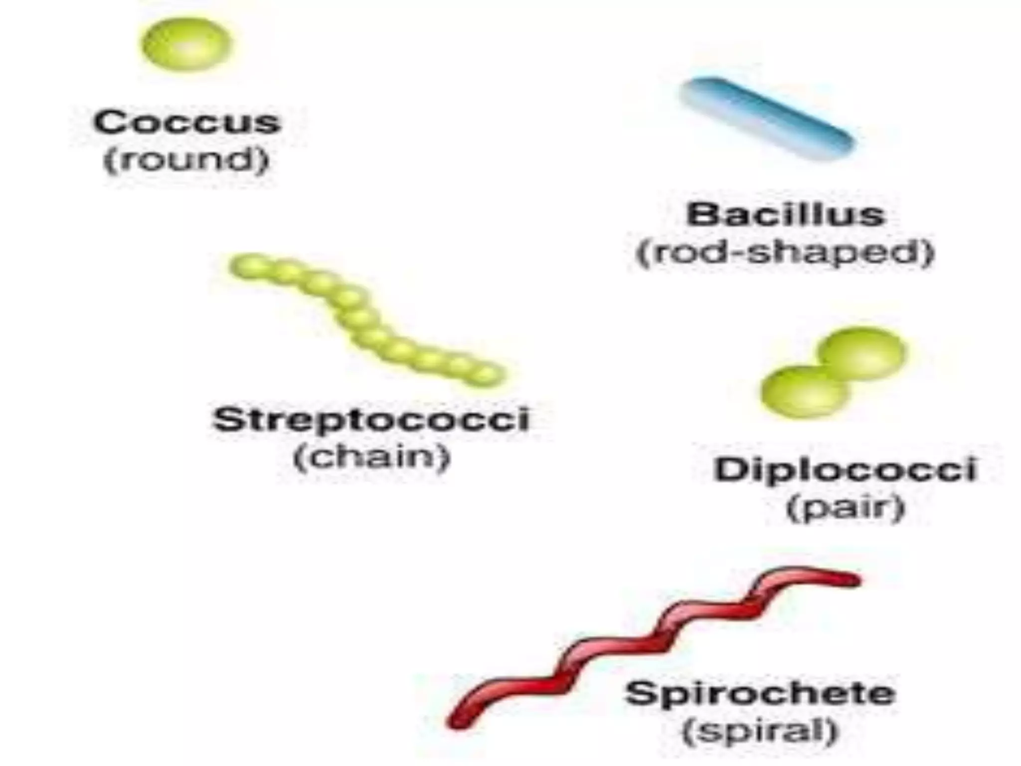
• Bacteriahave characteristic shape. The common
microscopic morphologies are :
spherical or ovoid ( cocci )
rod –shaped ( bacilli )
comma shaped ( vibrio )
spiral ( spirillum and spirochete )
• Some cocci characterristically grouped in pairs or
chains; some form grapelike clusters of sperical cells;
some round cocci form cubic packets.
• Bacterial cell of other species grow separately .
• The microscopic appearance is therefore valuable in
classification and diagnosis.
6.
Structure of Bacterialcell
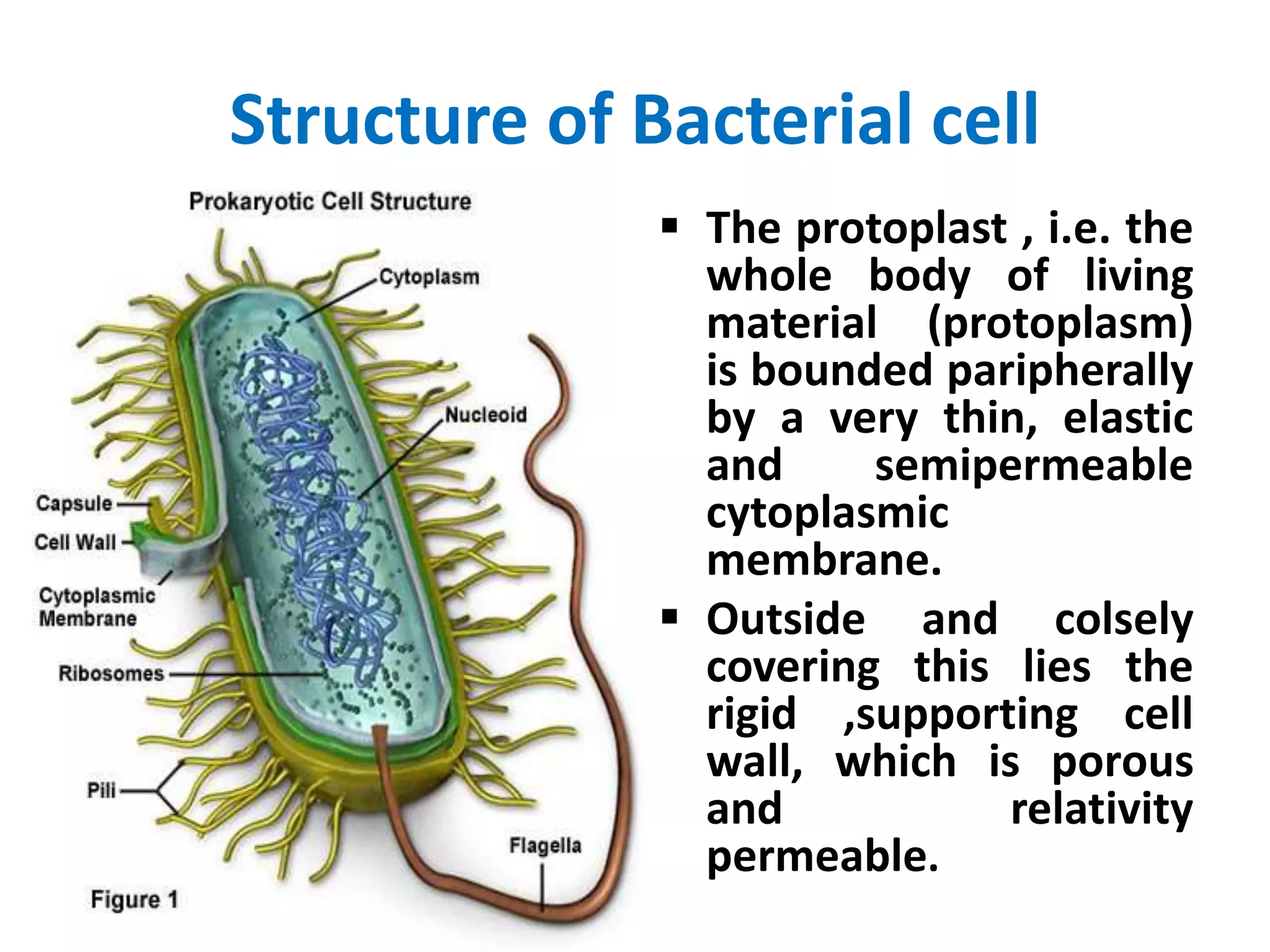
The protoplast , i.e. the
whole body of living
material (protoplasm)
is bounded paripherally
by a very thin, elastic
and semipermeable
cytoplasmic
membrane.
Outside and colsely
covering this lies the
rigid ,supporting cell
wall, which is porous
and relativity
permeable.
7.
1] cell wall
The cell wall encases the protoplast and lies
immediately external to the cytoplasmic
membrane.
It is 10-25 nm thick, strong and relatively rigid,
though with some elasticity and openly porous ,
being freely permeable to solute molecules
smaller then 10 kDa in mass and 1 nm in diameter
It shows granular structure and lacks microfibrils.
Gram Positive and Gram negative bacteria have
different type of bacterial cell wall
Gram positivecell wall :-
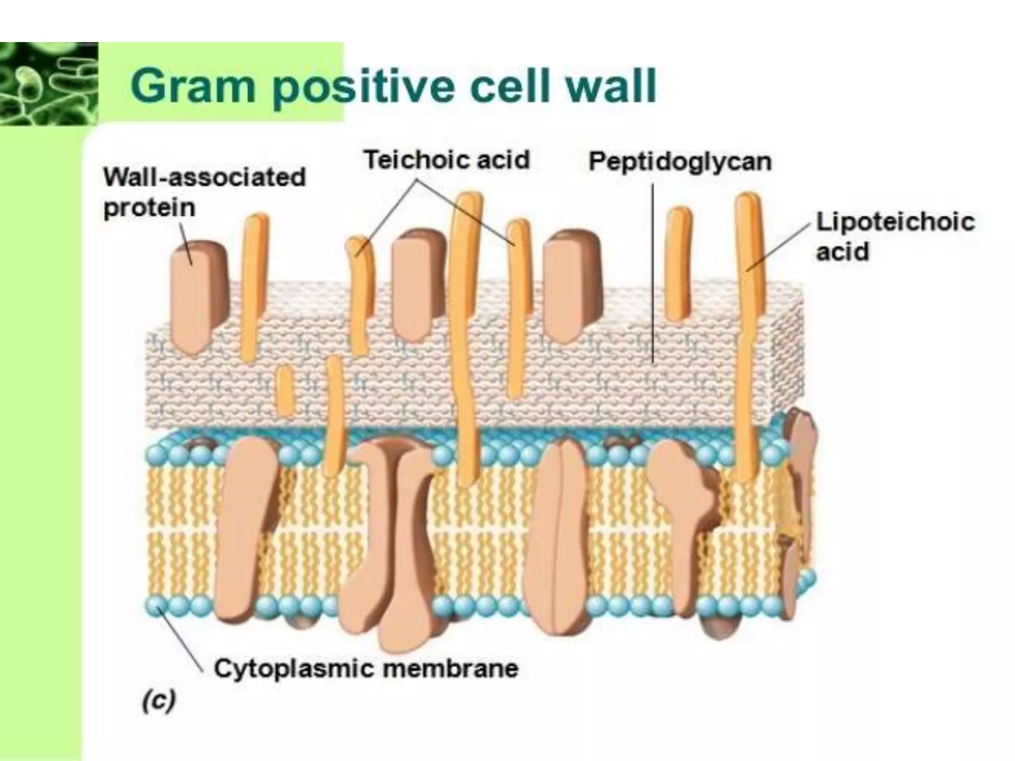
• The Gram positive type wall is relatively thick
(about 30-100 nm) and it generally has a simple,
uniform appearance under the electron
microscope.
• Some 40 – 80 % of wall is made of tough, complex
polymer , peptidoglycan .
• In this type of wall the sacculus consists of
multilayered peptidoglycan which, during growth,
develops by the “inside–to-outside” mechenism.
10.
continue…..
• Covalently boundto peptidoglycan are compounds
such as tichoic acid : typically ,substituted
polymers glycerol phosphate or ribitol phosphate.
• In some bacteria (e.g. mycobacterium ) the wall
contains lipids, while in others (strains of
streptococcus) it contains carbohydrates.
• The composition of the wall can vary with growth
condition ; for example in Bacillus , the availability
of phosphate affects the amount of cell wall
teichoic acids.
12.
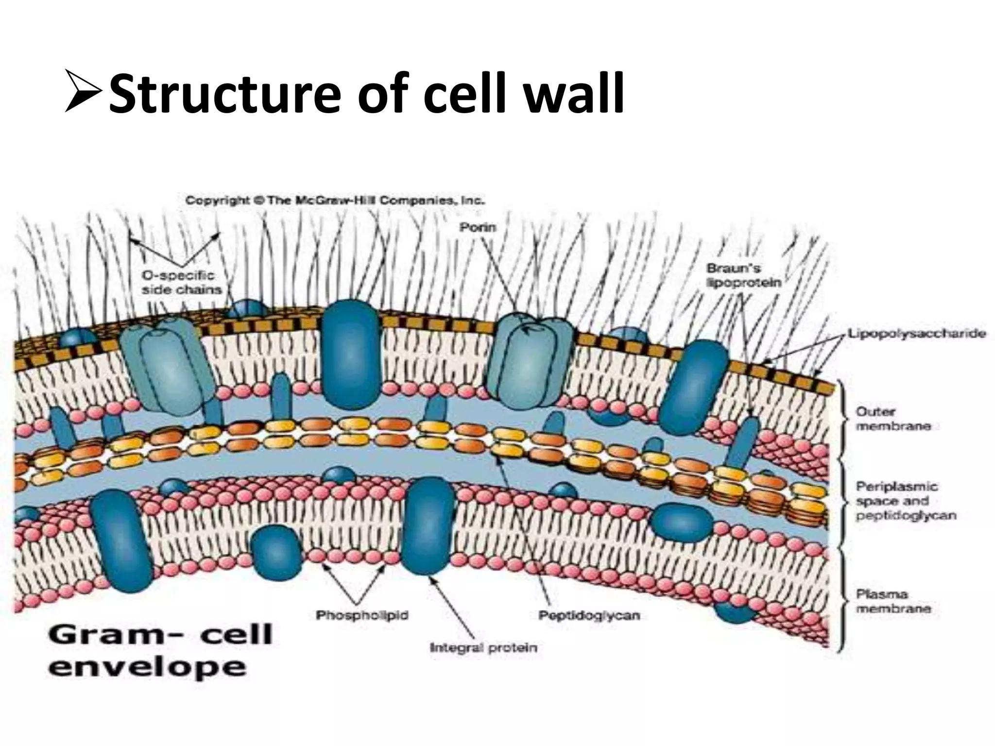
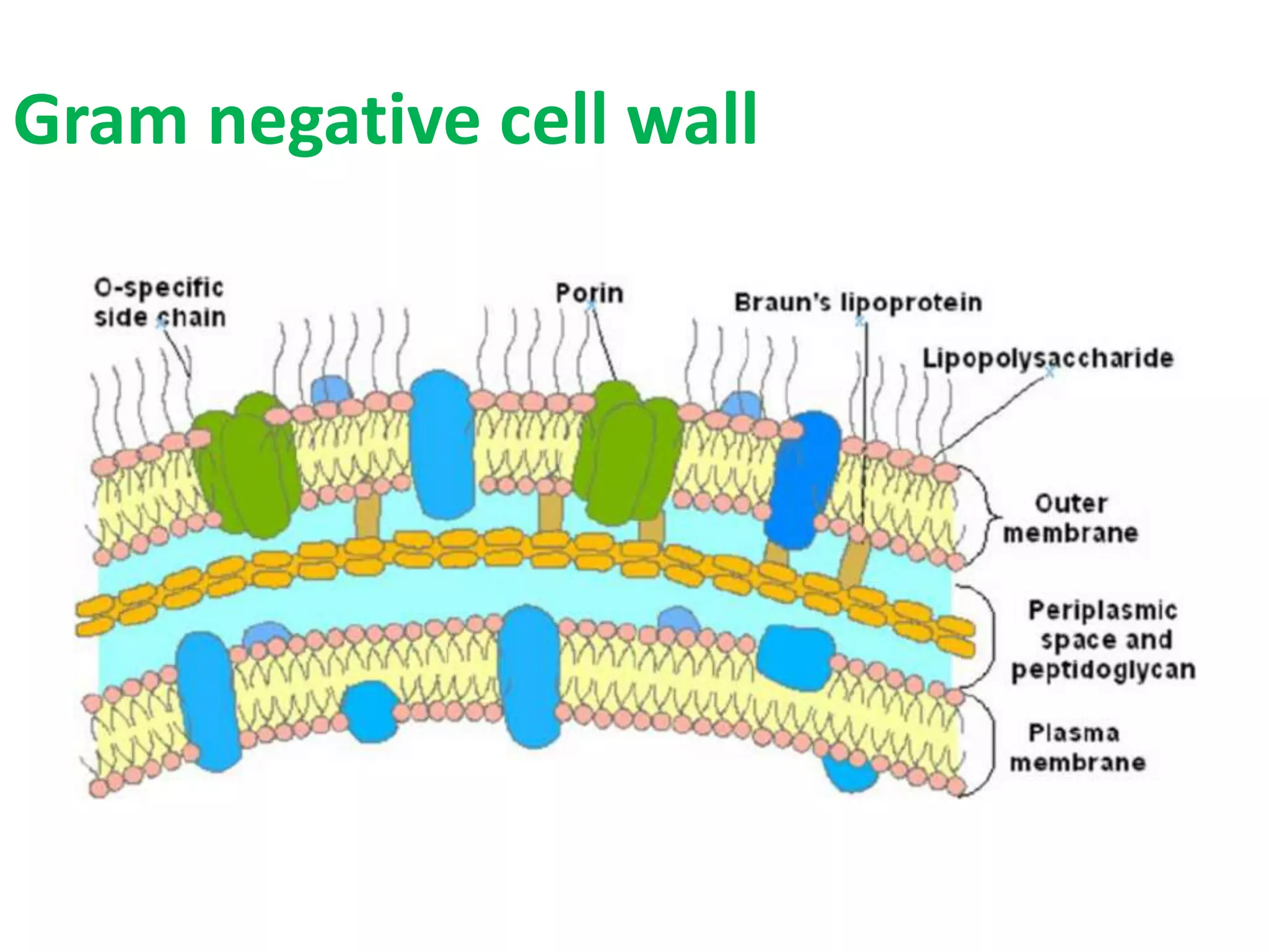
Gram negative cellwall :-
• The cell walls of Gram-negative bacteria are more
chemically complex, thinner and less compact.
• Peptidoglycan makes up only 5 – 20% of the cell
wall, and is not the outermost layer.
• The peptidoglycan of Gram-negative bacteria is
located between the plasma membrane and an
outer, LPS membrane.
• This LPS membrane is similar to the plasma
membrane, but is less permeable and is composed
of lipopolysaccharides (LPS).
2) Plasma membrane:-
• Plasma membranes in bacteria are composed of
phospholipids contain a polar group attached to a
3 carbon glycerol back bone.
• They are also two fatty acid chains dangling from
the other carbons of glycerol.
• The phosphate end of the molecule is hydrophilic
and is attracted to water.
• The fatty acids are hydrophobic.
• Membrane also contain protein.
15.
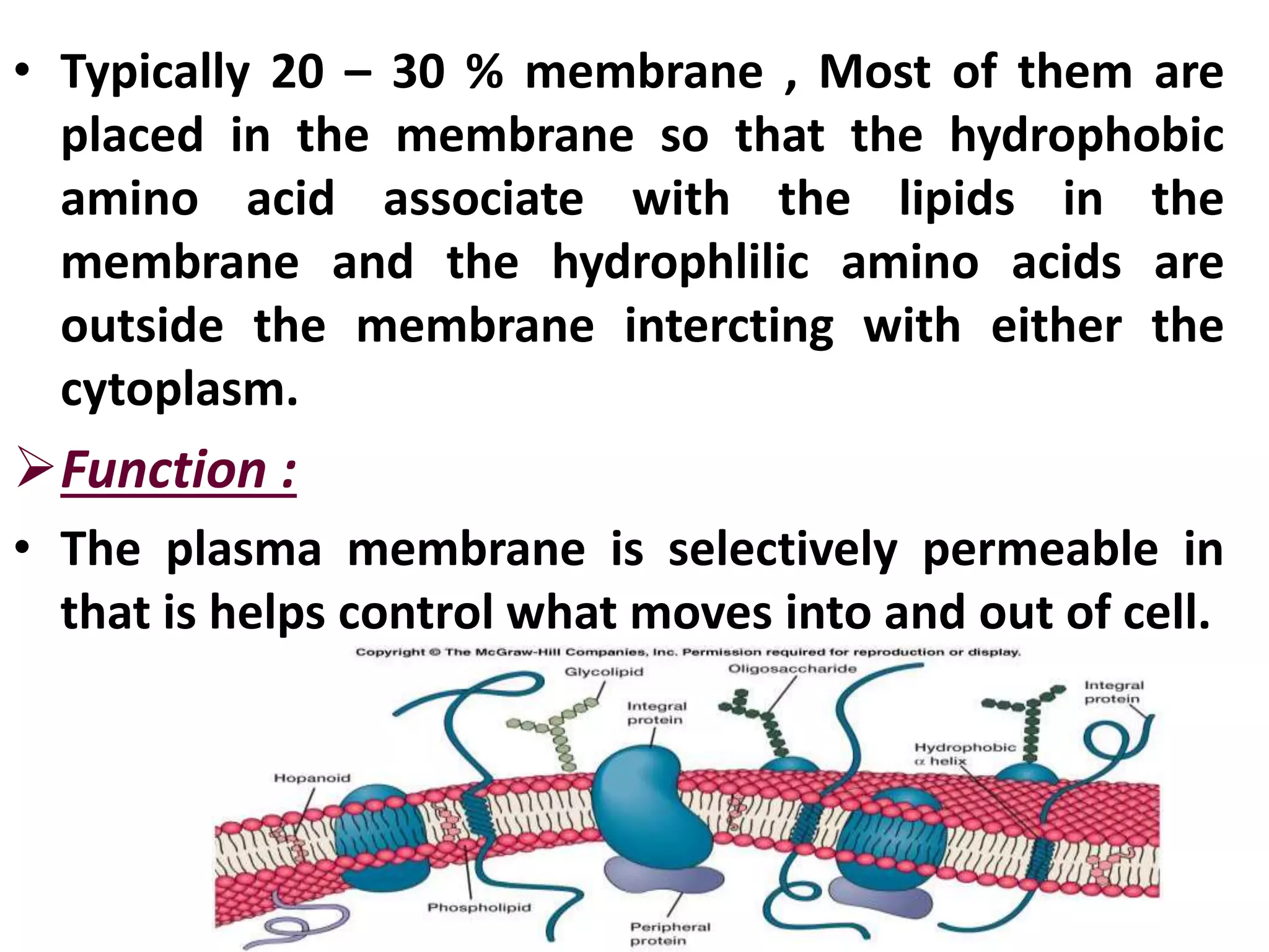
• Typically 20– 30 % membrane , Most of them are
placed in the membrane so that the hydrophobic
amino acid associate with the lipids in the
membrane and the hydrophlilic amino acids are
outside the membrane intercting with either the
cytoplasm.
Function :
• The plasma membrane is selectively permeable in
that is helps control what moves into and out of cell.
16.
3) Extracellular structure
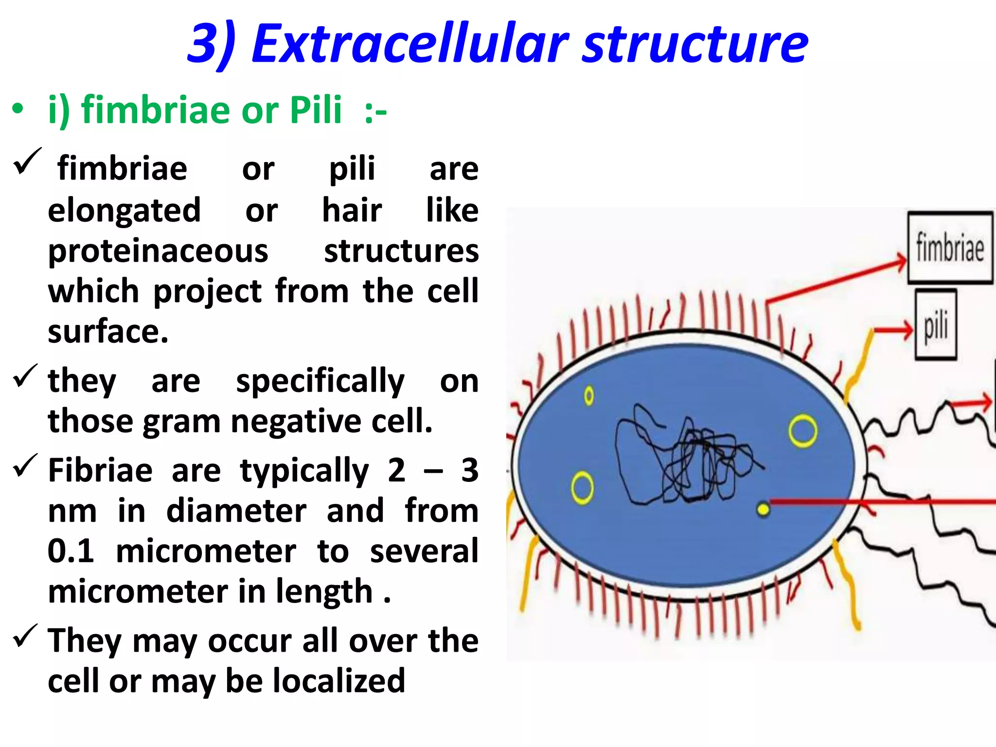
•i) fimbriae or Pili :-
fimbriae or pili are
elongated or hair like
proteinaceous structures
which project from the cell
surface.
they are specifically on
those gram negative cell.
Fibriae are typically 2 – 3
nm in diameter and from
0.1 micrometer to several
micrometer in length .
They may occur all over the
cell or may be localized
17.
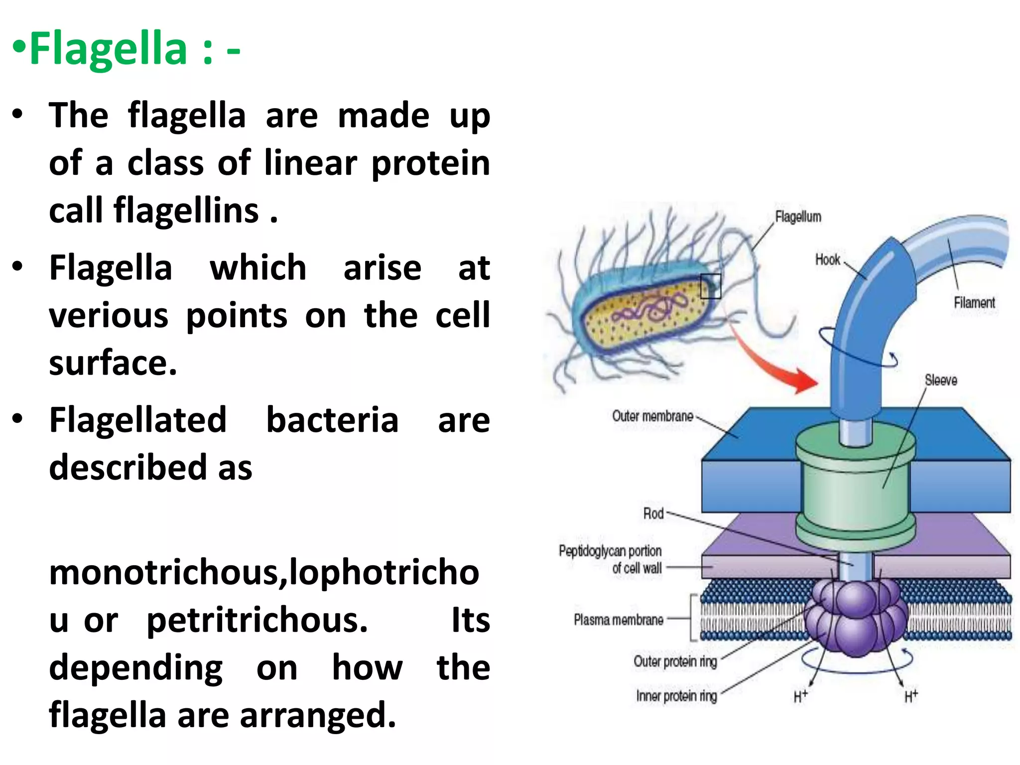
•Flagella : -
•The flagella are made up
of a class of linear protein
call flagellins .
• Flagella which arise at
verious points on the cell
surface.
• Flagellated bacteria are
described as
monotrichous,lophotricho
u or petritrichous. Its
depending on how the
flagella are arranged.
18.
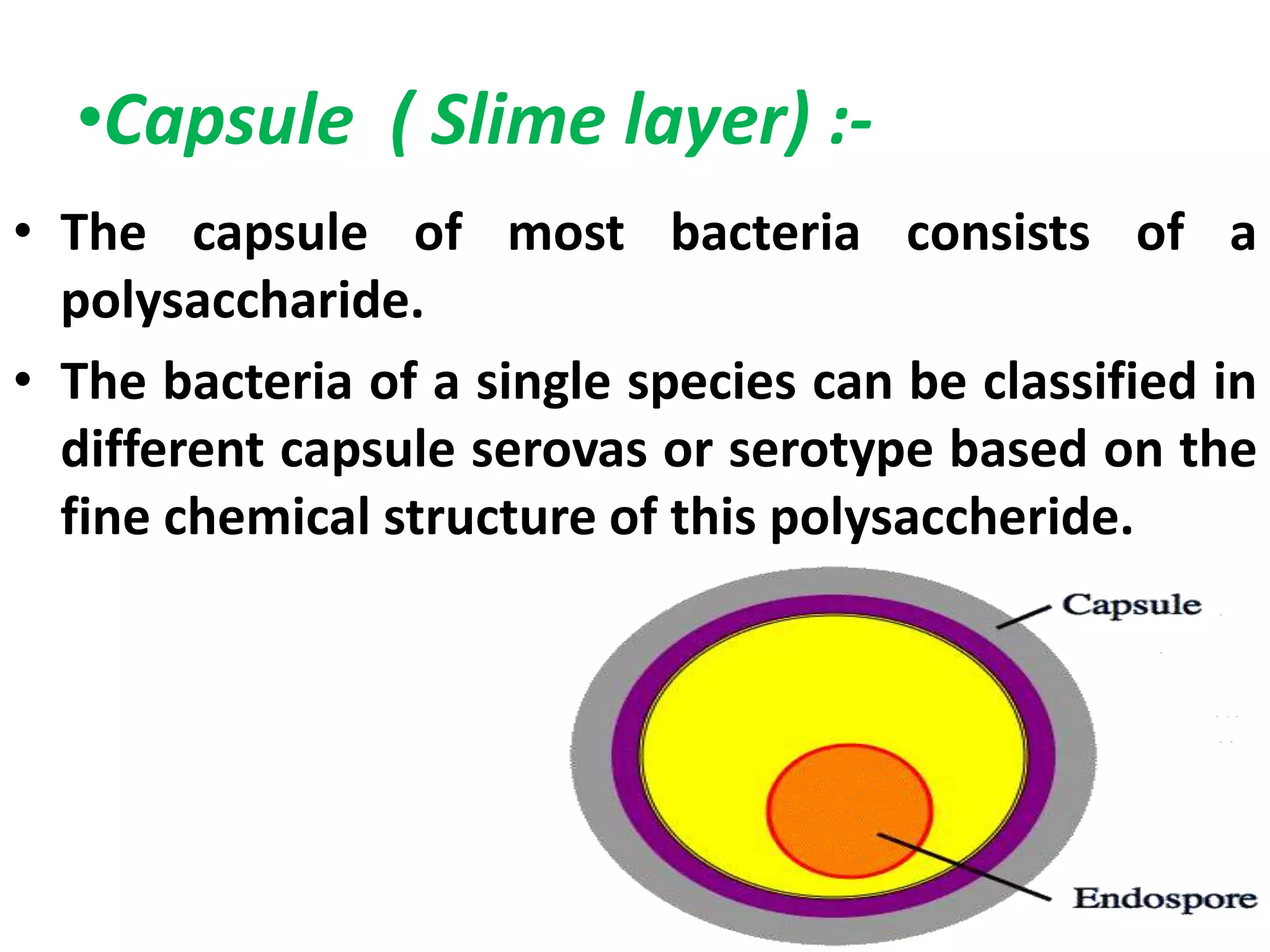
•Capsule ( Slimelayer) :-
• The capsule of most bacteria consists of a
polysaccharide.
• The bacteria of a single species can be classified in
different capsule serovas or serotype based on the
fine chemical structure of this polysaccheride.
19.
4)Intracellular structure
• Plasmids:-
• The term plasmid was first introduced by the
American molecular biologist Joshua lederberg in
1952.
• A plasmids is a short usually circular and double
stranded segment of DNA .
• That is found in the cytoplasm separate from the
main bacterial chromosome.
• Their size veries from 1 kbp to over 100 kilobase
pare.
20.
• Plasmids arecapable of autonomous replication.
• Plasmids can transfer genes from one cell to other
cell.
21.
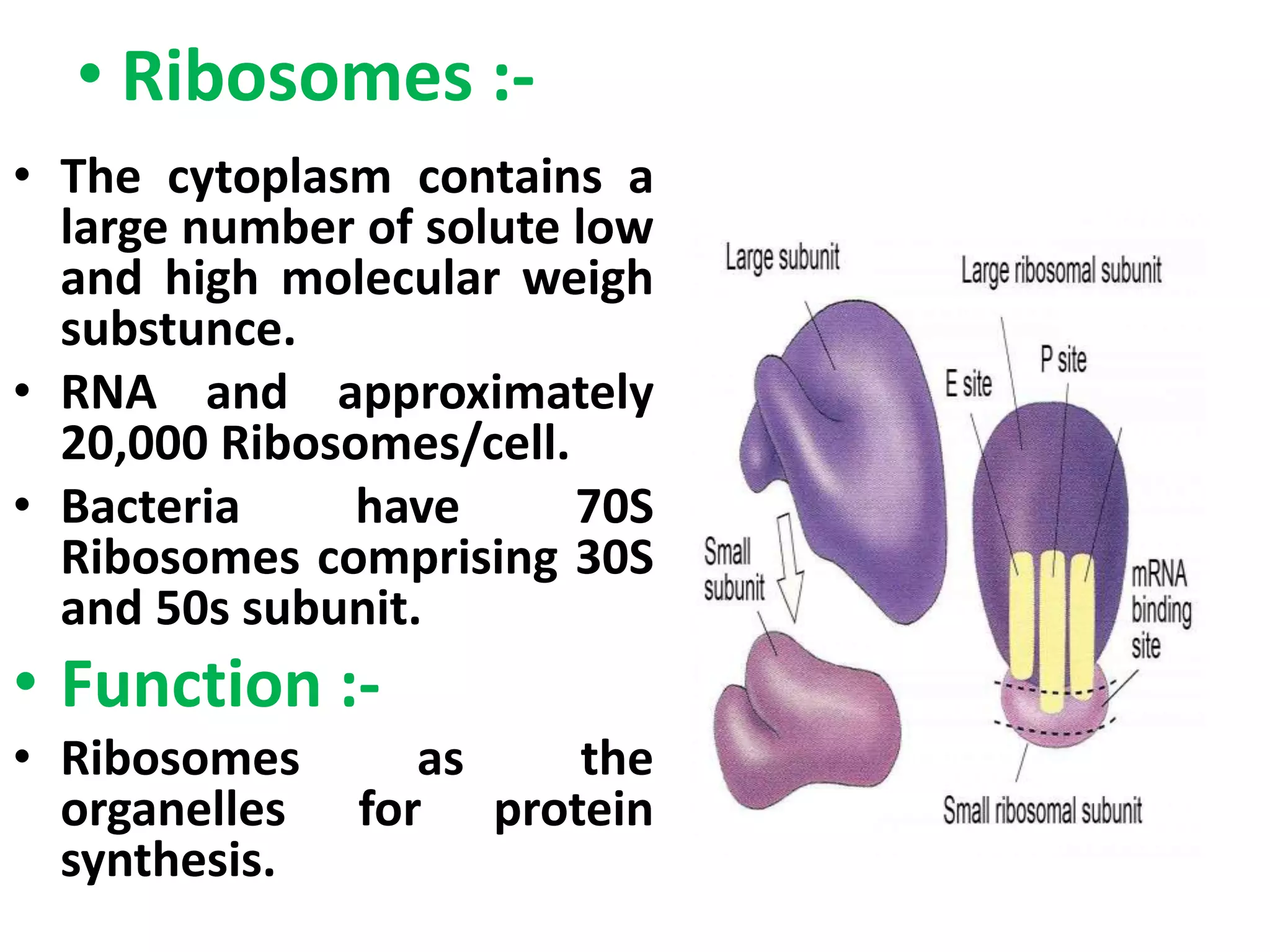
• Ribosomes :-
•The cytoplasm contains a
large number of solute low
and high molecular weigh
substunce.
• RNA and approximately
20,000 Ribosomes/cell.
• Bacteria have 70S
Ribosomes comprising 30S
and 50s subunit.
• Function :-
• Ribosomes as the
organelles for protein
synthesis.
22.
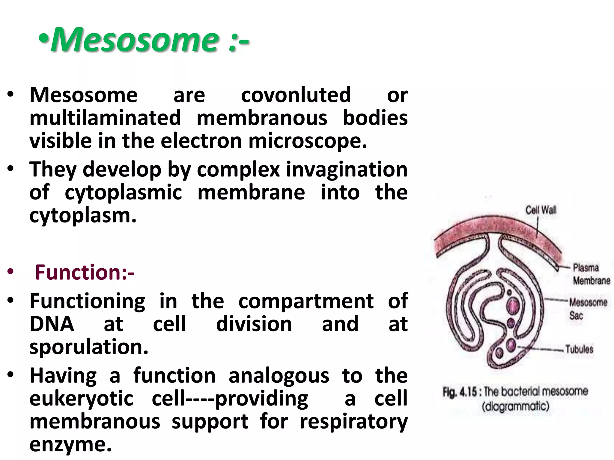
•Mesosome :-
• Mesosomeare covonluted or
multilaminated membranous bodies
visible in the electron microscope.
• They develop by complex invagination
of cytoplasmic membrane into the
cytoplasm.
• Function:-
• Functioning in the compartment of
DNA at cell division and at
sporulation.
• Having a function analogous to the
eukeryotic cell----providing a cell
membranous support for respiratory
enzyme.
23.
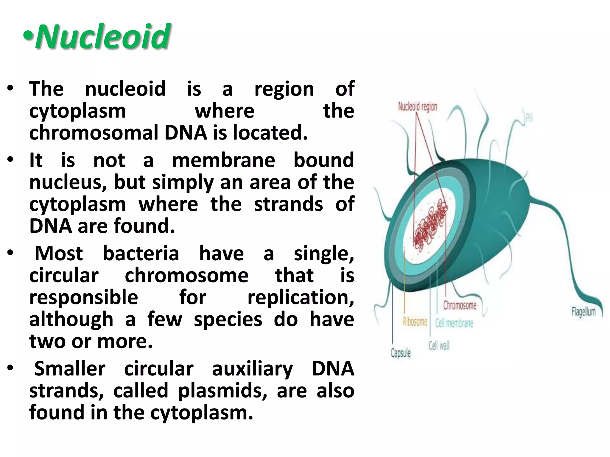
•Nucleoid
• The nucleoidis a region of
cytoplasm where the
chromosomal DNA is located.
• It is not a membrane bound
nucleus, but simply an area of the
cytoplasm where the strands of
DNA are found.
• Most bacteria have a single,
circular chromosome that is
responsible for replication,
although a few species do have
two or more.
• Smaller circular auxiliary DNA
strands, called plasmids, are also
found in the cytoplasm.
24.
Reference :-
• Bacteriain biology , biotechnology and
medicine 4th Edition
By Paul singleton
• www.structure of bacterial cell.com
![Content
• Introduction
• Cell Morphology
• Structure of bacterial cell
1] Cell Wall
2] Plasma membrane
3] Extracellular (external ) structure
4] Intracellular ( Internal) structure](https://image.slidesharecdn.com/structureofbacterialcell-180806181503/75/Structure-of-bacterial-cell-2-2048.jpg)
![1] cell wall
The cell wall encases the protoplast and lies
immediately external to the cytoplasmic
membrane.
It is 10-25 nm thick, strong and relatively rigid,
though with some elasticity and openly porous ,
being freely permeable to solute molecules
smaller then 10 kDa in mass and 1 nm in diameter
It shows granular structure and lacks microfibrils.
Gram Positive and Gram negative bacteria have
different type of bacterial cell wall](https://image.slidesharecdn.com/structureofbacterialcell-180806181503/75/Structure-of-bacterial-cell-7-2048.jpg)