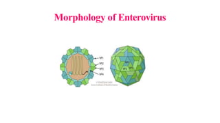
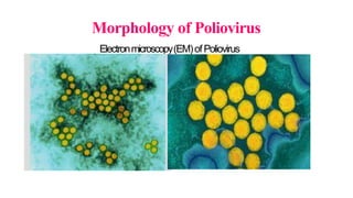
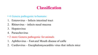
This document provides an overview of picornaviruses, with a focus on poliovirus. It begins by outlining the objectives of discussing picornavirus morphology, classification, pathogenesis, and the clinical manifestations, diagnosis, treatment and prevention of poliomyelitis and other diseases caused by echoviruses and rhinoviruses. It then provides details on picornavirus morphology, classification, the history of poliovirus research and discovery, epidemiology, characteristics, cultivation, pathogenesis, and clinical features of poliomyelitis. Key points covered include that poliovirus is an enterovirus that can cause paralysis, replication in the gastrointestinal tract and nervous system, and that infection may be asymptomatic, minor
A picornavirus is a virus belonging to the family Picornaviridae, a family of viruses in the order Picornavirales. Vertebrates, including humans, serve as natural hosts. Picornaviruses are nonenveloped viruses that represent a large family of small, cytoplasmic, plus-strand RNA viruses with a 30-nm icosahedral capsid.
The Paramyxoviridae is a family of single-stranded RNA viruses known to cause different types of infections in vertebrates. Examples of these infections in humans include the measles virus, mumps virus, parainfluenza virus, and respiratory syncytial virus (RSV).
Poxviruses are brick or oval-shaped viruses with large double-stranded DNA genomes. Poxviruses exist throughout the world and cause disease in humans and many other types of animals. Poxvirus infections typically result in the formation of lesions, skin nodules, or disseminated rash.
The rhinovirus (from the Greek ῥίς rhis "nose", gen ῥινός rhinos "of the nose", and the Latin vīrus) is the most common viral infectious agent in humans and is the predominant cause of the common cold. Rhinovirus infection proliferates in temperatures of 33–35 °C (91–95 °F), the temperatures found in the nose. Rhinoviruses belong to the genus Enterovirus in the family Picornaviridae.
The three species of rhinovirus (A, B, and C) include around 160 recognized types of human rhinovirus that differ according to their surface proteins (serotypes).[1] They are lytic in nature and are among the smallest viruses, with diameters of about 30 nanometers. By comparison, other viruses, such as smallpox and vaccinia, are around ten times larger at about 300 nanometers; while flu viruses are around 80–120 nm.
polio virus lecture for MBBS
The picornaviruses are small (22 to 30 nm) nonenveloped, single-stranded RNA viruses with cubic symmetry. The virus capsid is composed of 60 protein subunits, each consisting of four poly-peptides VP1–VP4. Because they contain no essential lipids, they are ether resistant. They replicate in the cytoplasm.
Picornaviruses presentation for medical student created by: Farhang Shapouran
References : Moray medical microbiology, Jawetz medical microbiology, ICTV
HOT NEW PRODUCT! BIG SALES FAST SHIPPING NOW FROM CHINA!! EU KU DB BK substit...GL Anaacs
Contact us if you are interested:
Email / Skype : kefaya1771@gmail.com
Threema: PXHY5PDH
New BATCH Ku !!! MUCH IN DEMAND FAST SALE EVERY BATCH HAPPY GOOD EFFECT BIG BATCH !
Contact me on Threema or skype to start big business!!
Hot-sale products:
NEW HOT EUTYLONE WHITE CRYSTAL!!
5cl-adba precursor (semi finished )
5cl-adba raw materials
ADBB precursor (semi finished )
ADBB raw materials
APVP powder
5fadb/4f-adb
Jwh018 / Jwh210
Eutylone crystal
Protonitazene (hydrochloride) CAS: 119276-01-6
Flubrotizolam CAS: 57801-95-3
Metonitazene CAS: 14680-51-4
Payment terms: Western Union,MoneyGram,Bitcoin or USDT.
Deliver Time: Usually 7-15days
Shipping method: FedEx, TNT, DHL,UPS etc.Our deliveries are 100% safe, fast, reliable and discreet.
Samples will be sent for your evaluation!If you are interested in, please contact me, let's talk details.
We specializes in exporting high quality Research chemical, medical intermediate, Pharmaceutical chemicals and so on. Products are exported to USA, Canada, France, Korea, Japan,Russia, Southeast Asia and other countries.
Flu Vaccine Alert in Bangalore Karnatakaaddon Scans
As flu season approaches, health officials in Bangalore, Karnataka, are urging residents to get their flu vaccinations. The seasonal flu, while common, can lead to severe health complications, particularly for vulnerable populations such as young children, the elderly, and those with underlying health conditions.
Dr. Vidisha Kumari, a leading epidemiologist in Bangalore, emphasizes the importance of getting vaccinated. "The flu vaccine is our best defense against the influenza virus. It not only protects individuals but also helps prevent the spread of the virus in our communities," he says.
This year, the flu season is expected to coincide with a potential increase in other respiratory illnesses. The Karnataka Health Department has launched an awareness campaign highlighting the significance of flu vaccinations. They have set up multiple vaccination centers across Bangalore, making it convenient for residents to receive their shots.
To encourage widespread vaccination, the government is also collaborating with local schools, workplaces, and community centers to facilitate vaccination drives. Special attention is being given to ensuring that the vaccine is accessible to all, including marginalized communities who may have limited access to healthcare.
Residents are reminded that the flu vaccine is safe and effective. Common side effects are mild and may include soreness at the injection site, mild fever, or muscle aches. These side effects are generally short-lived and far less severe than the flu itself.
Healthcare providers are also stressing the importance of continuing COVID-19 precautions. Wearing masks, practicing good hand hygiene, and maintaining social distancing are still crucial, especially in crowded places.
Protect yourself and your loved ones by getting vaccinated. Together, we can help keep Bangalore healthy and safe this flu season. For more information on vaccination centers and schedules, residents can visit the Karnataka Health Department’s official website or follow their social media pages.
Stay informed, stay safe, and get your flu shot today!
These lecture slides, by Dr Sidra Arshad, offer a quick overview of physiological basis of a normal electrocardiogram.
Learning objectives:
1. Define an electrocardiogram (ECG) and electrocardiography
2. Describe how dipoles generated by the heart produce the waveforms of the ECG
3. Describe the components of a normal electrocardiogram of a typical bipolar leads (limb II)
4. Differentiate between intervals and segments
5. Enlist some common indications for obtaining an ECG
Study Resources:
1. Chapter 11, Guyton and Hall Textbook of Medical Physiology, 14th edition
2. Chapter 9, Human Physiology - From Cells to Systems, Lauralee Sherwood, 9th edition
3. Chapter 29, Ganong’s Review of Medical Physiology, 26th edition
4. Electrocardiogram, StatPearls - https://www.ncbi.nlm.nih.gov/books/NBK549803/
5. ECG in Medical Practice by ABM Abdullah, 4th edition
6. ECG Basics, http://www.nataliescasebook.com/tag/e-c-g-basics
MANAGEMENT OF ATRIOVENTRICULAR CONDUCTION BLOCK.pdfJim Jacob Roy
Cardiac conduction defects can occur due to various causes.
Atrioventricular conduction blocks ( AV blocks ) are classified into 3 types.
This document describes the acute management of AV block.
Title: Sense of Taste
Presenter: Dr. Faiza, Assistant Professor of Physiology
Qualifications:
MBBS (Best Graduate, AIMC Lahore)
FCPS Physiology
ICMT, CHPE, DHPE (STMU)
MPH (GC University, Faisalabad)
MBA (Virtual University of Pakistan)
Learning Objectives:
Describe the structure and function of taste buds.
Describe the relationship between the taste threshold and taste index of common substances.
Explain the chemical basis and signal transduction of taste perception for each type of primary taste sensation.
Recognize different abnormalities of taste perception and their causes.
Key Topics:
Significance of Taste Sensation:
Differentiation between pleasant and harmful food
Influence on behavior
Selection of food based on metabolic needs
Receptors of Taste:
Taste buds on the tongue
Influence of sense of smell, texture of food, and pain stimulation (e.g., by pepper)
Primary and Secondary Taste Sensations:
Primary taste sensations: Sweet, Sour, Salty, Bitter, Umami
Chemical basis and signal transduction mechanisms for each taste
Taste Threshold and Index:
Taste threshold values for Sweet (sucrose), Salty (NaCl), Sour (HCl), and Bitter (Quinine)
Taste index relationship: Inversely proportional to taste threshold
Taste Blindness:
Inability to taste certain substances, particularly thiourea compounds
Example: Phenylthiocarbamide
Structure and Function of Taste Buds:
Composition: Epithelial cells, Sustentacular/Supporting cells, Taste cells, Basal cells
Features: Taste pores, Taste hairs/microvilli, and Taste nerve fibers
Location of Taste Buds:
Found in papillae of the tongue (Fungiform, Circumvallate, Foliate)
Also present on the palate, tonsillar pillars, epiglottis, and proximal esophagus
Mechanism of Taste Stimulation:
Interaction of taste substances with receptors on microvilli
Signal transduction pathways for Umami, Sweet, Bitter, Sour, and Salty tastes
Taste Sensitivity and Adaptation:
Decrease in sensitivity with age
Rapid adaptation of taste sensation
Role of Saliva in Taste:
Dissolution of tastants to reach receptors
Washing away the stimulus
Taste Preferences and Aversions:
Mechanisms behind taste preference and aversion
Influence of receptors and neural pathways
Impact of Sensory Nerve Damage:
Degeneration of taste buds if the sensory nerve fiber is cut
Abnormalities of Taste Detection:
Conditions: Ageusia, Hypogeusia, Dysgeusia (parageusia)
Causes: Nerve damage, neurological disorders, infections, poor oral hygiene, adverse drug effects, deficiencies, aging, tobacco use, altered neurotransmitter levels
Neurotransmitters and Taste Threshold:
Effects of serotonin (5-HT) and norepinephrine (NE) on taste sensitivity
Supertasters:
25% of the population with heightened sensitivity to taste, especially bitterness
Increased number of fungiform papillae
Anti ulcer drugs and their Advance pharmacology ||
Anti-ulcer drugs are medications used to prevent and treat ulcers in the stomach and upper part of the small intestine (duodenal ulcers). These ulcers are often caused by an imbalance between stomach acid and the mucosal lining, which protects the stomach lining.
||Scope: Overview of various classes of anti-ulcer drugs, their mechanisms of action, indications, side effects, and clinical considerations.
Factory Supply Best Quality Pmk Oil CAS 28578–16–7 PMK Powder in Stockrebeccabio
Factory Supply Best Quality Pmk Oil CAS 28578–16–7 PMK Powder in Stock
Telegram: bmksupplier
signal: +85264872720
threema: TUD4A6YC
You can contact me on Telegram or Threema
Communicate promptly and reply
Free of customs clearance, Double Clearance 100% pass delivery to USA, Canada, Spain, Germany, Netherland, Poland, Italy, Sweden, UK, Czech Republic, Australia, Mexico, Russia, Ukraine, Kazakhstan.Door to door service
Hot Selling Organic intermediates
TEST BANK for Operations Management, 14th Edition by William J. Stevenson, Ve...kevinkariuki227
TEST BANK for Operations Management, 14th Edition by William J. Stevenson, Verified Chapters 1 - 19, Complete Newest Version.pdf
Report Back from SGO 2024: What’s the Latest in Cervical Cancer?bkling
Are you curious about what’s new in cervical cancer research or unsure what the findings mean? Join Dr. Emily Ko, a gynecologic oncologist at Penn Medicine, to learn about the latest updates from the Society of Gynecologic Oncology (SGO) 2024 Annual Meeting on Women’s Cancer. Dr. Ko will discuss what the research presented at the conference means for you and answer your questions about the new developments.
2. Objectives of today’s class
Toknow and understand the morphology,classification,
pathogenesis of Picornaviruses
Tolearn about the clinical manifestations, lab diagnosis,
treatment, prevention and prophylaxis ofPoliomyelitis.
Tolearn about the clinical manifestations, lab diagnosis,
treatment, prevention and prophylaxis of ECHOand
Rhinoviruses.
3. Introduction to Picornaviruses
Family Picornaviridae
Consists of a large number of very small RNAviruses,
27-30 nm insize
Resistant to lipid solvents like Ether, Chloroform, Bile salts
8. Classification
• 4 Genera pathogenic to humans:
1. Enterovirus – Infects intestinal tract
2. Rhinovirus – infects nasal mucosa
3. Hepatovirus
4. Paraechovirus
• 2 more Genera pathogenic for animals
1. Aphthovirus – Foot and Mouth disease of catlle
2. Cardiovirus – Encephalomyocarditis virus that infects mice
9. ENTEROVIRUSES- History
• Infantile paralysis – Paralytic disease of children
• Landsteiner and Popper (1909) demonstrated
Fatal case of Poliomyelitis –
Spinal cord &Feces inoculation –
Experimental disease transmission - monkeys
10. ENTEROVIRUSES– History Continued
• Enders, Wellers &Robbins (1949) demonstrated
• Growth of Poliovirus
• Culture of non-neural cells from human embryos
• Produced Cytopathic effects (CPE)
• Major break through – Nobel prize for this discovery
• Development of Virology (Milestone)
11. ENTEROVIRUSES– History Continued….
• Dalldorf &Sickles (1948) demonstration:
• Anew type of virus isolated
• From feces of children with paralytic poliomyelitis
• Named Type 1 Poliovirus
• This virus caused paralysis on inoculation in suckling mice
• Was called the Coxsackie virus
• Aspatient was from village ‘Coxsackie’in NewYork
12. ENTEROVIRUSES– History Continued ………
• Introduction of Tissue culture technique
• Diagnostic virology
• Led to Fecal isolation of several cytopathogenic viruses from cases
• Were called ‘Orphan viruses’
• Not associated with any particular clinical disease
• Was known by descriptive term “ECHO”
• ECHO– Entero cytopathogenic human orphan viruses
13. ENTEROVIRUSES– Characteristics
• Entero viruses are ‘Host specific’
• Infects only one or a few related species
• No common group antigen
• Antigen cross reactions observed in few
14. ENTEROVIRUSES– Classification
Group Serotype
Poliovirus (3) Type 1, Type 2, Type 3
Coxsackie virus A(24) 1-22, 24
Coxsackie virus (6) 1-6
Echovirus (34) 1-9, 11-27, 29-31
Numbered enterovirus
(EVsince 1969 – Numbered 68 andso)
68-71
15. Poliovirus
• Clinical case scenarion:
• A12 years old boy from Kovilpalayam presented to the emergency
department of KFMS&Rwith a h/o mild fever and sore throat X8
days, condition worsened and became severe accompanied by neck
rigidity and vomiting for last 2 days after a brief asymptomatic
period of 2 days.
• HOPI: On morning, boy experienced pain in lower limbs which
increased and progressed as weakness at the time of presentation.
• Mother’s history: Childhood (<5 years) vaccination not remembered
16. Poliovirus
• Diagnostic &Management strategy of Case scenarion:
• Throat swab, Stool, CSFsent to Diagnostic Microbiology Dept. for
viral studies
• Throat swab and stool specimen tested positive for Cytopathic effect
(CPE)in tissueculture.
• Virus was confirmed as Poliovirus type 1 by Neutralization test
• ReverseTranscriptase PCRwas positive for Poliovirus in CSF
• Patient improved on supportive treatment.
17. Poliovirus - Introduction
• Group IV;FamilyPicornaviridae; Genus Enterovirus; Species
Poliovirus
• The virus is composed of an RNAgenome and a protein capsule. The
genome is single-stranded positive-sense RNA genome that is about
7500 nucleotides long.
• Often called the simplest significant virus - First isolated in 1909by
Karl Landsteiner and Erwin Popper
• Egyptian paintings depicted the effects of polio by showing otherwise
healthy individuals with withered limbs.
18. Morphology
• Spherical Virion – Icosahedral symmetry
• 27 nm in diameter insize
• Composed of 60 subunits
• Consists of 4 Viral Proteins (VP1,VP2,VP3 &VP4)
• VP1 faces outside – Major antigenic site for combination
• VP1has type-specific neutralizing antibodies
• Viral Genome: Single stranded positive strand (ss RNA+ve sense)
• Virus can be crystallized – seen in cytoplasm of infected cells
19. Resistance
• Resistant to lipid solvents – ether, chloroform, bile, proteolytic
enzymes of intestinal contents and detergents
• Stable at a pH of 3
• In feces, it can survive for 4 months at 4ºC and for years at-20ºC
• Room temperature survival of virus in feces vary (one day to several
weeks) and It depends on temperature, moisture, pH and amount of
virus
• Readily inactivated by heat (55ºC X30 minutes)
• Molar MgCl2 , Milk or Icecream protects virus against heat
inactivation
20. Resistance
• Formaldehyde and Oxidising disinfectants destroy the virus
• Chlorination destroy the virus in water
• Organic matter present delays inactivation of virus
• Phenolic disinfectants not effective
• Does not survive lyophilisation well
21. Antigenic properties
• Based on Neutralisation test
• Poliovirus (PV)classified into 3 types: Type 1, Type 2 &Type 3
• Prototype strains:
Brunhilde &Mahoney strains for Type1
Lansing and MEFIfor Type2
Leon and Saukett for Type 3
Type 1 Most common, causes most epidemics
Type 2 Usually cause endemic disease
Type 3 Strains causeepidemic.
22. Antigenic properties
• Based on Complement Fixation Test(CFT)or Enzyme Linked
Immuno SorbentAssay (ELISA)or Precipitation tests
• Two antigens recognized: C&D
1. Cantigen - Capsid, Coreless, also called Heated or Hantigen
2. D antigen – Dense, also called Native or N antigen
• D antigen / N antigen associated with whole virion – Type specific
• Cantigen / H antigen associated with ‘empty’non-infectious virus
and Less specific and reacts with heterotypic sera
23. Antigenic properties
• D antigen – converted to Cantigen by heating the virus at 56⁰C
• Anti D antibody is protective. So,Potency of Injectable Polio Vaccine
can be measured in terms of D antigen units.
• Anti-C antibody does not neutralize virus infectivity
24. Host range &Cultivation
• Natural infection occurs only in humans
• Experimental transmission in monkeys by intracerebral or
intraspinal inoculation
• Chimpanzees and Cynomolgus monkeys – can be infected orally
• Established non-fresh strains can be grown in rodents, chick
embryos
• Virus grows readily in Tissue cultures of primate origin
• Primary Monkey Kidney cultures are used for Diagnostic purpose
and for Vaccination
25. Host range &Cultivation
• Cytopathic effect (CPE):Infected cells round up and become refractile
and pyknotic.
• Eosinophilic intranuclear inclusion bodies – may be demonstrated in
stained preparations.
• Well-formed plaques develop in infected monolayers with agar
overlay.
26. Pathogenesis
• The polio virus infects human cells by binding to an
immunoglobubin-like receptor called CD155 (poliovirus receptor).
• The exact mechanism that poliovirus uses for entering the cell is
unknown.
• However, the interaction of poliovirus and CD155 causes a change
in the shape of the viral particle that is needed to enter thecell
• There are two thesis' for the way the viral nucleic acid to enters the
cell.
1. RNAof poliovirus is injected into the host cell through a pore in the
membrane of the host cell.
2. Thepoliovirus is taken in by the host cell through endocytosis.
27. Pathogenesis
• Thegenome inside poliovirus can be used as mRNAand immediately
translated by the host cell.
• The poliovirus mRNAis then translated into a long polypeptide
which is cleaved into 10 individual viral proteins.
• Translation of the viral RNAoccurs by an IRES-mediated (internal
ribosome entry site) mechanism. The IRES is the extremely long 5’
end of the poliovirus’ mRNA. The assembly of viral particles is not
fully understood.
• The particles leave the host cell 4-6 hours after the initialinfection.
Each dying host cell can release 10,000 polio virions making
poliovirus lytic.
28. Pathogenicity
1. Virus transmitted by Fecal-oral route through ingestion. Other
possible modes in close contacts in patients of early stages:
Inhalation or Entry through conjunctiva of droplets of respiratory
secretions.
2. Virus multiplies initially in the epithelial cells of the alimentary
canal and in the lymphatic tissues, from the tonsils to peyer’s
patches
3. Spreads to regional lymph nodes and enters blood stream (Primary
viremia)
4. Further multiplication takes place in reticulo-endothelial system
30. Pathogenicity - Continues
5. Virus enters the Blood stream again (Secondary viremia)
6. Virus is now carried to Central Nervous System (CNS) Spinal cord
and Brain.
7. In CNS,Virus multiplies in selective neurons and destroys them
8. Earliest change: Degeneration of Nissl’s or bodies (Chromatolysis)
9. Nuclear changes follows.
10.When degeneration becomes irreversible, the necrotic cells lyses or
is phagocytosed by leucocytes or macrophages
31. Pathogenicity - Continued
11. Lesions are mostly in the anterior horns of the spinal cord causing
Flaccid paralysis, but posterior and intermediate horns can also be
involved.
12. Pathological changes will be more than distribution of paralysis
13. Encephalitis primarily involving the brain stem and extending up
to motor and pre-motor areas can occur insome cases.
14. Special circumstances: Direct neural transmission of Virus to
Central Nervous System (CNS) as in poliomyelitis following
Tonsillectomy through glossopharyngeal nerve present in
tonsillar fossa.
15. Poliomyelitis: Polio= gray matter; Myelitis= Spinal cord
inflammation
33. Clinical features
Inapparent infection:
90-95% susceptible individuals develop only inapparent polioinfection
with Seroconversion alone.
Only 5-10% among them develop clinical infection. Incubationperiod:
About 10 days on average (Range:4 days –4weeks)
Minor illness:
Early manifestation is fever, headache, sore throat and malaise (Phaseof
primary viremia) lasting 1-5days called Minor illness or Abortive
poliomyelitis
Paralytic poliomyelitis or major illness:
Progression of infection 3-4 dyas after minor illness results in major
illness. Fever returns (Biphasic fever), along with headache, stiff neck and
other features of meningitis due to viral invasion of CNS(Polio case).
35. Poliomyelitis types and Complication
• Non- Paralytic polio: Disease does not progress beyond stage of
aseptic meningitis
• So,Types of polio: Nonparalytic polio &Paralytic polio.
• Paralytic polio can further be divided into: Spinal polio, bulbar polio,
and bulbospinal polio based on distribution of paralysis.
• Complication: Post-polio syndrome may also occur in which
symptoms ranging form breathing and swallowing problems to joint
pain, start many decades after the initial sickeness.
• Vaccine associated Poliomyelitis
36.
37. Laboratory diagnosis
• Samples to be collected: Blood, CSF,Throat swab, Feces
• Transport: Immediately to lab in viral transport media (Hank’s
Balanced Salt Solution)
• Storage: 4⁰C (Days),-20⁰C(monthsto years)
• Laboratory Diagnostic tests available:
Viral isolation
Serodiagnosis
Molecular diagnosis
38. 1. Viral isolation
• In tissue culture – during primary Viremia 3-5 days afterinfection
before neutralizing antibodies appear (from blood)
• Early stages – isolation from throat swabs
• First week of infection: 80-85% viral isolation from feces
• Second week of infection: 50% viral isolation from feces
• Third week of infection: 25% viral isolation from feces
• Fecal excretion – intermittent (So two samples needed to be tested)
• Prolonged fecal excretion in immunocompromised, but no
permanent carriers,
39. 1. Viral isolation - continues
• Seldom poliovirus isolated from CSF,but it can be isolated from
spinal cord and brain, post-mortem diagnosis. (Unlike enteroviruses)
• Primary monkey kidney cells are employed commonly
• Human or Simian kidney cells can also be used.
• Inference: Viral growth indicated by Cytopathic effect (CPE)in 2-3
days.
• Mere isolation does not confirm the diagnosis
40. 2. Serodiagnosis
• Antibody rise appears after the onset of paralysis – demonstrated
by Neutralisation tests or Complement Fixation Tests (CFT).
• Neutralising antibodies appear early and persista for life
• In CFT,Anti-C antibodies appear first and disappear in few
months.
• Anti-D antibodies rise after few weeks and lasts for 5 years.
•CFTis useful to identify the exposure to Poliovirus but
not for type-specific diagnosis
44. Immuno-prophylaxis
Liveoral polio vaccine (OPV) -four
doses in endemiccountries
or Inactivated polio vaccine (IPV)
given by injection - two-threedoses
depending on countryschedule
49. Treatment
• Currently there is no treatment to cure Polio. Treatment is focused on
supportive care. Moderate exercise - Anutritious diet
• Medication and rest to lower the fever and to reduce the painand
improve the strength. Breathing assistance with a ventilator
• ToPrevent Poliovirus: The most effective and most commonly used is
the Polio vaccine. This vaccine is given to young children in specific
increments.
• Vaccine works by strengthening and preparing the immune system
to a future encounter with the Poliovirus.
50. Coxsackie Group Avirus
• 24 serotypes
• Causes:
1. Herpangina (Vesicular pharyngitis),
2. Hand, Foot and Mouth disease (HFMD),
3. Aseptic meningitis,
4. Minor respiratory infection
51. Coxsackie Group Bvirus
• 6 serotypes
• Causes:
1. Epidemic pleurodynia or Bornholm disease
2. Myocarditis and pericarditis
3. Juvenile Diabetes – Coxsackie B4 ?
4. Orchitis
5. Transplacental and neonatal transmission
6. Post-viral fatigue syndrome
52. Coxsackie virus
• Labdiagnosis:
1. Animal inoculation – inoculating in suckling mice
2. Tissue culture – not useful
3. Serodiagnosis not practicable – due to several antigenic types
53. ECHOvirus
Enterocytopathogenic Human orphan virus
Grow in Human and simian kidney culture
Fever with rash, Aseptic Meningitis (most common cause)
Lab diagnosis: Feces, throat swab, CSF- Culture
54. New Entero virus
Acute hemorrhagic conjunctivitis – EV68,69,70,71
Radiculopathy – EV-70
Grows in Human embryonic kidney or HeLa cell lines.
55. Rhinoviruses
Common cold virus – 100 serotypes by neutralization
Transmitted by droplet infection
Culture – Just like for ECHO and other new entero viruses
56. Summary
• Polio virus – Belong to Entero virus – ss RNAvirus +sense
• Polio virus: 3 types – 1, 2 &3
• CD 155 receptor – Feco-oral transmission
• Paralytic or Non –paralytic polio
• CSF,Blood, Throat swab &feces
• Neutralisation tests, ELISA,CFT,RT-PCR
• OPV,IPV–Salk and Sabin
• Pulse polio immunization programme.