Downloaded 141 times


![CLINICAL PRESENTATION:
Most cases go through three stages. [classical
rabies]:
I. Prodromal Stage :
lasts from 2-10 days and presents in the form of
fever, headache, malaise, fatigue, and also
localized pain around area of initial
infection.
I. Sensory Excitation:
hyperactivity, hallucinations, disorientation, seizur
es and bizarre behavior.](https://image.slidesharecdn.com/rhabdovirus-120725062246-phpapp01/85/Rhabdo-virus-14-320.jpg)
![CLINICAL PRESENTATION:
About 20% of cases have only two phases [dump
rabies]:
patient skips the sensory excitation phase and
progresses right to the coma and paralysis phase.
Dumb rabies is almost 100% fatal with only three
known cases of survival.
In each of these cases the patients had high titers of
antibody in the CSF.](https://image.slidesharecdn.com/rhabdovirus-120725062246-phpapp01/85/Rhabdo-virus-16-320.jpg)
![LABORATORY DIAGNOSIS:
Diagnosis is perform best by FA staining or RT-PCR
of infected cells or tissues.
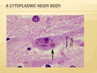
Animals are diagnosed through histological
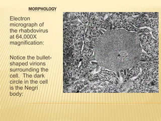
examination of the CNS for Negri bodies.
The cytoplasmic Negri body is a diagnostic of rabies
encephalitis. size (7 micrometers) and color as a
mature RBC.
brain biopsy.
Indirect immunofluorescence is most often used to
detect rabies antigen [impression smear, oronasal
mucosa scrapings, or hair follicles of the neck].](https://image.slidesharecdn.com/rhabdovirus-120725062246-phpapp01/85/Rhabdo-virus-17-320.jpg)


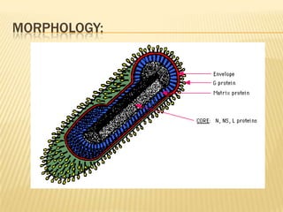
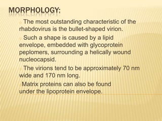
Rhabdoviruses are a family of viruses that contain negative-stranded RNA and infect vertebrates including humans. They are transmitted primarily through animal bites. The most common rhabdovirus that infects humans is the rabies virus. Rabies virus causes an acute viral infection of the central nervous system that is nearly always fatal if post-exposure prophylaxis is not administered.


