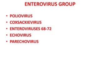
Picornaviruses
- DR. ANKUR KUMAR
Picornaviruses
TOPICS for discussion-
• Classification
• Morphology
• Enteroviruses
• Poliovirus
• Coxsackievirus
• Enteroviruses 68-72
• Echovirus
• Parechovirus
• Rhinoviruses
CLASSIFICATION
FAMILY Picronaviruses
GENUS Enteroviruses (Infects the
enteric tract)
Rhinoviruses
(Infects the
nasal
mucosa)
Hepatovir
us
Parec
hovir
us
Aphthoviru
s(foot-and-
mouth ds.
in cattle)
Cardiovi
rus(Infe
cts
mice)
GROUP
(SEROTYPES)
•Poliovirus (1-3)
•Coxsackie A (1-24)
•Coxsackie B ( 1-6)
•Echovirus (1-33)
•Parechovirus ( 1-3)
•Enteroviruses (68-72)
•34 newly identified
enteroviruses (beginning
with enterovirus 73).
NOTE-
serotype 72 is reclassified
as Hepatitis A virus
PICORNAVIRUSES- MORPHOLOGYPicornaviruses
• Simple in structure, very small size
(28-30 nm size= So, named pico)
• Nonenveloped virus.
• Spherical shaped and have
icosahedral symmetry
• Single-stranded positive sense linear
RNA (ss RNA).
• Capsid is composed of 60 subunits,
each consisting of four viral proteins
(VP1-VP4 ) -except Parechoviruses
(have three proteins).
• Resistance:
 Acid- Enteroviruses are stable at acidic
pH, whereas rhinoviruses are acid-labile.
 Heat labile: All enteroviruses and some
rhinoviruses are inactivated by heating
at 550C for 30 minutes, which can be
prevented by magnesium chloride.
ENTEROVIRUS GROUP
• POLIOVIRUS
• COXSACKIEVIRUS
• ENTEROVIRUSES 68-72
• ECHOVIRUS
• PARECHOVIRUS
POLIOVIRUSES
Antigenic Types-
• Three antigenic types of polioviruses= Type-1,
Type-2 and Type-3
Type-1 (Brunhilde and Malioney strains): Most
common serotype to cause epidemics of
poliomyelitis.
Type-2 (Lansing and MEF-1 strain): Most antigenic
serotype and most common serotype found
among the VDPV strains (vaccine derived
poliovirus)
Type-3 (Leon andSaukeustrain)- Considered as
the most common serotype to cause VAPP
(vaccine-associated paralytic poliomyelitis).
Poliovirus has two antigens: C and D
• The D antigen (dense):
 associated with the whole virion and is type-specific.
 Anti-D antibodies are protective
 Potency of the injectable polio vaccine(IPV) can be
measured in terms of D antigen units.
• C antigen (capsid):
it is associated with the noninfectious virus and is
less specific.
Anti-C antibody does not neutralize virus
infectivity.
POLIOVIRUSES
Pathogenesis
• Transmission: by feco-oral route (most common),
followed by respiratory droplets via inhalation or rarely
by conjunctival contact.
• Multiply locally: multiples in intestinal epithelial cells,
sub mucosal lymphoid tissues, tonsils and Peyer's
patches.
• Receptor: Viral entry into the host via CD155 receptors
present on the host cell surface.
• Spread to CNS/spinal cord:
Hematogenous spread (most common): Virus spreads to
the regional lymph nodes and spills over to the
bloodstream- primary viremia. After further multiplying in
the reticuloendothelial system, the virus enters the
bloodstream again, causing secondary viremia. Then it is
carried to the spinal cord and brain.
Neural spread: Virus may also spread directly through
nerves.
Pathogenesis
• Site of location:
Motor nerve ending i.e. anterior horn cells of the
spinal cord which leads to muscle weakness and
flaccid paralysis.
• Neuron degeneration
 Virus-infected neurons undergo degeneration. Earliest
change in neuron is the degeneration of Nissl body
(aggregated ribosomes, normally found in the cytoplasm
of neurons).
• Pathological changes are always more extensive than
the distribution of paralysis.
Clinical Manifestations
• Incubation period - 7-14 days.
• Manifestations- Range from asymptomatic stage to the
most severe paralytic stage.
Inapparent infection: asymptomatic cases, Most common
91-96% .
Abortive infection: About 5% of patients develop minor
symptoms such as fever, malaise, sore throat, anorexia,
myalgia and headache.
Nonparalytic poliomyelilis: seen in 1% of patients,
presented as aseptic meningitis.
Paralytic poliomyelitis- least common form < 1%. it is
characterized by descending asymmetric acute flaccid
paralysis (AFP). Proximal muscles are affected earlier than
the distal muscles; paralysis starts at hip-proceeds towards
extremities; which leads to the characteristic tripod sign
(child sits with flexed hip, both arms are extended towards
the back for support)
Clinical Manifestations
• Sites involved- spinal, bulbospinal and bulbar.
 Respiratory insufficiency or dysphagia are
common in bulbar involvement
• Biphasic course: In children, the disease
progression is typically biphasic
Aseptic meningitis occurs first---recovery----return
of fever with paralytic features 1-2days later.
Risk factors:
• Paralytic disease is more common among:
Older children and adults.
Pregnant women.
Following heavy muscular exercise.
Persons undergoing trauma at the time of CNS
symptoms.
Tonsillectomy: predisposes to bulbar poliomyelitis.
H/o injections: increase the risk of paralysis in the
involved limb.
• Postpolio muscle atrophy syndrome:
 A recrudescence of paralysis and muscle wasting has been
observed in individuals, usually decades (20-40 years)
after the episode of paralytic poliomyelitis.
Laboratory Diagnosis
Virus Isolation
• Specimen:
Throat swabs (up to 1 week of illness)
Rectal swabs or stool samples (up to 6-8 weeks)
CSF or blood- very rare.
• Transport: Specimens should be kept frozen during
transport to the laboratory (Reverse cold chain).
• Cell line: Primary monkey kidney cells (PMKC) are the
most recommended cell lines. Virus growth can be
identified by various methods.
Cytopathogenic effects- appears in 3-6days; described as
crenation and degeneration of the entire cell sheet.
Antigen detection: Isolated virus can be identified and
serotyped by neutalization with specific antiserum.
Specific gene detection by PCR assays.
Laboratory Diagnosis
Antibody Detection
• Rising antibody titer in paired sera collected 1-2
weeks interval is recommended.
• Both neutralizing antibodies (neutralization test)
and complement fixing antibodies (complement
fixation test) can be detected.
• Only first infection with poliovirus produces
strictly type-specific responses.
• Subsequent infections induce antibodies against
group specific antigen common to all the three
serotypes.
LABORATORY DIAGNOSIS- SUMMARY
Polioviruses
• Virus isolation in primary monkey kidney cell
(PMKC) line from throat swabs, rectal swabs or
stool samples-
viral growth is detected by CPE, viral antigen or
viral gene in cell line.
• Antibody detection- by neutralization test and
CFT
Vaccine
• Both inactivated(IPV) and live attenuated polio
vaccines(OPV) are available-
• Injectable Polio Vaccine (IPV, Salk Vaccine)
Discovery: Jonas Salk prepared IPV in Hela cells in 1952. An
outbreak of vaccine induced paralytic poliomyelitis had
occurred in America (1955) that had killed more than 100
people. It was due to improper inactivation of IPV. Vaccine
was modified later, after which it has been completely
safe.
Preparation: Virus is grown in monkey kidney cell line and
inactivated by formalin. Each dose (0.5 ml) of vaccine
contains 80 units of D-antigen of all the three poliovirus
serotypes (40 units of type 1, 8 units of type 2, and 32 units of type
3).
Schedule: Administered by intramuscular route in four
doses- first three doses with 1-2 months gap, 4th booster
dose is given 6- 12 months after the 3rd dose.
Efficacy- 80-90% after the full course of vaccination.
Injectable Polio Vaccine (IPV, Salk Vacdne)
Advantages:
• IPV is much safer than OPV, safer even in
immunocompromized people.
• it does not cause vaccine-associated paralytic polio (VAPP).
• more stable, does not require stringent storage conditions.
Disadvantages:
• it does not provide herd immunity: Being inactivated
vaccine, it can not spread by feco-oral rute.
• it does not useful during epidemics; as there is no
community protection. lnstead, it can precipitate paralysis.
• it does not induce mucosal lg-A production, hence the local
immunity is absent.
• it is relatively expensive than OPV.
Oral Polio Vaccine (OPV, Sabin Vaccine)
• Discovery: developed by Albert Sabin, Koprowski and
Cox (1955).
• Preparation: Each dose (0.5 mL) contains type 1 virus
(3 lakh), type 2 virus (1 lakh), type 3 virus (3 lakh) of
TCID50 (tissue culture infective dose-50).
• Schedule: OPV recommended under national
immunization schedule of India, administered orally (
2 drops/dose).
 Total five doses are given
 Zero dose: given at birth
 1st/2nd/3rd doses: given at 6/ 10/ 14 weeks
 Booster dose: Given at 16-24 months of birth
• Efficacy- 90-100%, which is achieved much faster
with one or two doses than IPV.
Advantages:
• Herd immunity: OPV strains being live, can shed in the feces and spread in
the community by feco-oral route, hence it can induce herd immunity. It
can provide both individual and community protection. OPV is the vaccine
of choice during epidemics.
• Local immunity: OPV induces mucosal lgA production, hence provides
local or mucosal immunity. Cheaper than IPV Easy to administer (given by
oral route).
Disadvantages: -
• Safety: Risky to give in immunocompromised people, pregnancy,
and old age.
• Stability: OPV is unstable vaccine, requires stringent conditions such
as: • Storage at (-200C), • Stabilized in MgCl2, • pH <7
• Efficacy of OPV decreases by:
 lnterference by other enreroviruses
 Diarrhea: OPV gets washed away in diarrheal stool.
 Breastfeeding: OPV gets washed away in stool if given immediately
before or after breast feeding. Hence, breast feeding should be
avoided before or after administration of OPV.
• OPV can cause vaccine-associated paralytic poliomyelitis (VAPP) and
vaccine-derived polioviruses (VDPV)
Oral Polio Vaccine (OPV, Sabin Vaccine)
Differences b/n injectable and oral polio vaccines-
Epidemiology
• Reservoir: Man is the only known reservoir. Most cases
are subclinical.
• Clinical-subclinical ratio: For every clinical case, there
may be 1 ,000 children and 75 adults of subclinical
cases.
• There are no chronic carriers. However,
immunodeficient individuals may excrete the virus for
longer periods.
• Source: Infective materials such as stool and
oropbaryngeal secretions are the sources of infection.
• Age: Younger children and infants are more susceptible
to infection than adults.
• Period of communicability: Patients are infectious,
shedding the virus in the feces from 7- 10 days before
the onset of symptoms up to 2-3 weeks thereafter,
sometimes as long as 3-4 months.
Epidemiology
• Polio Eradication-
 Poliomyelitis is now at the verge of eradication. its is attributed
to the extensive immunization programme being conducted
globally. Pulse Polio lmmunization (PPI) was initiated globally to
eradicate poliomyelitis. In India, it was in operation since 1995-
96.
 Two rounds of PPI (6 weeks apart) are scheduled every year
during the winter season, where all children under the age of 5
years are vaccinated with OPV irrespective of their OPV
vaccination status.
• Currently, all natural cases due to wild polio virus(wPV) are
caused by type-1.
• No natural cases due to Type-2 and 3 wPW have been
reported since 1999 and 2013 respectively and Type-2 and
3 declared eradicated in 2015 & 2019 respectively.
• Endemic (PAN) countries: Currently polio is endemic only in
three countries- Pakistan, Afghanistan and Nigeria
(abbreviated as PAN countries).
• India has been declared polio-free since January 2014, the
last natural case was detected three years back (2011).
OTHER ENTEROVIRUSES-
COXSACKIEVIRUSES
• Coxsackieviruses (named after the place of
discovery; Coxsackie village in USA) divided
into 2 groups- A and B, based on their
pathogenic potentiials in suckling mice.
• Serorypes:
Group A coxsackieviruses - serotypes 1-24 (there is
no serorype 23)
Group B - serorypes 1- 6.
Clinical Manifestations
• Incubation period – 2 to 9 days.
• Aseptic meningitis: caused by all types of group B
coxsackieviruses and by many group-A coxsackieviruses
(most commonly A7 and A9).
• Herpangina: It is a severe febrile vesicular pharyngitis
that is caused by certain group A viruses (type 2-6, 8,
10).
• Hand-foot-and-mouth disease: characterized by oral
and pharyngeal ulcerations and vesicular rashes of the
palms and soles which heal without crusting. It is
particularly associated with coxsackievirus A16.
• Pleurodynia (Bornholm disease or epidemic myalgia):
caused by coxsackie B viruses. It is characterized by
fever and abrupt onset of stabbing chest pain.
Clinical Manifestations
• Cardiac: Myocarditis and pericarditis are caused by
coxsackievirus B types 1-5.
• Respiratory: Coxsackieviruses A and B have been
associated with common colds. Pneumonia may be
caused by coxsackieviruses B 4 and 5.
• Acute hemorrhagic conjunctivitis: caused by coxsackie-
A24 and enrerovirus 70. It is a self-limiting
subconjunctival hemorrhage. Incubation period is
about 1 day. Complete recovery occurs withn 8- 10
days.
• Generalized disease of infants: It is an extremely
serious disease involving multiple organs, caused by
group B coxsackieviruses .
• Pancreatitis leading to juvenile diabetes mellitus is
caused by coxsackie B4.
Laboratory Diagnosis
• Specimen collection- depends on the type of
infection, include throat swabs, stool and CSF
• Isolation of the virus: Coxsackieviruses can be
recovered by:
 intracerebral inoculation into suckling mice-
 Coxsackie-A produce= Flaccid paralysis
 Coxsackie-B produce= Spastic paralysis
Inoculating into tissue culture: Cytopathic effect can
be observed within 5- 14 days.
• PCR - Rapid, more sensitive and serotype-specific.
• Serology is performed to detect neutralizing
antibodies.
OTHER ENTEROVIRUSES-
Enteroviruses 68-71
• Enrerovirus 68- causes pneumonia.
• Enterovirus 70- causes acute hemorrhagic
conjunctivitis. It uses CD55 as, host cell
receptor.
• Enterovirus 71- causes aseptic meningitis,
encephalitis, hand-foot and mouth disease,
herpangina, pulmonary edema and paralysis
resembling poliomyelitis.
• Enterovirus72- is reclassified as hepatitis A
virus.
OTHER ENTEROVIRUSES-
ECHOVIRUSES
• Echoviruses (Enteric Cytopathogenic Human
Orphan viruses)
Infect the human intestine and
They can be isolated in certain tissue cultures.
They were named 'orphan' viruses because at the
time of their discovery, they were not attributed to
any disease.
Serotypes 1-33 (there are no types- 10, 22, 23or28),
but not all cause human illness.
They are associated with aseptic meningitis,
encephalitis, rashes, common cold, and ocular
disease.
Disease is confirmed if the, virus is isolated from body
fluids (such as CSF) and antibodies are found in
patient's sera.
OTHER ENTEROVIRUSES-
PARECHOVIRUSES
Parechoviruses have three serotypes:
Serotype 1 and 2 were previously classified as
echoviruses 22 and 23 respectively.
Their capsid consists of three viral proteins (in
contrast to four proteins in most picornaviruses).
They have been rarely associated with aseptic
meningitis, respiratory and neonatal diseases.
RHINOVIRUS GROUP
• Rhinoviruses are the most common cause of
common cold.
• Use host cell intercellular adhesion molecule-1
(ICAM-1) as receptor.
• More than 100 serotypes have been identified.
• They are similar to enteroviruses in structure and
properties except that:
Buoyant density in cesium chloride is of 1.40 g/mL (in
contrast to 1.34 for enteroviruses).
Acid-labile (unstable below pH 6).
Transmission is by close respiratory contact via
infected secretions.
Optimal temperature for growth is 330C (in contrast to
370C for enteroviruses ).
RHINOVIRUSES
Clinical features:-
• Incubation period = 2- 4days.
• Common cold syndrome:
Rhinoviral symptoms are similar to that of any other
viruses causing common cold syndrome such as
coronaviruses, adenoviruses, enteroviruses, parainfluenza
viruses and influenza viruses.
Usual symptoms in adults include sneezing, nasal
obstruction, nasal discharge and sore throat, but no fever.
• Secondary bacterial infection may produce otitis
media, sinusitis, bronchitis, or pneumonitis, especially
in children.
• Relapse: average adult gets 1-2 attacks each year
• Infectious only for humans, gibbons and chimpanzees.
RHINOVIRUSES
Laboratory diagnosis -
• Rhinoviruses can be grown in human diploid
cell lines such as WI-38 and MRC-5 cell lines.
• Organ cultures of ferret and human tracheal
epithelium may be necessary for some
fastidious strains.
• Most of the strains grow better at 330 C
(nasopharynx temperature) but not at 370C.
Treatment -
• Supportive (i.e. symptomatic treatment).
THANK
YOU

Picornavirus

  • 1.
  • 2.
    Picornaviruses TOPICS for discussion- •Classification • Morphology • Enteroviruses • Poliovirus • Coxsackievirus • Enteroviruses 68-72 • Echovirus • Parechovirus • Rhinoviruses
  • 3.
    CLASSIFICATION FAMILY Picronaviruses GENUS Enteroviruses(Infects the enteric tract) Rhinoviruses (Infects the nasal mucosa) Hepatovir us Parec hovir us Aphthoviru s(foot-and- mouth ds. in cattle) Cardiovi rus(Infe cts mice) GROUP (SEROTYPES) •Poliovirus (1-3) •Coxsackie A (1-24) •Coxsackie B ( 1-6) •Echovirus (1-33) •Parechovirus ( 1-3) •Enteroviruses (68-72) •34 newly identified enteroviruses (beginning with enterovirus 73). NOTE- serotype 72 is reclassified as Hepatitis A virus
  • 4.
    PICORNAVIRUSES- MORPHOLOGYPicornaviruses • Simplein structure, very small size (28-30 nm size= So, named pico) • Nonenveloped virus. • Spherical shaped and have icosahedral symmetry • Single-stranded positive sense linear RNA (ss RNA). • Capsid is composed of 60 subunits, each consisting of four viral proteins (VP1-VP4 ) -except Parechoviruses (have three proteins). • Resistance:  Acid- Enteroviruses are stable at acidic pH, whereas rhinoviruses are acid-labile.  Heat labile: All enteroviruses and some rhinoviruses are inactivated by heating at 550C for 30 minutes, which can be prevented by magnesium chloride.
  • 5.
    ENTEROVIRUS GROUP • POLIOVIRUS •COXSACKIEVIRUS • ENTEROVIRUSES 68-72 • ECHOVIRUS • PARECHOVIRUS
  • 6.
    POLIOVIRUSES Antigenic Types- • Threeantigenic types of polioviruses= Type-1, Type-2 and Type-3 Type-1 (Brunhilde and Malioney strains): Most common serotype to cause epidemics of poliomyelitis. Type-2 (Lansing and MEF-1 strain): Most antigenic serotype and most common serotype found among the VDPV strains (vaccine derived poliovirus) Type-3 (Leon andSaukeustrain)- Considered as the most common serotype to cause VAPP (vaccine-associated paralytic poliomyelitis).
  • 7.
    Poliovirus has twoantigens: C and D • The D antigen (dense):  associated with the whole virion and is type-specific.  Anti-D antibodies are protective  Potency of the injectable polio vaccine(IPV) can be measured in terms of D antigen units. • C antigen (capsid): it is associated with the noninfectious virus and is less specific. Anti-C antibody does not neutralize virus infectivity. POLIOVIRUSES
  • 8.
    Pathogenesis • Transmission: byfeco-oral route (most common), followed by respiratory droplets via inhalation or rarely by conjunctival contact. • Multiply locally: multiples in intestinal epithelial cells, sub mucosal lymphoid tissues, tonsils and Peyer's patches. • Receptor: Viral entry into the host via CD155 receptors present on the host cell surface. • Spread to CNS/spinal cord: Hematogenous spread (most common): Virus spreads to the regional lymph nodes and spills over to the bloodstream- primary viremia. After further multiplying in the reticuloendothelial system, the virus enters the bloodstream again, causing secondary viremia. Then it is carried to the spinal cord and brain. Neural spread: Virus may also spread directly through nerves.
  • 9.
    Pathogenesis • Site oflocation: Motor nerve ending i.e. anterior horn cells of the spinal cord which leads to muscle weakness and flaccid paralysis. • Neuron degeneration  Virus-infected neurons undergo degeneration. Earliest change in neuron is the degeneration of Nissl body (aggregated ribosomes, normally found in the cytoplasm of neurons). • Pathological changes are always more extensive than the distribution of paralysis.
  • 10.
    Clinical Manifestations • Incubationperiod - 7-14 days. • Manifestations- Range from asymptomatic stage to the most severe paralytic stage. Inapparent infection: asymptomatic cases, Most common 91-96% . Abortive infection: About 5% of patients develop minor symptoms such as fever, malaise, sore throat, anorexia, myalgia and headache. Nonparalytic poliomyelilis: seen in 1% of patients, presented as aseptic meningitis. Paralytic poliomyelitis- least common form < 1%. it is characterized by descending asymmetric acute flaccid paralysis (AFP). Proximal muscles are affected earlier than the distal muscles; paralysis starts at hip-proceeds towards extremities; which leads to the characteristic tripod sign (child sits with flexed hip, both arms are extended towards the back for support)
  • 11.
    Clinical Manifestations • Sitesinvolved- spinal, bulbospinal and bulbar.  Respiratory insufficiency or dysphagia are common in bulbar involvement • Biphasic course: In children, the disease progression is typically biphasic Aseptic meningitis occurs first---recovery----return of fever with paralytic features 1-2days later.
  • 12.
    Risk factors: • Paralyticdisease is more common among: Older children and adults. Pregnant women. Following heavy muscular exercise. Persons undergoing trauma at the time of CNS symptoms. Tonsillectomy: predisposes to bulbar poliomyelitis. H/o injections: increase the risk of paralysis in the involved limb. • Postpolio muscle atrophy syndrome:  A recrudescence of paralysis and muscle wasting has been observed in individuals, usually decades (20-40 years) after the episode of paralytic poliomyelitis.
  • 13.
    Laboratory Diagnosis Virus Isolation •Specimen: Throat swabs (up to 1 week of illness) Rectal swabs or stool samples (up to 6-8 weeks) CSF or blood- very rare. • Transport: Specimens should be kept frozen during transport to the laboratory (Reverse cold chain). • Cell line: Primary monkey kidney cells (PMKC) are the most recommended cell lines. Virus growth can be identified by various methods. Cytopathogenic effects- appears in 3-6days; described as crenation and degeneration of the entire cell sheet. Antigen detection: Isolated virus can be identified and serotyped by neutalization with specific antiserum. Specific gene detection by PCR assays.
  • 14.
    Laboratory Diagnosis Antibody Detection •Rising antibody titer in paired sera collected 1-2 weeks interval is recommended. • Both neutralizing antibodies (neutralization test) and complement fixing antibodies (complement fixation test) can be detected. • Only first infection with poliovirus produces strictly type-specific responses. • Subsequent infections induce antibodies against group specific antigen common to all the three serotypes.
  • 15.
    LABORATORY DIAGNOSIS- SUMMARY Polioviruses •Virus isolation in primary monkey kidney cell (PMKC) line from throat swabs, rectal swabs or stool samples- viral growth is detected by CPE, viral antigen or viral gene in cell line. • Antibody detection- by neutralization test and CFT
  • 16.
    Vaccine • Both inactivated(IPV)and live attenuated polio vaccines(OPV) are available- • Injectable Polio Vaccine (IPV, Salk Vaccine) Discovery: Jonas Salk prepared IPV in Hela cells in 1952. An outbreak of vaccine induced paralytic poliomyelitis had occurred in America (1955) that had killed more than 100 people. It was due to improper inactivation of IPV. Vaccine was modified later, after which it has been completely safe. Preparation: Virus is grown in monkey kidney cell line and inactivated by formalin. Each dose (0.5 ml) of vaccine contains 80 units of D-antigen of all the three poliovirus serotypes (40 units of type 1, 8 units of type 2, and 32 units of type 3). Schedule: Administered by intramuscular route in four doses- first three doses with 1-2 months gap, 4th booster dose is given 6- 12 months after the 3rd dose. Efficacy- 80-90% after the full course of vaccination.
  • 17.
    Injectable Polio Vaccine(IPV, Salk Vacdne) Advantages: • IPV is much safer than OPV, safer even in immunocompromized people. • it does not cause vaccine-associated paralytic polio (VAPP). • more stable, does not require stringent storage conditions. Disadvantages: • it does not provide herd immunity: Being inactivated vaccine, it can not spread by feco-oral rute. • it does not useful during epidemics; as there is no community protection. lnstead, it can precipitate paralysis. • it does not induce mucosal lg-A production, hence the local immunity is absent. • it is relatively expensive than OPV.
  • 18.
    Oral Polio Vaccine(OPV, Sabin Vaccine) • Discovery: developed by Albert Sabin, Koprowski and Cox (1955). • Preparation: Each dose (0.5 mL) contains type 1 virus (3 lakh), type 2 virus (1 lakh), type 3 virus (3 lakh) of TCID50 (tissue culture infective dose-50). • Schedule: OPV recommended under national immunization schedule of India, administered orally ( 2 drops/dose).  Total five doses are given  Zero dose: given at birth  1st/2nd/3rd doses: given at 6/ 10/ 14 weeks  Booster dose: Given at 16-24 months of birth • Efficacy- 90-100%, which is achieved much faster with one or two doses than IPV.
  • 19.
    Advantages: • Herd immunity:OPV strains being live, can shed in the feces and spread in the community by feco-oral route, hence it can induce herd immunity. It can provide both individual and community protection. OPV is the vaccine of choice during epidemics. • Local immunity: OPV induces mucosal lgA production, hence provides local or mucosal immunity. Cheaper than IPV Easy to administer (given by oral route). Disadvantages: - • Safety: Risky to give in immunocompromised people, pregnancy, and old age. • Stability: OPV is unstable vaccine, requires stringent conditions such as: • Storage at (-200C), • Stabilized in MgCl2, • pH <7 • Efficacy of OPV decreases by:  lnterference by other enreroviruses  Diarrhea: OPV gets washed away in diarrheal stool.  Breastfeeding: OPV gets washed away in stool if given immediately before or after breast feeding. Hence, breast feeding should be avoided before or after administration of OPV. • OPV can cause vaccine-associated paralytic poliomyelitis (VAPP) and vaccine-derived polioviruses (VDPV) Oral Polio Vaccine (OPV, Sabin Vaccine)
  • 20.
    Differences b/n injectableand oral polio vaccines-
  • 21.
    Epidemiology • Reservoir: Manis the only known reservoir. Most cases are subclinical. • Clinical-subclinical ratio: For every clinical case, there may be 1 ,000 children and 75 adults of subclinical cases. • There are no chronic carriers. However, immunodeficient individuals may excrete the virus for longer periods. • Source: Infective materials such as stool and oropbaryngeal secretions are the sources of infection. • Age: Younger children and infants are more susceptible to infection than adults. • Period of communicability: Patients are infectious, shedding the virus in the feces from 7- 10 days before the onset of symptoms up to 2-3 weeks thereafter, sometimes as long as 3-4 months.
  • 22.
    Epidemiology • Polio Eradication- Poliomyelitis is now at the verge of eradication. its is attributed to the extensive immunization programme being conducted globally. Pulse Polio lmmunization (PPI) was initiated globally to eradicate poliomyelitis. In India, it was in operation since 1995- 96.  Two rounds of PPI (6 weeks apart) are scheduled every year during the winter season, where all children under the age of 5 years are vaccinated with OPV irrespective of their OPV vaccination status. • Currently, all natural cases due to wild polio virus(wPV) are caused by type-1. • No natural cases due to Type-2 and 3 wPW have been reported since 1999 and 2013 respectively and Type-2 and 3 declared eradicated in 2015 & 2019 respectively. • Endemic (PAN) countries: Currently polio is endemic only in three countries- Pakistan, Afghanistan and Nigeria (abbreviated as PAN countries). • India has been declared polio-free since January 2014, the last natural case was detected three years back (2011).
  • 23.
    OTHER ENTEROVIRUSES- COXSACKIEVIRUSES • Coxsackieviruses(named after the place of discovery; Coxsackie village in USA) divided into 2 groups- A and B, based on their pathogenic potentiials in suckling mice. • Serorypes: Group A coxsackieviruses - serotypes 1-24 (there is no serorype 23) Group B - serorypes 1- 6.
  • 24.
    Clinical Manifestations • Incubationperiod – 2 to 9 days. • Aseptic meningitis: caused by all types of group B coxsackieviruses and by many group-A coxsackieviruses (most commonly A7 and A9). • Herpangina: It is a severe febrile vesicular pharyngitis that is caused by certain group A viruses (type 2-6, 8, 10). • Hand-foot-and-mouth disease: characterized by oral and pharyngeal ulcerations and vesicular rashes of the palms and soles which heal without crusting. It is particularly associated with coxsackievirus A16. • Pleurodynia (Bornholm disease or epidemic myalgia): caused by coxsackie B viruses. It is characterized by fever and abrupt onset of stabbing chest pain.
  • 25.
    Clinical Manifestations • Cardiac:Myocarditis and pericarditis are caused by coxsackievirus B types 1-5. • Respiratory: Coxsackieviruses A and B have been associated with common colds. Pneumonia may be caused by coxsackieviruses B 4 and 5. • Acute hemorrhagic conjunctivitis: caused by coxsackie- A24 and enrerovirus 70. It is a self-limiting subconjunctival hemorrhage. Incubation period is about 1 day. Complete recovery occurs withn 8- 10 days. • Generalized disease of infants: It is an extremely serious disease involving multiple organs, caused by group B coxsackieviruses . • Pancreatitis leading to juvenile diabetes mellitus is caused by coxsackie B4.
  • 26.
    Laboratory Diagnosis • Specimencollection- depends on the type of infection, include throat swabs, stool and CSF • Isolation of the virus: Coxsackieviruses can be recovered by:  intracerebral inoculation into suckling mice-  Coxsackie-A produce= Flaccid paralysis  Coxsackie-B produce= Spastic paralysis Inoculating into tissue culture: Cytopathic effect can be observed within 5- 14 days. • PCR - Rapid, more sensitive and serotype-specific. • Serology is performed to detect neutralizing antibodies.
  • 27.
    OTHER ENTEROVIRUSES- Enteroviruses 68-71 •Enrerovirus 68- causes pneumonia. • Enterovirus 70- causes acute hemorrhagic conjunctivitis. It uses CD55 as, host cell receptor. • Enterovirus 71- causes aseptic meningitis, encephalitis, hand-foot and mouth disease, herpangina, pulmonary edema and paralysis resembling poliomyelitis. • Enterovirus72- is reclassified as hepatitis A virus.
  • 28.
    OTHER ENTEROVIRUSES- ECHOVIRUSES • Echoviruses(Enteric Cytopathogenic Human Orphan viruses) Infect the human intestine and They can be isolated in certain tissue cultures. They were named 'orphan' viruses because at the time of their discovery, they were not attributed to any disease. Serotypes 1-33 (there are no types- 10, 22, 23or28), but not all cause human illness. They are associated with aseptic meningitis, encephalitis, rashes, common cold, and ocular disease. Disease is confirmed if the, virus is isolated from body fluids (such as CSF) and antibodies are found in patient's sera.
  • 29.
    OTHER ENTEROVIRUSES- PARECHOVIRUSES Parechoviruses havethree serotypes: Serotype 1 and 2 were previously classified as echoviruses 22 and 23 respectively. Their capsid consists of three viral proteins (in contrast to four proteins in most picornaviruses). They have been rarely associated with aseptic meningitis, respiratory and neonatal diseases.
  • 30.
    RHINOVIRUS GROUP • Rhinovirusesare the most common cause of common cold. • Use host cell intercellular adhesion molecule-1 (ICAM-1) as receptor. • More than 100 serotypes have been identified. • They are similar to enteroviruses in structure and properties except that: Buoyant density in cesium chloride is of 1.40 g/mL (in contrast to 1.34 for enteroviruses). Acid-labile (unstable below pH 6). Transmission is by close respiratory contact via infected secretions. Optimal temperature for growth is 330C (in contrast to 370C for enteroviruses ).
  • 31.
    RHINOVIRUSES Clinical features:- • Incubationperiod = 2- 4days. • Common cold syndrome: Rhinoviral symptoms are similar to that of any other viruses causing common cold syndrome such as coronaviruses, adenoviruses, enteroviruses, parainfluenza viruses and influenza viruses. Usual symptoms in adults include sneezing, nasal obstruction, nasal discharge and sore throat, but no fever. • Secondary bacterial infection may produce otitis media, sinusitis, bronchitis, or pneumonitis, especially in children. • Relapse: average adult gets 1-2 attacks each year • Infectious only for humans, gibbons and chimpanzees.
  • 32.
    RHINOVIRUSES Laboratory diagnosis - •Rhinoviruses can be grown in human diploid cell lines such as WI-38 and MRC-5 cell lines. • Organ cultures of ferret and human tracheal epithelium may be necessary for some fastidious strains. • Most of the strains grow better at 330 C (nasopharynx temperature) but not at 370C. Treatment - • Supportive (i.e. symptomatic treatment).
  • 33.