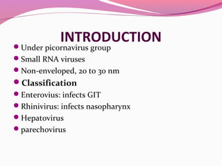
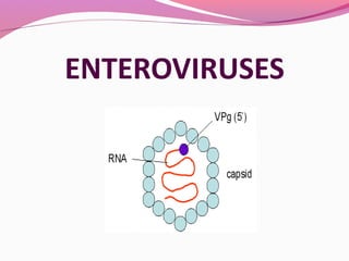
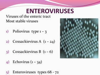
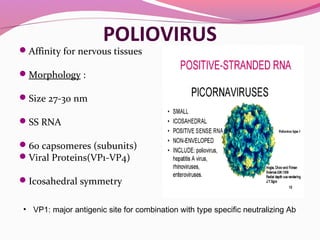
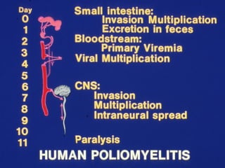
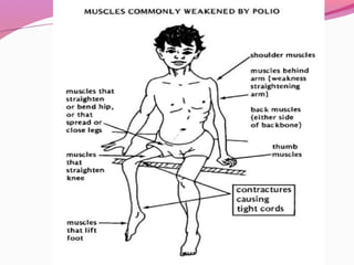
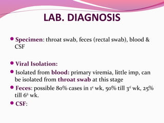
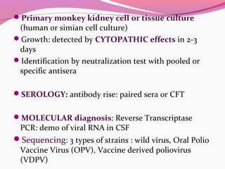
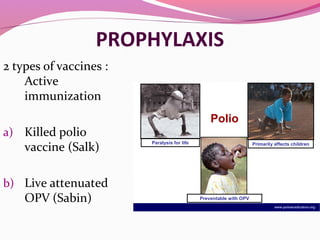
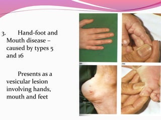
Dr V S Vatkar
Asso Prof
Microbiology Department
D Y Patil Medical College, Kolhapur
INTRODUCTIONUnder picornavirus group
Small RNA viruses
Non-enveloped, 20 to 30 nm
Classification
Enterovius: infects GIT
Rhinivirus: infects nasopharynx
Hepatovirus
parechovirus
HISTORY
Paralytic Ds of children
Landsteiner & Popper: demonstrated experimental
transmission of ds in monkeys
Ender, Weller & Robbins (1948) Nobel Prize:
demonstrated growth in non neural cell culture like:
human embryo cells , shows cytopathic changes
ENTEROVIRUSES
Viruses of the enteric tract
Most stable viruses
1) Poliovirus type 1 – 3
2) Coxsackievirus A (1 – 24)
3) Coxsackievirus B (1 – 6)
4) Echovirus (1 – 34)
5) Enteroviruses types 68 - 72
POLIOVIRUS
Affinity for nervous tissues
Morphology :
Size 27-30 nm
SS RNA
60 capsomeres (subunits)
Viral Proteins(VP1-VP4)
Icosahedral symmetry
• VP1: major antigenic site for combination with type specific neutralizing Ab
RESISTANCE
Resistant to ether ,chloroform , bile &
proteolytic enzymes of the intestinal tract,
detergents
In feces : can survive up to month at 4 0 C
Stable at pH 3
years at -20°C
55°C 30 mins
MgCL2 protects the virus from heat inactivation.
Does not survive lyophilisation
Formaldehyde & oxidising disinfectants: destroy
virus
ANTIGENIC PROPERTIES
Poliovirus strains classified into 3 types by
neutralization test – 1,2, and 3
Type 1: the commonest & causes most epidemics
Type 2: causes endemic infections
Type 3: causes epidemics, type specific
2 Antigens are recognized by CFT , ELIZA or
PPTn test -
Antigen C : heated or H Ag, associated with
empty or non - infectious virus, anti C Ab : not
protective, not neutralizes viral infectivity
Antigen D: native or N Ag, associated with whole
virion, type specific
Antigen D is converted to Antigen C by heating
the virus at 56°C, anti D Ab protective
Potency of injectable polio vaccine : measured in
terms of D Ag units.
PATHOGENESIS
Transmission: Virus enters by oral route through
ingestion food & water contaminated with human
feces.
Colonizes the nasopharynx & multiply initially
Found in throat & feces in initial phases
Then passes down to Peyer’s patches & epithelial cells
of alimentary canal & lymphatic system
Inhalation of droplets can also be a mode of entry
Goes to regional gr of LN
Enters into blood stream (Primary Viremia)
Further multiplication in reticuloendothelial system
Virus again re-enters into blood stream (secondary
Viremia)
Virus goes to spinal cord & brain (pass along axons of
peripheral nerves to CNS)
In CNS: virus multiplies selectively in neurons &
destroys them.
Degeneration of Nissl body (earliest change)
Degeneration : irreversible, necrotic cell lyses or
phagocytosis by leucocytes or macrophages
Anterior horn cells of spinal cord : mostly
involved, causes flaccid paralysis, post horn cells may
also involved
Direct neural transmission to the CNS may occur
in special cases as in poliomyelitis, following
tonsillectomy
 Some cases encephalitis may occur , involves
brainstem extending up to motor & premotor
areas of cerebral cortex
CLINICAL FEATURES
Inapparent infection: exposed to polio virus, 90-
95% susceptible individuals develop inapparent
infection, causes seroconversion only.
Only 5-10% cases show clinical illness
I P : 10 days (range 4 days – 4 weeks)
Abortive poliomyelitis: (minor illness)- earliest
manifestations like fever, headache, sore throat,
malaise , last for 1-5 days
CLINICAL FEATURES
Paralytic poliomyelitis: (Major illness)-
If the infection progresses ,the minor illness is followed by
major illness in 3-4 days
 Fever again (biphasic fever) with headache ,stiff neck and
other signs of meningitis- stage of viral invasion of the CNS
Flaccid paralysis : depending on distribution of paralysis,
classified as SPINAL, BULBAR or SPINOBULBAR
Mortality is 5-10 % mainly due to respiratory failure
Recovery of paralised ms : next 4-8 wks, complete after 6
mnths, leaving behind varying degrees of residual paralysis
Non paralytic poliomyelitis: ds dose not progress beyond
aseptic meningitis.
LAB. DIAGNOSIS
Specimen: throat swab, feces (rectal swab), blood &
CSF
Viral Isolation:
Isolated from blood: primary viremia, little imp, can
be isolated from throat swab at this stage
Feces: possible 80% cases in 1st
wk, 50% till 3rd
wk, 25%
till 6th
wk.
CSF:
Primary monkey kidney cell or tissue culture
(human or simian cell culture)
Growth: detected by CYTOPATHIC effects in 2-3
days
Identification by neutralization test with pooled or
specific antisera
SEROLOGY: antibody rise: paired sera or CFT
MOLECULAR diagnosis: Reverse Transcriptase
PCR: demo of viral RNA in CSF
Sequencing: 3 types of strains : wild virus, Oral Polio
Vaccine Virus (OPV), Vaccine derived poliovirus
(VDPV)
PROPHYLAXIS
2 types of vaccines :
Active
immunization
a) Killed polio
vaccine (Salk)
b) Live attenuated
OPV (Sabin)
www.polioeradication.org
Polio
Paralysis for life Primarily affects children
Preventable with OPV
SALKS KILLED POLIO VACCINE/INACTIVATED
POLIO VACCINE
Dev.by Salk in 1953
Formalin inactivated prep. of 3 t/o polio viruses, grown on
monkey kidney tissue culture, inactivated with formaline
(1:4000) at 37 0 C for 12-15 days
Deep subcut.or IM injection
Produces long lasting immunity
Induces serum antibodies IgM & IgG
Does not induce IgA in the intestine and hence not able to
prevent alimentary tract infections
Vaccination schedule
Three doses, given 4-6 weeks apart
Booster dose six months later
1st
dose: after 6 mnths: maternal Ab does not interfere
2nd
dose: 4 -6 wks after 1st
dose, induses better
seroconversion
3rd
dose: 6-12 mnths later,
immunity lasts for 3-5 yrs, after booster dose
LIVE ATTENUATED OPV/SABIN VACCINE
Dev.by Koprowsky, Cox & Sabin in 1962
Contains live attenuated strains of polio viruses 1,2 and 3
Shelf life 4 M at 4 - 8°C and 2 yrs.at -20°C
Improper storage conditions and cold chain failure –
problem in dev.countries
Administered orally
Stimulates both IgA and humoral antibodies IgG and IgM
OPV used in INDIA: contains
Type 1 virus : 10 lakhs
Type 2 virus : 2 lakhs
Type 3 virus : 3 lakhs
TC ID50 per dose ( 0.5 ml)
Stabilized by MgCl2
pH: 7.0
Safety
OPV : tends to acquire neurovirulence on serial
enteric passages, may seen following vaccination.
Incidence ; very low.
Vaccine associated paralytic polio or Vaccine
Derived Polio Virus (VDPV): rare strain of polio virus,
very rarely OPV strain converts into VDPV strain,
polio myelitis following vaccination
Contraindication:
immunodeficient/immunocompromised person
Vaccination schedule
Live attenuated OPV
3 doses orally
Zero dose (0 dose): at birth
First dose along with DPT at
4-6 weeks (1 & 1/2mnth)
2nd and 3rd – at an interval
of 4-6 weeks
Booster dose at the age of
16 – 24 months
Issues related to vaccine failure
Interference by other entero V, may be
Coxsackie B
Frequent diarrhoea : prevents colonization
of virus in gut
Breast feeding before & after vaccination
(maternal Ab present in breast milk)
Inhibitors of polio V : present in saliva
Proper maintenance of cold chain
PULSE POLIO IMMUNIZATION
To eradicate polio myelitis from India
Mass immunization of OPV on a single day to all
children aged 0-5 years in the community
Irrespective , whether they are vaccinated or not,
through Universal Immunization Program (UPI)
Two doses at the interval of 4-6 wks during low
polio transmission period (Nov to Feb)
Herd immunity: produced in the community
EPIDEMIOLOGY
Exclusively a Human
disease
Feces- imp. source of
spread of virus in the
community
Warm weather –
conducive to virus
spread
Type 1 – causes
epidemics
Type 2 – paralytic
poliomyelitis
WHO- 13th
May 1988-
Global eradication by
COXSACKIEVIRUS
1949 in Coxsackie village of NY
Resemble polio viruses in properties & epi.
Infects suckling mice but not the adult mice
Classified in 2 groups, based on the patho.changes they
produce
Group A and Group B
By neutralization test –
Group A – divided into 24 types
Group B - divided into 6 types
GROUP A COXSACKIE VIRUSES
1. Aseptic meningitis
2. Herpangina
(Vesicular pharyngitis) –
caused by types
2,4,5,6,8 and 10
Abrupt onset of
fever,pharyngitis ,pain in
abdomen,
headache
3. Hand-foot and
Mouth disease –
caused by types 5
and 16
Presents as a
vesicular lesion
involving hands,
mouth and feet
GROUP B COXSACKIE VIRUSES
1. Epidemic myalgia – Bornholm disease
Fever, stitch like pain in chest & abdo.
2. Myocarditis and Pericarditis – newborn
3. Aseptic meningitis with paralysis
4. Juvenile diabetes –
5. Neonatal infections –
Hepatitis,meningoencephalitis
Lab. diagnosis
Virus isolation from feces or the lesions
a. Inoculation into suckling mice
b. Tissue culture – kidney cells of monkeys
ECHOVIRUSES
Enteric Cytopathogenic Human Orphan (ECHO) viruses
34 types – 1 to 34 except 10 and 28 (Rhinoviruses)
Alimentary tract
Faeco-oral route
Some echoviruses agglutinate human RBC’s
Resistant to 20% ether
CLINICAL FEATURES
Most infections are asymptomatic
Rash with fever
Resp.diseases
Paralysis
Infantile diarrhoea
Pericarditis and
Aseptic meningitis
Lab. diagnosis
Specimens are :
1. Throat secretions
2. Stool
3. CSF
Isolation of the virus – inoculation into
kidney cells of monkey
ENTEROVIRUSES 68 - 72
68 : Isolated from pharyngeal secretions of children with
pneumonia and bronchitis
69 : Not asso. with any human disease
70 : Acute hemorrhagic conjunctivitis
71 : Isolated from cases of meningitis & encephalitis
72 : Hepatitis virus type A
Enteroviruses

Enteroviruses

  • 1.
    Dr V SVatkar Asso Prof Microbiology Department D Y Patil Medical College, Kolhapur
  • 2.
    INTRODUCTIONUnder picornavirus group SmallRNA viruses Non-enveloped, 20 to 30 nm Classification Enterovius: infects GIT Rhinivirus: infects nasopharynx Hepatovirus parechovirus
  • 4.
    HISTORY Paralytic Ds ofchildren Landsteiner & Popper: demonstrated experimental transmission of ds in monkeys Ender, Weller & Robbins (1948) Nobel Prize: demonstrated growth in non neural cell culture like: human embryo cells , shows cytopathic changes
  • 5.
    ENTEROVIRUSES Viruses of theenteric tract Most stable viruses 1) Poliovirus type 1 – 3 2) Coxsackievirus A (1 – 24) 3) Coxsackievirus B (1 – 6) 4) Echovirus (1 – 34) 5) Enteroviruses types 68 - 72
  • 6.
    POLIOVIRUS Affinity for nervoustissues Morphology : Size 27-30 nm SS RNA 60 capsomeres (subunits) Viral Proteins(VP1-VP4) Icosahedral symmetry • VP1: major antigenic site for combination with type specific neutralizing Ab
  • 7.
    RESISTANCE Resistant to ether,chloroform , bile & proteolytic enzymes of the intestinal tract, detergents In feces : can survive up to month at 4 0 C Stable at pH 3 years at -20°C 55°C 30 mins MgCL2 protects the virus from heat inactivation. Does not survive lyophilisation Formaldehyde & oxidising disinfectants: destroy virus
  • 8.
    ANTIGENIC PROPERTIES Poliovirus strainsclassified into 3 types by neutralization test – 1,2, and 3 Type 1: the commonest & causes most epidemics Type 2: causes endemic infections Type 3: causes epidemics, type specific 2 Antigens are recognized by CFT , ELIZA or PPTn test -
  • 9.
    Antigen C :heated or H Ag, associated with empty or non - infectious virus, anti C Ab : not protective, not neutralizes viral infectivity Antigen D: native or N Ag, associated with whole virion, type specific Antigen D is converted to Antigen C by heating the virus at 56°C, anti D Ab protective Potency of injectable polio vaccine : measured in terms of D Ag units.
  • 10.
    PATHOGENESIS Transmission: Virus entersby oral route through ingestion food & water contaminated with human feces. Colonizes the nasopharynx & multiply initially Found in throat & feces in initial phases Then passes down to Peyer’s patches & epithelial cells of alimentary canal & lymphatic system Inhalation of droplets can also be a mode of entry
  • 11.
    Goes to regionalgr of LN Enters into blood stream (Primary Viremia) Further multiplication in reticuloendothelial system Virus again re-enters into blood stream (secondary Viremia) Virus goes to spinal cord & brain (pass along axons of peripheral nerves to CNS)
  • 12.
    In CNS: virusmultiplies selectively in neurons & destroys them. Degeneration of Nissl body (earliest change) Degeneration : irreversible, necrotic cell lyses or phagocytosis by leucocytes or macrophages Anterior horn cells of spinal cord : mostly involved, causes flaccid paralysis, post horn cells may also involved
  • 13.
    Direct neural transmissionto the CNS may occur in special cases as in poliomyelitis, following tonsillectomy  Some cases encephalitis may occur , involves brainstem extending up to motor & premotor areas of cerebral cortex
  • 14.
    CLINICAL FEATURES Inapparent infection:exposed to polio virus, 90- 95% susceptible individuals develop inapparent infection, causes seroconversion only. Only 5-10% cases show clinical illness I P : 10 days (range 4 days – 4 weeks) Abortive poliomyelitis: (minor illness)- earliest manifestations like fever, headache, sore throat, malaise , last for 1-5 days
  • 15.
    CLINICAL FEATURES Paralytic poliomyelitis:(Major illness)- If the infection progresses ,the minor illness is followed by major illness in 3-4 days  Fever again (biphasic fever) with headache ,stiff neck and other signs of meningitis- stage of viral invasion of the CNS Flaccid paralysis : depending on distribution of paralysis, classified as SPINAL, BULBAR or SPINOBULBAR Mortality is 5-10 % mainly due to respiratory failure Recovery of paralised ms : next 4-8 wks, complete after 6 mnths, leaving behind varying degrees of residual paralysis Non paralytic poliomyelitis: ds dose not progress beyond aseptic meningitis.
  • 19.
    LAB. DIAGNOSIS Specimen: throatswab, feces (rectal swab), blood & CSF Viral Isolation: Isolated from blood: primary viremia, little imp, can be isolated from throat swab at this stage Feces: possible 80% cases in 1st wk, 50% till 3rd wk, 25% till 6th wk. CSF:
  • 20.
    Primary monkey kidneycell or tissue culture (human or simian cell culture) Growth: detected by CYTOPATHIC effects in 2-3 days Identification by neutralization test with pooled or specific antisera SEROLOGY: antibody rise: paired sera or CFT MOLECULAR diagnosis: Reverse Transcriptase PCR: demo of viral RNA in CSF Sequencing: 3 types of strains : wild virus, Oral Polio Vaccine Virus (OPV), Vaccine derived poliovirus (VDPV)
  • 21.
    PROPHYLAXIS 2 types ofvaccines : Active immunization a) Killed polio vaccine (Salk) b) Live attenuated OPV (Sabin) www.polioeradication.org Polio Paralysis for life Primarily affects children Preventable with OPV
  • 22.
    SALKS KILLED POLIOVACCINE/INACTIVATED POLIO VACCINE Dev.by Salk in 1953 Formalin inactivated prep. of 3 t/o polio viruses, grown on monkey kidney tissue culture, inactivated with formaline (1:4000) at 37 0 C for 12-15 days Deep subcut.or IM injection Produces long lasting immunity Induces serum antibodies IgM & IgG Does not induce IgA in the intestine and hence not able to prevent alimentary tract infections
  • 23.
    Vaccination schedule Three doses,given 4-6 weeks apart Booster dose six months later 1st dose: after 6 mnths: maternal Ab does not interfere 2nd dose: 4 -6 wks after 1st dose, induses better seroconversion 3rd dose: 6-12 mnths later, immunity lasts for 3-5 yrs, after booster dose
  • 24.
    LIVE ATTENUATED OPV/SABINVACCINE Dev.by Koprowsky, Cox & Sabin in 1962 Contains live attenuated strains of polio viruses 1,2 and 3 Shelf life 4 M at 4 - 8°C and 2 yrs.at -20°C Improper storage conditions and cold chain failure – problem in dev.countries Administered orally Stimulates both IgA and humoral antibodies IgG and IgM
  • 25.
    OPV used inINDIA: contains Type 1 virus : 10 lakhs Type 2 virus : 2 lakhs Type 3 virus : 3 lakhs TC ID50 per dose ( 0.5 ml) Stabilized by MgCl2 pH: 7.0
  • 26.
    Safety OPV : tendsto acquire neurovirulence on serial enteric passages, may seen following vaccination. Incidence ; very low. Vaccine associated paralytic polio or Vaccine Derived Polio Virus (VDPV): rare strain of polio virus, very rarely OPV strain converts into VDPV strain, polio myelitis following vaccination Contraindication: immunodeficient/immunocompromised person
  • 27.
    Vaccination schedule Live attenuatedOPV 3 doses orally Zero dose (0 dose): at birth First dose along with DPT at 4-6 weeks (1 & 1/2mnth) 2nd and 3rd – at an interval of 4-6 weeks Booster dose at the age of 16 – 24 months
  • 28.
    Issues related tovaccine failure Interference by other entero V, may be Coxsackie B Frequent diarrhoea : prevents colonization of virus in gut Breast feeding before & after vaccination (maternal Ab present in breast milk) Inhibitors of polio V : present in saliva Proper maintenance of cold chain
  • 29.
    PULSE POLIO IMMUNIZATION Toeradicate polio myelitis from India Mass immunization of OPV on a single day to all children aged 0-5 years in the community Irrespective , whether they are vaccinated or not, through Universal Immunization Program (UPI) Two doses at the interval of 4-6 wks during low polio transmission period (Nov to Feb) Herd immunity: produced in the community
  • 30.
    EPIDEMIOLOGY Exclusively a Human disease Feces-imp. source of spread of virus in the community Warm weather – conducive to virus spread Type 1 – causes epidemics Type 2 – paralytic poliomyelitis WHO- 13th May 1988- Global eradication by
  • 31.
    COXSACKIEVIRUS 1949 in Coxsackievillage of NY Resemble polio viruses in properties & epi. Infects suckling mice but not the adult mice Classified in 2 groups, based on the patho.changes they produce Group A and Group B By neutralization test – Group A – divided into 24 types Group B - divided into 6 types
  • 32.
    GROUP A COXSACKIEVIRUSES 1. Aseptic meningitis 2. Herpangina (Vesicular pharyngitis) – caused by types 2,4,5,6,8 and 10 Abrupt onset of fever,pharyngitis ,pain in abdomen, headache
  • 33.
    3. Hand-foot and Mouthdisease – caused by types 5 and 16 Presents as a vesicular lesion involving hands, mouth and feet
  • 34.
    GROUP B COXSACKIEVIRUSES 1. Epidemic myalgia – Bornholm disease Fever, stitch like pain in chest & abdo. 2. Myocarditis and Pericarditis – newborn 3. Aseptic meningitis with paralysis 4. Juvenile diabetes – 5. Neonatal infections – Hepatitis,meningoencephalitis
  • 35.
    Lab. diagnosis Virus isolationfrom feces or the lesions a. Inoculation into suckling mice b. Tissue culture – kidney cells of monkeys
  • 36.
    ECHOVIRUSES Enteric Cytopathogenic HumanOrphan (ECHO) viruses 34 types – 1 to 34 except 10 and 28 (Rhinoviruses) Alimentary tract Faeco-oral route Some echoviruses agglutinate human RBC’s Resistant to 20% ether
  • 37.
    CLINICAL FEATURES Most infectionsare asymptomatic Rash with fever Resp.diseases Paralysis Infantile diarrhoea Pericarditis and Aseptic meningitis
  • 39.
    Lab. diagnosis Specimens are: 1. Throat secretions 2. Stool 3. CSF Isolation of the virus – inoculation into kidney cells of monkey
  • 40.
    ENTEROVIRUSES 68 -72 68 : Isolated from pharyngeal secretions of children with pneumonia and bronchitis 69 : Not asso. with any human disease 70 : Acute hemorrhagic conjunctivitis 71 : Isolated from cases of meningitis & encephalitis 72 : Hepatitis virus type A