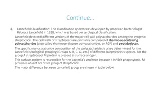
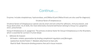
Streptococcus
By: Raju Yadav
M.Sc. Medical Microbiology
Introduction
Streptococcus pyogenes, also known as Group A Streptococcus (GAS), is a highly contagious, gram-positive
bacterium primarily found in the human respiratory tract and on the skin.
It is a major human pathogen responsible for a wide spectrum of diseases, ranging from common mild
infections like strep throat to life-threatening conditions like "flesh-eating" disease.
Some Key characteristics are as follows:-
Shape and Arrangement: Spherical cocci that grow in pairs or long, berry-like chains.
Hemolysis: Classified as beta-hemolytic, meaning it causes complete destruction of red blood cells when
grown on blood agar, creating a clear zone around colonies.
Biochemical Profile: Catalase-negative (distinguishes it from Staphylococcus) and sensitive to the
antibiotic bacitracin.
S. pyogens can be found as a commensal in the upper respiratory tract, particularly of children.
Classification of Streptococcus
The classification of Streptococci is based on the following ways:
1. Oxygen requirements:
I. Aerobic or facultative anaerobes: E.g. Streptococcus spp
II. Obligate anaerobes: E.g. Peptostreptococcus
2. Brown Classification: This classification was described by J.H
Brown in 1919. He classified Streptococci on the basis of
hemolytic pattern on blood agar.
i. Alpha-hemolysis group:
Form incomplete hemolysis on blood agar
Shows greenish discoloration around colony and widespread of
some unhemolysed RBCs. E.g. Streptococcus pneumoniae and
Viridians Streptococci
Continue…
II. Beta-hemolysis group:
Form complete hemolysis on blood agar
Give 2-3mm diameter zone of hemolysis. E.g. Streptococcus pyogens
III. Non-hemolysis group
Does not cause hemolysis at all
They simply form the colony on the blood agar. E.g. Streptococcus faecalis
3. Shermann’s Classification: This classification is based on the basis of physiological characteristics into four groups:
i. Pyogenic: Includes disease-causing streptococci (e.g., Group A, B, C), often β-hemolytic.
ii. Viridans: Found in the mouth and upper respiratory tract, typically non-β-hemolytic, less tolerant to salt/high
pH.
iii. Lactic: Dairy-related streptococci, non-pathogenic, growing at 10°C but not 45°C.
iv. Enterococci: Salt-tolerant (6.5% NaCl) and grow at high pH (9.6), now often classified as the
genus Enterococcus.
Continue…
4. Lancefield Classification: This classification system was developed by American bacteriologist
Rebecca Lancefield in 1928, which was based on serological classification.
Lancefield detected different versions of the major cell wall polysaccharides among the pyogenic
streptococci. The cell walls of streptococci are primarily composed of rhamnose-containing
polysaccharides (also called rhamnose-glucose polysaccharides, or RGP) and peptidoglycan.
The specific monosaccharide composition of the polysaccharides is a key determinant for the
Lancefield serological grouping (Groups A, B, C, G, etc.) of different Streptococcus species. For the
group A streptococci M protein is present as surface antigen.
This surface antigen is responsible for the bacteria’s virulence because it inhibit phagocytosis. M
protein is absent on other group of streptococci.
The major difference between Lancefield group are shown in table below.
Lancefield Group Characteristic Carbohydrate(s)
Group A (S.
pyogenes)
Polyrhamnose backbone with N-acetylglucosamine ( -linked GlcNAc) side
𝛽
chains.
Group B (S.
agalactiae)
Rhamnose-rich polysaccharide with galactose and N-acetylglucosamine side
chains.
Group C (S.
dysgalactiae)
Primarily rhamnose and N-acetylgalactosamine (GalNAc).
Group
D (Enterococcus
spp.)
Contains rhamnose, glucose, galactose, N-acetylglucosamine, and N-
acetylgalactosamine; the group antigen is an intracellular teichoic acid.
Group G (S.
dysgalactiae)
Composed of rhamnose, N-acetylgalactosamine, and galactose, where
rhamnose is a major antigenic determinant.
Group H (S. sanguis) Contains a group-specific antigen made of glycerol, phosphate, and glucose,
often with α-glucosidic linkages.
S. mutans (serotype
c)
Polyrhamnose backbone with glucose side chains, often with glycerol
phosphate modifications.
Continue…
Group F streptococci are primarily members of the Streptococcus milleri group (like S. anginosus, S.
constellatus, S. intermedius), common commensals of the mouth, gut, and perineum that cause
abscesses and purulent infections in various body sites (brain, lungs, abdomen, tissues) when they
cause disease, often linked to trauma or underlying issues.
Group F antigen is composed of rhamnose, glucose, and galactosamine, with a small percentage of
glucosamine. There are over 20 Lancefield groups.
Most of the Lancefield group organism shows the beta hemolysis on blood agar. Groups (A, C, F, G, L)
are primarily beta-hemolytic, Group B (S. agalactiae) and certain Group D (like Enterococci), can be
non-hemolytic or weakly hemolytic (alpha).
Extracellular enzymes and toxin produced by S. pyogens
Streptolysins (toxins that haemolysed red cells):-
Streotolysin S that is active aerobically (beta-hemolysis on blood agar). It is non-antigenic.
Stroptolysin O that hemolyzes red cells under anaerobic conditions, e.g. sub-surface agar stabs.
It is antigenic, stimulating the production of antistreptolysin O antibody.
Streptokinase- a protease that lyzes fibrin
Hyaluronidase: Facilitates spread in the tissues by destroying hyaluronic acid. Streptococcal
hyaluronidase is antigenic (antibody formed after infection).
Leukocidin: Destroys leucocytes
Lipoteichoic acid: Facilitates adherence to pharyngeal epithelial cells.
M-protein (antigens): Anti-phagocytic virulence factors (different for different strains). Antibodies to
M antigens are protective. Selected M serotypes appear to be associated with rheumatic fever,
acute glomerulonephritis and severe S.pyogens infection.
Continue…
NADase (nicotinamide adenine dinucleotidease): Kills leukocytes. Antibody formed after infection.
DNA-ase (deoxyribonuclease) A,B,C,D that break down DNA and stimulate an antibody response,
particularly against DNA-ase B, Anti-DNA-ase B tests are available.
Erythrogenic toxin: Responsible for the rash seen in scarlet fever and is also associated with streptococcal
toxic shock syndrome. It is produced as a result of a lysogenic phage present in the streptococci.
Cultural characteristics of Streptococcus pyogens
The media used for culture of S. pyogens is the blood agar. S. pyogens produces beta haemolytic colonies,
i.e. the colonies are surrounded by a zone of complete hemolysis with decolorization of the hemoglobin.
Colonies are usually small (0.5-1 mm), dry, shiny or mucoid. Hemolysis is more marked under anaerobic
conditions.
Continue…
Choice of blood
To isolate beta-haemolytic streptococci, use sheep blood (1st
choice), horse, rabbit or goat blood to prepare blood
agar plates. Do not use human blood because this may contain unwanted substances such as citrate, antibiotics or
antibodies such as ASO or anti-M protein that could interfere with the growth or hemolytic activity of S. pyogens.
Other selective agents used in blood agar are:-
Neomycin & Polymyxin B: Common antibiotics in Strep Selective Agar to inhibit Gram-negative rods, staphylococci,
and other background flora.
Crystal Violet: Used in some blood agars to inhibit Staphylococci and Gram-negatives.
Colistin & Nalidixic Acid: Another antibiotic combination for selectivity.
Nutrient Enhancers: Ribonucleic acid (RNA) and maltose can be added to boost Streptolysin S production,
enhancing the visible hemolysis.
Continue…
PNF Agar (Phenylethyl Alcohol with Neomycin & Fucidin):PNF Agar is a selective blood agar
medium containing Phenylethyl alcohol, Neomycin, and Fusidic acid, used for the selective isolation
of Streptococcus pyogenes and other hemolytic streptococci, especially from clinical specimens with
mixed bacterial flora like burn swabs.
The medium combines several agents to achieve selectivity:
Phenylethyl Alcohol (PEA): The primary selective agent, it largely inhibits the growth of gram-
negative bacteria by interfering with their DNA synthesis and damaging the cell membrane. It also
prevents the swarming phenomenon often seen with Proteus species.
Neomycin and Fusidic Acid: These antibiotics further inhibit a range of common contaminants,
including Staphylococcus aureus, Pseudomonas pyocyanea, and E. coli, while allowing the growth of
streptococci.
Colonies are tiny, translucent to grayish-white colonies that are pinpoint in size (0.5-1mm) and
surrounded by a clear, wide halo of beta-hemolysis (complete red blood cell lysis) due to
streptolysin S and O, often appearing more distinct in stabbed areas where oxygen is limited
Continue…
Strep Selective Agar is a specialized culture medium used in microbiology to isolate and grow
various Streptococcus species (like Group A, B, C, D) from mixed samples, especially from the
respiratory tract, by inhibiting competing bacteria (Gram-negative rods, Staphylococci) with selective
agents like sodium azide, crystal violet, colistin, or neomycin,
while blood or nutrients support streptococcal growth and hemolysis.
Selective Agents:
Sodium Azide/Sulphite: Inhibits Gram-negative bacteria.
Crystal Violet: Suppresses Staphylococci.
Colistin/Neomycin/Polymyxin B: Target Gram-negative flora, Pseudomonas etc.
On Strep Selective Agar, Streptococcus pyogenes (Group A Strep) colonies appear as tiny, pinpoint-
sized, gray-white, dome-shaped, smooth, and moist, often displaying distinct β-hemolysis.
Biochemical test foe streptococcus
Biochemical identification of Streptococcus species involves confirming they are Gram-positive, chain-forming
cocci that are catalase-negative.
Core Biochemical Tests for Streptococcus:
Catalase Test: Negative (no bubbles when mixed with 3% H2O2). This is the primary test to distinguish
Streptococcus (negative) from Staphylococcus (positive)
Hemolysis on Blood Agar:
Beta-hemolysis ( ): Complete lysis of red blood cells (e.g.,
𝛽 S. pyogenes, S. agalactiae).
Alpha-hemolysis ( ): Partial/greenish lysis (e.g.,
𝛼 S. pneumoniae, viridans group).
Gamma-hemolysis ( ): No lysis.
𝛾
PYR Test (Pyrrolidonyl aminopeptidase): Positive (cherry red color) for Group A Strep (S. pyogenes) and
Enterococcus.
Continue…
Bacitracin Susceptibility (Taxo A): Susceptible for S.
pyogenes (Group A), resistant for others.
CAMP Test: Positive for S. agalactiae (Group B), showing an
arrowhead-shaped zone of increased hemolysis.
Optochin Susceptibility (Taxo P): Susceptible for S. pneumoniae.
Bile Solubility Test: S. pneumoniae colonies dissolve in bile salt
solution.
6.5% NaCl Growth: Used to identify Enterococcus (positive) and
distinguish it from Streptococcus.
Esculin Hydrolysis (Bile Esculin Agar): Positive for Group D
streptococci and Enterococcus (blackening of the medium.
Species-Specific Biochemical Tests
Test Target Species Result Clinical Use
Bacitracin Sensitivity S. pyogenes (Group A) Sensitive (Zone of inhibition) Differentiates Group A from
other -hemolytic streps.
𝛽
PYR Test S. pyogenes (Group A) Positive (Red color) Rapidly identifies S. pyogenes and
Enterococcus.
CAMP Test S. agalactiae (Group B) Positive (Arrowhead hemolysis) Presumptive identification of
Group B Strep.
Optochin Sensitivity S. pneumoniae Sensitive Distinguishes S. pneumoniae from
other -hemolytic streptococci.
𝛼
Bile Solubility S. pneumoniae Soluble (Clearing of turbidity) Gold standard for identifying S.
pneumoniae.
Hippurate Hydrolysis S. agalactiae (Group B) Positive (Deep purple/precipitate) Differentiates S. agalactiae from
other -hemolytic streps.
𝛽
Bile Esculin Test Group D Strep / Enterococcus Positive (Blackening of media) Differentiates Group D from other
viridans streptococci.
Antigenic property of streptococcus
The antigenic properties of Streptococcus are primarily defined by the complex chemical structures found
in their cell walls and the exotoxins they secrete. These antigens are the basis for medical classification
(Lancefield grouping) and are central to the pathogenesis of diseases like rheumatic fever and scarlet fever.
1. Cell Wall Antigens
Group-Specific Carbohydrate (C-Substance): Most streptococci are classified into Lancefield Groups (A-
V) based on these carbohydrate antigens in the cell wall.
Group A (S. pyogenes): Contains a polymer of rhamnose and N-acetylglucosamine.
Group B (S. agalactiae): Identified by a specific polysaccharide capsule.
M Protein (Type-Specific Antigen): A major virulence factor found in Group A Streptococci (GAS). It is
strongly antigenic and determines the specific serotype (over 150 types).
Function: It resists phagocytosis by inhibiting the complement pathway.
Continue…..
Molecular Mimicry: Parts of the M protein share structural similarities with human cardiac muscle, which can
trigger an autoimmune response leading to rheumatic fever.
T and R Proteins: These are surface antigens used for serological typing but, unlike M protein, are not directly
linked to virulence.
Polysaccharide Capsule: In S. pneumoniae (which lacks a Lancefield antigen), the capsule is the primary
antigen, with over 90 distinct serotypes.
2. Extracellular (Secreted) Antigens: Streptococci secrete various proteins that act as antigens and are used
for clinical diagnosis:
Streptolysin O (SLO): A strongly antigenic oxygen-labile toxin. Elevated Antistreptolysin O (ASO) titers in blood
indicate a recent streptococcal infection.
Erythrogenic Toxins (Pyrogenic Exotoxins): Responsible for the characteristic rash of scarlet fever. These often
act as superantigens, causing a massive, non-specific release of cytokines.
Continue…..
Enzymes: Includes streptokinase, hyaluronidase, and DNAse B (anti-DNAse B tests are also used for diagnosis).
Virulence factor of streptococcus:
Virulence factors of Streptococcus species vary by strain and are critical for adhesion, immune evasion, and
tissue destruction. As of 2026, the most extensively characterized factors belong to Group A (S. pyogenes)
and S. pneumoniae.
Group A Streptococcus (S. pyogenes): The primary virulence factor for Group A Streptococcus is the M protein,
which is essential for survival in human blood.
1. Adhesion & Invasion:
M Protein: Inhibits opsonization by binding complement regulators and fibrinogen.
Lipoteichoic Acid & Protein F: Mediate attachment to host cells.
FbaA & FbaB: Fibronectin-binding proteins that aid in tissue invasion.
Continue….
2. Immune Evasion:
Hyaluronic Acid Capsule: Masks the bacteria to prevent phagocytosis.
C5a Peptidase (ScpA): Cleaves the chemotaxin C5a to prevent neutrophil recruitment.
SpyCEP: A serine protease that inactivates IL-8, further impeding neutrophil migration.
DNases (e.g., Sda1): Degrade Neutrophil Extracellular Traps (NETs) to escape killing.
3. Toxins & Enzymes:
Streptolysin O (SLO) & S (SLS): Pore-forming toxins that lyse red blood cells (hemolysis) and
immune cells.
Streptokinase: Activates plasminogen to plasmin, promoting the spread of bacteria through
tissues.
Superantigens (SpeA, SpeC): Induce massive cytokine release, leading to toxic shock-like syndrome.
Continue….
Streptococcus pneumoniae
Polysaccharide Capsule: The most critical factor for S. pneumoniae; it provides antiphagocytic activity and is used for
serotyping.
Pneumolysin (PLY): A cholesterol-dependent cytotoxin that forms pores in host cell membranes and activates
complement.
Autolysin (LytA): Breaks down the bacterial cell wall to release internal toxins like pneumolysin.
PspA & PspC: Choline-binding proteins that interfere with the complement system.
PsaA: A surface lipoprotein involved in manganese transport and adherence.
Group B Streptococcus (S. agalactiae)
Capsular Polysaccharide (CPS): Inhibits phagocytosis and resists killing by host cells.
β-hemolysin/Cytolysin (CylE): Causes tissue damage and helps cross the blood-brain barrier.
Continue…
CAMP Factor: Works synergistically with Staphylococcus aureus β-toxin to form pores in target cells.
HvgA: A hypervirulent adhesin that enables crossing of the intestinal and blood-brain barriers.
Disease caused by Streptococcus
Streptococcal bacteria cause a wide range of diseases in 2026, varying from mild throat and skin infections
to life-threatening invasive conditions. These bacteria are primarily categorized into Group A, Group B,
and Streptococcus pneumoniae, each associated with specific illnesses.
Common Streptococcal Diseases
Strep Throat (Pharyngitis): A common Group A infection causing sudden sore throat, fever, and swollen
tonsils often with white patches.
Scarlet Fever: A red, sandpaper-like rash and "strawberry tongue" that often follows a strep throat infection.
Fig: Scarlet fever.
Continue…
Skin Infections:
Impetigo: Contagious sores that break open and
develop honey-colored crusts.
Cellulitis: Infection of the deeper skin layers
causing redness, warmth, and swelling.
Erysipelas: A superficial skin infection involving a
raised, bright red rash.
Pneumonia: Inflammation of the lungs, most
commonly caused by Streptococcus pneumoniae.
Meningitis: Infection of the membranes covering
the brain and spinal cord, frequently seen in
newborns due to Group B strep.
Figure: Impetigo, Cellulitis and Erysipelas.
Continue….
Severe Invasive Diseases (iGAS): These occur when bacteria enter parts of the body that are normally
sterile, such as the blood or deep tissue.
Necrotizing Fasciitis: Also known as "flesh-eating disease," this is a rapidly progressing infection that
destroys skin, fat, and muscle.
Streptococcal Toxic Shock Syndrome (STSS): A life-threatening condition causing rapid drop in blood
pressure and multiple organ failure.
Bacteremia/Sepsis: Bloodstream infections that can lead to systemic inflammation and shock.
Post-Infectious Complications: Delayed immune-mediated reactions can occur weeks after the initial
infection.
Rheumatic Fever: An inflammatory disease that can affect the heart, joints, and brain, potentially leading
to permanent heart valve damage.
Post-Streptococcal Glomerulonephritis (PSGN): A kidney disease causing blood in the urine, facial swelling,
and high blood pressure.
PANDAS: A controversial link between strep infections and sudden onset of neuropsychiatric symptoms like
OCD or tics in children.
Diagnostic, Prevention and Control
Diagnostic methods for Streptococcus (most commonly Group A Strep or Strep Throat) vary
depending on whether the infection is acute or a post-streptococcal complication. As of 2026, the
primary diagnostic approach combines clinical screening with rapid testing or gold-standard
laboratory cultures.
1. Clinical Screening: Healthcare providers first assess symptoms using standardized clinical
decision rules like the Centor Criteria or McIsaac Score to determine if testing is necessary.
Key Indicators: Sudden fever (≥38°C), absence of cough, swollen tonsils with white patches
(exudates), and tender neck glands.
Guideline: Patients with a low clinical score (0–1) typically do not require testing.
2. Acute Infection Testing: If strep is suspected, one or more of the following laboratory tests are
used:
Rapid Antigen Detection Test (RADT): A throat swab that provides results in 10–20 minutes.
While highly specific (positive results are definitive), it can have varying sensitivity, sometimes
missing infections.
Continue…
Throat Culture (Gold Standard): A swab sample is grown on a blood agar plate for 24–48 hours. This
is the most accurate method and is often used to confirm negative rapid tests in children.
Molecular Tests (NAAT/PCR): These advanced tests (e.g., cobas Strep A) detect bacterial DNA. They
are faster than cultures and more sensitive than rapid antigen tests, but are generally more
expensive.
3. Laboratory Identification (Microbiology): In a lab setting, Streptococcus pyogenes (Group A) is
identified by specific biochemical markers:
Gram Stain: Shows Gram-positive cocci in chains.
Hemolysis: On blood agar, it shows beta-hemolysis (complete destruction of red blood cells,
creating a clear zone around colonies).
Bacitracin Sensitivity: Most Group A Strep are sensitive to the antibiotic bacitracin, which helps
distinguish it from other groups.
PYR Test: A rapid colorimetric test that turns red in the presence of specific enzymes found in
Group A Strep.
Continue….
4. Serological Testing (Antibodies): Serology is used to diagnose post-streptococcal
complications (like rheumatic fever or kidney disease) rather than acute infections.
ASO Titer (Antistreptolysin O): Measures antibodies against a toxin produced by the bacteria;
levels peak 3–6 weeks after infection.
Anti-DNase B: Often more reliable than ASO for confirming recent skin infections.
5. Other Streptococcal Species:
Group B Strep (GBS): Diagnosed via vaginal-rectal cultures in pregnant individuals at 36–37 weeks.
Streptococcus pneumoniae: Identified through sputum culture, urine antigen tests, or optochin
sensitivity (unlike Group A, it is sensitive to optochin).
Prevention and control
Prevention and control of Streptococcus infections—including Group A Strep (GAS), which causes strep
throat and skin infections, and Streptococcus pneumoniae that rely on a combination of hygiene, medical
treatment, and vaccination.
A. Personal Prevention and Hygiene: The most effective way to stop the spread of Streptococcus is through
consistent hygiene practices.
Hygiene: Wash hands frequently with soap and water for at least 20 seconds, especially after coughing
or sneezing, and before preparing food or eating.
Respiratory Etiquette: Cover mouth and nose with a tissue when coughing or sneezing, and dispose of
used tissues immediately.
Personal Item Hygiene: Avoid sharing cups, utensils, and personal items (e.g., towels) with individuals
who are sick.
Wound Care: Clean and cover skin infections or open wounds, as Streptococcus can cause infections
such as cellulitis.
Continue….
B. Medical Control and Treatment: Early diagnosis and full completion of treatment are vital for
both individual recovery and public safety.
Antibiotics: The common antibiotics recommended for the streptococcal infection are
Penicillin or Amoxicillin, Cephalexin (if the allergy is not severe), Azithromycin, or Clindamycin.
Stay Home While Sick: To prevent community spread, individuals with strep infections should
stay home from school or work for at least 12 to 24 hours after starting antibiotic treatment and
until they are fever-free.
C. Vaccination:
Pneumococcal Vaccines: Vaccines are available to prevent infections caused by Streptococcus
pneumoniae (such as pneumonia and meningitis).
Group A Strep: There is currently no vaccine available for Group A Streptococcus, though several are
in development. Staying up to date with influenza, COVID-19, and chickenpox vaccines may
indirectly reduce the risk of secondary invasive GAS infections.
Continue….
D. Environmental and Institutional Control:
Surface Disinfection: Regularly clean frequently touched surfaces like door handles and light
switches.
Ventilation: Improve indoor airflow by opening windows and doors to reduce the concentration of
respiratory droplets.
Healthcare Precautions: In hospitals, control measures include using personal protective
equipment (PPE), isolating infected patients, and screening staff if an outbreak is suspected.

Streptococcus pathogenic species....pptx

  • 1.
  • 2.
    Introduction Streptococcus pyogenes, alsoknown as Group A Streptococcus (GAS), is a highly contagious, gram-positive bacterium primarily found in the human respiratory tract and on the skin. It is a major human pathogen responsible for a wide spectrum of diseases, ranging from common mild infections like strep throat to life-threatening conditions like "flesh-eating" disease. Some Key characteristics are as follows:- Shape and Arrangement: Spherical cocci that grow in pairs or long, berry-like chains. Hemolysis: Classified as beta-hemolytic, meaning it causes complete destruction of red blood cells when grown on blood agar, creating a clear zone around colonies. Biochemical Profile: Catalase-negative (distinguishes it from Staphylococcus) and sensitive to the antibiotic bacitracin. S. pyogens can be found as a commensal in the upper respiratory tract, particularly of children.
  • 3.
    Classification of Streptococcus Theclassification of Streptococci is based on the following ways: 1. Oxygen requirements: I. Aerobic or facultative anaerobes: E.g. Streptococcus spp II. Obligate anaerobes: E.g. Peptostreptococcus 2. Brown Classification: This classification was described by J.H Brown in 1919. He classified Streptococci on the basis of hemolytic pattern on blood agar. i. Alpha-hemolysis group: Form incomplete hemolysis on blood agar Shows greenish discoloration around colony and widespread of some unhemolysed RBCs. E.g. Streptococcus pneumoniae and Viridians Streptococci
  • 4.
    Continue… II. Beta-hemolysis group: Formcomplete hemolysis on blood agar Give 2-3mm diameter zone of hemolysis. E.g. Streptococcus pyogens III. Non-hemolysis group Does not cause hemolysis at all They simply form the colony on the blood agar. E.g. Streptococcus faecalis 3. Shermann’s Classification: This classification is based on the basis of physiological characteristics into four groups: i. Pyogenic: Includes disease-causing streptococci (e.g., Group A, B, C), often β-hemolytic. ii. Viridans: Found in the mouth and upper respiratory tract, typically non-β-hemolytic, less tolerant to salt/high pH. iii. Lactic: Dairy-related streptococci, non-pathogenic, growing at 10°C but not 45°C. iv. Enterococci: Salt-tolerant (6.5% NaCl) and grow at high pH (9.6), now often classified as the genus Enterococcus.
  • 5.
    Continue… 4. Lancefield Classification:This classification system was developed by American bacteriologist Rebecca Lancefield in 1928, which was based on serological classification. Lancefield detected different versions of the major cell wall polysaccharides among the pyogenic streptococci. The cell walls of streptococci are primarily composed of rhamnose-containing polysaccharides (also called rhamnose-glucose polysaccharides, or RGP) and peptidoglycan. The specific monosaccharide composition of the polysaccharides is a key determinant for the Lancefield serological grouping (Groups A, B, C, G, etc.) of different Streptococcus species. For the group A streptococci M protein is present as surface antigen. This surface antigen is responsible for the bacteria’s virulence because it inhibit phagocytosis. M protein is absent on other group of streptococci. The major difference between Lancefield group are shown in table below.
  • 6.
    Lancefield Group CharacteristicCarbohydrate(s) Group A (S. pyogenes) Polyrhamnose backbone with N-acetylglucosamine ( -linked GlcNAc) side 𝛽 chains. Group B (S. agalactiae) Rhamnose-rich polysaccharide with galactose and N-acetylglucosamine side chains. Group C (S. dysgalactiae) Primarily rhamnose and N-acetylgalactosamine (GalNAc). Group D (Enterococcus spp.) Contains rhamnose, glucose, galactose, N-acetylglucosamine, and N- acetylgalactosamine; the group antigen is an intracellular teichoic acid. Group G (S. dysgalactiae) Composed of rhamnose, N-acetylgalactosamine, and galactose, where rhamnose is a major antigenic determinant. Group H (S. sanguis) Contains a group-specific antigen made of glycerol, phosphate, and glucose, often with α-glucosidic linkages. S. mutans (serotype c) Polyrhamnose backbone with glucose side chains, often with glycerol phosphate modifications.
  • 7.
    Continue… Group F streptococciare primarily members of the Streptococcus milleri group (like S. anginosus, S. constellatus, S. intermedius), common commensals of the mouth, gut, and perineum that cause abscesses and purulent infections in various body sites (brain, lungs, abdomen, tissues) when they cause disease, often linked to trauma or underlying issues. Group F antigen is composed of rhamnose, glucose, and galactosamine, with a small percentage of glucosamine. There are over 20 Lancefield groups. Most of the Lancefield group organism shows the beta hemolysis on blood agar. Groups (A, C, F, G, L) are primarily beta-hemolytic, Group B (S. agalactiae) and certain Group D (like Enterococci), can be non-hemolytic or weakly hemolytic (alpha).
  • 8.
    Extracellular enzymes andtoxin produced by S. pyogens Streptolysins (toxins that haemolysed red cells):- Streotolysin S that is active aerobically (beta-hemolysis on blood agar). It is non-antigenic. Stroptolysin O that hemolyzes red cells under anaerobic conditions, e.g. sub-surface agar stabs. It is antigenic, stimulating the production of antistreptolysin O antibody. Streptokinase- a protease that lyzes fibrin Hyaluronidase: Facilitates spread in the tissues by destroying hyaluronic acid. Streptococcal hyaluronidase is antigenic (antibody formed after infection). Leukocidin: Destroys leucocytes Lipoteichoic acid: Facilitates adherence to pharyngeal epithelial cells. M-protein (antigens): Anti-phagocytic virulence factors (different for different strains). Antibodies to M antigens are protective. Selected M serotypes appear to be associated with rheumatic fever, acute glomerulonephritis and severe S.pyogens infection.
  • 9.
    Continue… NADase (nicotinamide adeninedinucleotidease): Kills leukocytes. Antibody formed after infection. DNA-ase (deoxyribonuclease) A,B,C,D that break down DNA and stimulate an antibody response, particularly against DNA-ase B, Anti-DNA-ase B tests are available. Erythrogenic toxin: Responsible for the rash seen in scarlet fever and is also associated with streptococcal toxic shock syndrome. It is produced as a result of a lysogenic phage present in the streptococci. Cultural characteristics of Streptococcus pyogens The media used for culture of S. pyogens is the blood agar. S. pyogens produces beta haemolytic colonies, i.e. the colonies are surrounded by a zone of complete hemolysis with decolorization of the hemoglobin. Colonies are usually small (0.5-1 mm), dry, shiny or mucoid. Hemolysis is more marked under anaerobic conditions.
  • 10.
    Continue… Choice of blood Toisolate beta-haemolytic streptococci, use sheep blood (1st choice), horse, rabbit or goat blood to prepare blood agar plates. Do not use human blood because this may contain unwanted substances such as citrate, antibiotics or antibodies such as ASO or anti-M protein that could interfere with the growth or hemolytic activity of S. pyogens. Other selective agents used in blood agar are:- Neomycin & Polymyxin B: Common antibiotics in Strep Selective Agar to inhibit Gram-negative rods, staphylococci, and other background flora. Crystal Violet: Used in some blood agars to inhibit Staphylococci and Gram-negatives. Colistin & Nalidixic Acid: Another antibiotic combination for selectivity. Nutrient Enhancers: Ribonucleic acid (RNA) and maltose can be added to boost Streptolysin S production, enhancing the visible hemolysis.
  • 11.
    Continue… PNF Agar (PhenylethylAlcohol with Neomycin & Fucidin):PNF Agar is a selective blood agar medium containing Phenylethyl alcohol, Neomycin, and Fusidic acid, used for the selective isolation of Streptococcus pyogenes and other hemolytic streptococci, especially from clinical specimens with mixed bacterial flora like burn swabs. The medium combines several agents to achieve selectivity: Phenylethyl Alcohol (PEA): The primary selective agent, it largely inhibits the growth of gram- negative bacteria by interfering with their DNA synthesis and damaging the cell membrane. It also prevents the swarming phenomenon often seen with Proteus species. Neomycin and Fusidic Acid: These antibiotics further inhibit a range of common contaminants, including Staphylococcus aureus, Pseudomonas pyocyanea, and E. coli, while allowing the growth of streptococci. Colonies are tiny, translucent to grayish-white colonies that are pinpoint in size (0.5-1mm) and surrounded by a clear, wide halo of beta-hemolysis (complete red blood cell lysis) due to streptolysin S and O, often appearing more distinct in stabbed areas where oxygen is limited
  • 12.
    Continue… Strep Selective Agaris a specialized culture medium used in microbiology to isolate and grow various Streptococcus species (like Group A, B, C, D) from mixed samples, especially from the respiratory tract, by inhibiting competing bacteria (Gram-negative rods, Staphylococci) with selective agents like sodium azide, crystal violet, colistin, or neomycin, while blood or nutrients support streptococcal growth and hemolysis. Selective Agents: Sodium Azide/Sulphite: Inhibits Gram-negative bacteria. Crystal Violet: Suppresses Staphylococci. Colistin/Neomycin/Polymyxin B: Target Gram-negative flora, Pseudomonas etc. On Strep Selective Agar, Streptococcus pyogenes (Group A Strep) colonies appear as tiny, pinpoint- sized, gray-white, dome-shaped, smooth, and moist, often displaying distinct β-hemolysis.
  • 13.
    Biochemical test foestreptococcus Biochemical identification of Streptococcus species involves confirming they are Gram-positive, chain-forming cocci that are catalase-negative. Core Biochemical Tests for Streptococcus: Catalase Test: Negative (no bubbles when mixed with 3% H2O2). This is the primary test to distinguish Streptococcus (negative) from Staphylococcus (positive) Hemolysis on Blood Agar: Beta-hemolysis ( ): Complete lysis of red blood cells (e.g., 𝛽 S. pyogenes, S. agalactiae). Alpha-hemolysis ( ): Partial/greenish lysis (e.g., 𝛼 S. pneumoniae, viridans group). Gamma-hemolysis ( ): No lysis. 𝛾 PYR Test (Pyrrolidonyl aminopeptidase): Positive (cherry red color) for Group A Strep (S. pyogenes) and Enterococcus.
  • 14.
    Continue… Bacitracin Susceptibility (TaxoA): Susceptible for S. pyogenes (Group A), resistant for others. CAMP Test: Positive for S. agalactiae (Group B), showing an arrowhead-shaped zone of increased hemolysis. Optochin Susceptibility (Taxo P): Susceptible for S. pneumoniae. Bile Solubility Test: S. pneumoniae colonies dissolve in bile salt solution. 6.5% NaCl Growth: Used to identify Enterococcus (positive) and distinguish it from Streptococcus. Esculin Hydrolysis (Bile Esculin Agar): Positive for Group D streptococci and Enterococcus (blackening of the medium.
  • 15.
    Species-Specific Biochemical Tests TestTarget Species Result Clinical Use Bacitracin Sensitivity S. pyogenes (Group A) Sensitive (Zone of inhibition) Differentiates Group A from other -hemolytic streps. 𝛽 PYR Test S. pyogenes (Group A) Positive (Red color) Rapidly identifies S. pyogenes and Enterococcus. CAMP Test S. agalactiae (Group B) Positive (Arrowhead hemolysis) Presumptive identification of Group B Strep. Optochin Sensitivity S. pneumoniae Sensitive Distinguishes S. pneumoniae from other -hemolytic streptococci. 𝛼 Bile Solubility S. pneumoniae Soluble (Clearing of turbidity) Gold standard for identifying S. pneumoniae. Hippurate Hydrolysis S. agalactiae (Group B) Positive (Deep purple/precipitate) Differentiates S. agalactiae from other -hemolytic streps. 𝛽 Bile Esculin Test Group D Strep / Enterococcus Positive (Blackening of media) Differentiates Group D from other viridans streptococci.
  • 16.
    Antigenic property ofstreptococcus The antigenic properties of Streptococcus are primarily defined by the complex chemical structures found in their cell walls and the exotoxins they secrete. These antigens are the basis for medical classification (Lancefield grouping) and are central to the pathogenesis of diseases like rheumatic fever and scarlet fever. 1. Cell Wall Antigens Group-Specific Carbohydrate (C-Substance): Most streptococci are classified into Lancefield Groups (A- V) based on these carbohydrate antigens in the cell wall. Group A (S. pyogenes): Contains a polymer of rhamnose and N-acetylglucosamine. Group B (S. agalactiae): Identified by a specific polysaccharide capsule. M Protein (Type-Specific Antigen): A major virulence factor found in Group A Streptococci (GAS). It is strongly antigenic and determines the specific serotype (over 150 types). Function: It resists phagocytosis by inhibiting the complement pathway.
  • 17.
    Continue….. Molecular Mimicry: Partsof the M protein share structural similarities with human cardiac muscle, which can trigger an autoimmune response leading to rheumatic fever. T and R Proteins: These are surface antigens used for serological typing but, unlike M protein, are not directly linked to virulence. Polysaccharide Capsule: In S. pneumoniae (which lacks a Lancefield antigen), the capsule is the primary antigen, with over 90 distinct serotypes. 2. Extracellular (Secreted) Antigens: Streptococci secrete various proteins that act as antigens and are used for clinical diagnosis: Streptolysin O (SLO): A strongly antigenic oxygen-labile toxin. Elevated Antistreptolysin O (ASO) titers in blood indicate a recent streptococcal infection. Erythrogenic Toxins (Pyrogenic Exotoxins): Responsible for the characteristic rash of scarlet fever. These often act as superantigens, causing a massive, non-specific release of cytokines.
  • 18.
    Continue….. Enzymes: Includes streptokinase,hyaluronidase, and DNAse B (anti-DNAse B tests are also used for diagnosis). Virulence factor of streptococcus: Virulence factors of Streptococcus species vary by strain and are critical for adhesion, immune evasion, and tissue destruction. As of 2026, the most extensively characterized factors belong to Group A (S. pyogenes) and S. pneumoniae. Group A Streptococcus (S. pyogenes): The primary virulence factor for Group A Streptococcus is the M protein, which is essential for survival in human blood. 1. Adhesion & Invasion: M Protein: Inhibits opsonization by binding complement regulators and fibrinogen. Lipoteichoic Acid & Protein F: Mediate attachment to host cells. FbaA & FbaB: Fibronectin-binding proteins that aid in tissue invasion.
  • 19.
    Continue…. 2. Immune Evasion: HyaluronicAcid Capsule: Masks the bacteria to prevent phagocytosis. C5a Peptidase (ScpA): Cleaves the chemotaxin C5a to prevent neutrophil recruitment. SpyCEP: A serine protease that inactivates IL-8, further impeding neutrophil migration. DNases (e.g., Sda1): Degrade Neutrophil Extracellular Traps (NETs) to escape killing. 3. Toxins & Enzymes: Streptolysin O (SLO) & S (SLS): Pore-forming toxins that lyse red blood cells (hemolysis) and immune cells. Streptokinase: Activates plasminogen to plasmin, promoting the spread of bacteria through tissues. Superantigens (SpeA, SpeC): Induce massive cytokine release, leading to toxic shock-like syndrome.
  • 20.
    Continue…. Streptococcus pneumoniae Polysaccharide Capsule:The most critical factor for S. pneumoniae; it provides antiphagocytic activity and is used for serotyping. Pneumolysin (PLY): A cholesterol-dependent cytotoxin that forms pores in host cell membranes and activates complement. Autolysin (LytA): Breaks down the bacterial cell wall to release internal toxins like pneumolysin. PspA & PspC: Choline-binding proteins that interfere with the complement system. PsaA: A surface lipoprotein involved in manganese transport and adherence. Group B Streptococcus (S. agalactiae) Capsular Polysaccharide (CPS): Inhibits phagocytosis and resists killing by host cells. β-hemolysin/Cytolysin (CylE): Causes tissue damage and helps cross the blood-brain barrier.
  • 21.
    Continue… CAMP Factor: Workssynergistically with Staphylococcus aureus β-toxin to form pores in target cells. HvgA: A hypervirulent adhesin that enables crossing of the intestinal and blood-brain barriers. Disease caused by Streptococcus Streptococcal bacteria cause a wide range of diseases in 2026, varying from mild throat and skin infections to life-threatening invasive conditions. These bacteria are primarily categorized into Group A, Group B, and Streptococcus pneumoniae, each associated with specific illnesses. Common Streptococcal Diseases Strep Throat (Pharyngitis): A common Group A infection causing sudden sore throat, fever, and swollen tonsils often with white patches. Scarlet Fever: A red, sandpaper-like rash and "strawberry tongue" that often follows a strep throat infection.
  • 22.
  • 23.
    Continue… Skin Infections: Impetigo: Contagioussores that break open and develop honey-colored crusts. Cellulitis: Infection of the deeper skin layers causing redness, warmth, and swelling. Erysipelas: A superficial skin infection involving a raised, bright red rash. Pneumonia: Inflammation of the lungs, most commonly caused by Streptococcus pneumoniae. Meningitis: Infection of the membranes covering the brain and spinal cord, frequently seen in newborns due to Group B strep. Figure: Impetigo, Cellulitis and Erysipelas.
  • 24.
    Continue…. Severe Invasive Diseases(iGAS): These occur when bacteria enter parts of the body that are normally sterile, such as the blood or deep tissue. Necrotizing Fasciitis: Also known as "flesh-eating disease," this is a rapidly progressing infection that destroys skin, fat, and muscle. Streptococcal Toxic Shock Syndrome (STSS): A life-threatening condition causing rapid drop in blood pressure and multiple organ failure. Bacteremia/Sepsis: Bloodstream infections that can lead to systemic inflammation and shock. Post-Infectious Complications: Delayed immune-mediated reactions can occur weeks after the initial infection. Rheumatic Fever: An inflammatory disease that can affect the heart, joints, and brain, potentially leading to permanent heart valve damage. Post-Streptococcal Glomerulonephritis (PSGN): A kidney disease causing blood in the urine, facial swelling, and high blood pressure. PANDAS: A controversial link between strep infections and sudden onset of neuropsychiatric symptoms like OCD or tics in children.
  • 25.
    Diagnostic, Prevention andControl Diagnostic methods for Streptococcus (most commonly Group A Strep or Strep Throat) vary depending on whether the infection is acute or a post-streptococcal complication. As of 2026, the primary diagnostic approach combines clinical screening with rapid testing or gold-standard laboratory cultures. 1. Clinical Screening: Healthcare providers first assess symptoms using standardized clinical decision rules like the Centor Criteria or McIsaac Score to determine if testing is necessary. Key Indicators: Sudden fever (≥38°C), absence of cough, swollen tonsils with white patches (exudates), and tender neck glands. Guideline: Patients with a low clinical score (0–1) typically do not require testing. 2. Acute Infection Testing: If strep is suspected, one or more of the following laboratory tests are used: Rapid Antigen Detection Test (RADT): A throat swab that provides results in 10–20 minutes. While highly specific (positive results are definitive), it can have varying sensitivity, sometimes missing infections.
  • 26.
    Continue… Throat Culture (GoldStandard): A swab sample is grown on a blood agar plate for 24–48 hours. This is the most accurate method and is often used to confirm negative rapid tests in children. Molecular Tests (NAAT/PCR): These advanced tests (e.g., cobas Strep A) detect bacterial DNA. They are faster than cultures and more sensitive than rapid antigen tests, but are generally more expensive. 3. Laboratory Identification (Microbiology): In a lab setting, Streptococcus pyogenes (Group A) is identified by specific biochemical markers: Gram Stain: Shows Gram-positive cocci in chains. Hemolysis: On blood agar, it shows beta-hemolysis (complete destruction of red blood cells, creating a clear zone around colonies). Bacitracin Sensitivity: Most Group A Strep are sensitive to the antibiotic bacitracin, which helps distinguish it from other groups. PYR Test: A rapid colorimetric test that turns red in the presence of specific enzymes found in Group A Strep.
  • 27.
    Continue…. 4. Serological Testing(Antibodies): Serology is used to diagnose post-streptococcal complications (like rheumatic fever or kidney disease) rather than acute infections. ASO Titer (Antistreptolysin O): Measures antibodies against a toxin produced by the bacteria; levels peak 3–6 weeks after infection. Anti-DNase B: Often more reliable than ASO for confirming recent skin infections. 5. Other Streptococcal Species: Group B Strep (GBS): Diagnosed via vaginal-rectal cultures in pregnant individuals at 36–37 weeks. Streptococcus pneumoniae: Identified through sputum culture, urine antigen tests, or optochin sensitivity (unlike Group A, it is sensitive to optochin).
  • 28.
    Prevention and control Preventionand control of Streptococcus infections—including Group A Strep (GAS), which causes strep throat and skin infections, and Streptococcus pneumoniae that rely on a combination of hygiene, medical treatment, and vaccination. A. Personal Prevention and Hygiene: The most effective way to stop the spread of Streptococcus is through consistent hygiene practices. Hygiene: Wash hands frequently with soap and water for at least 20 seconds, especially after coughing or sneezing, and before preparing food or eating. Respiratory Etiquette: Cover mouth and nose with a tissue when coughing or sneezing, and dispose of used tissues immediately. Personal Item Hygiene: Avoid sharing cups, utensils, and personal items (e.g., towels) with individuals who are sick. Wound Care: Clean and cover skin infections or open wounds, as Streptococcus can cause infections such as cellulitis.
  • 29.
    Continue…. B. Medical Controland Treatment: Early diagnosis and full completion of treatment are vital for both individual recovery and public safety. Antibiotics: The common antibiotics recommended for the streptococcal infection are Penicillin or Amoxicillin, Cephalexin (if the allergy is not severe), Azithromycin, or Clindamycin. Stay Home While Sick: To prevent community spread, individuals with strep infections should stay home from school or work for at least 12 to 24 hours after starting antibiotic treatment and until they are fever-free. C. Vaccination: Pneumococcal Vaccines: Vaccines are available to prevent infections caused by Streptococcus pneumoniae (such as pneumonia and meningitis). Group A Strep: There is currently no vaccine available for Group A Streptococcus, though several are in development. Staying up to date with influenza, COVID-19, and chickenpox vaccines may indirectly reduce the risk of secondary invasive GAS infections.
  • 30.
    Continue…. D. Environmental andInstitutional Control: Surface Disinfection: Regularly clean frequently touched surfaces like door handles and light switches. Ventilation: Improve indoor airflow by opening windows and doors to reduce the concentration of respiratory droplets. Healthcare Precautions: In hospitals, control measures include using personal protective equipment (PPE), isolating infected patients, and screening staff if an outbreak is suspected.