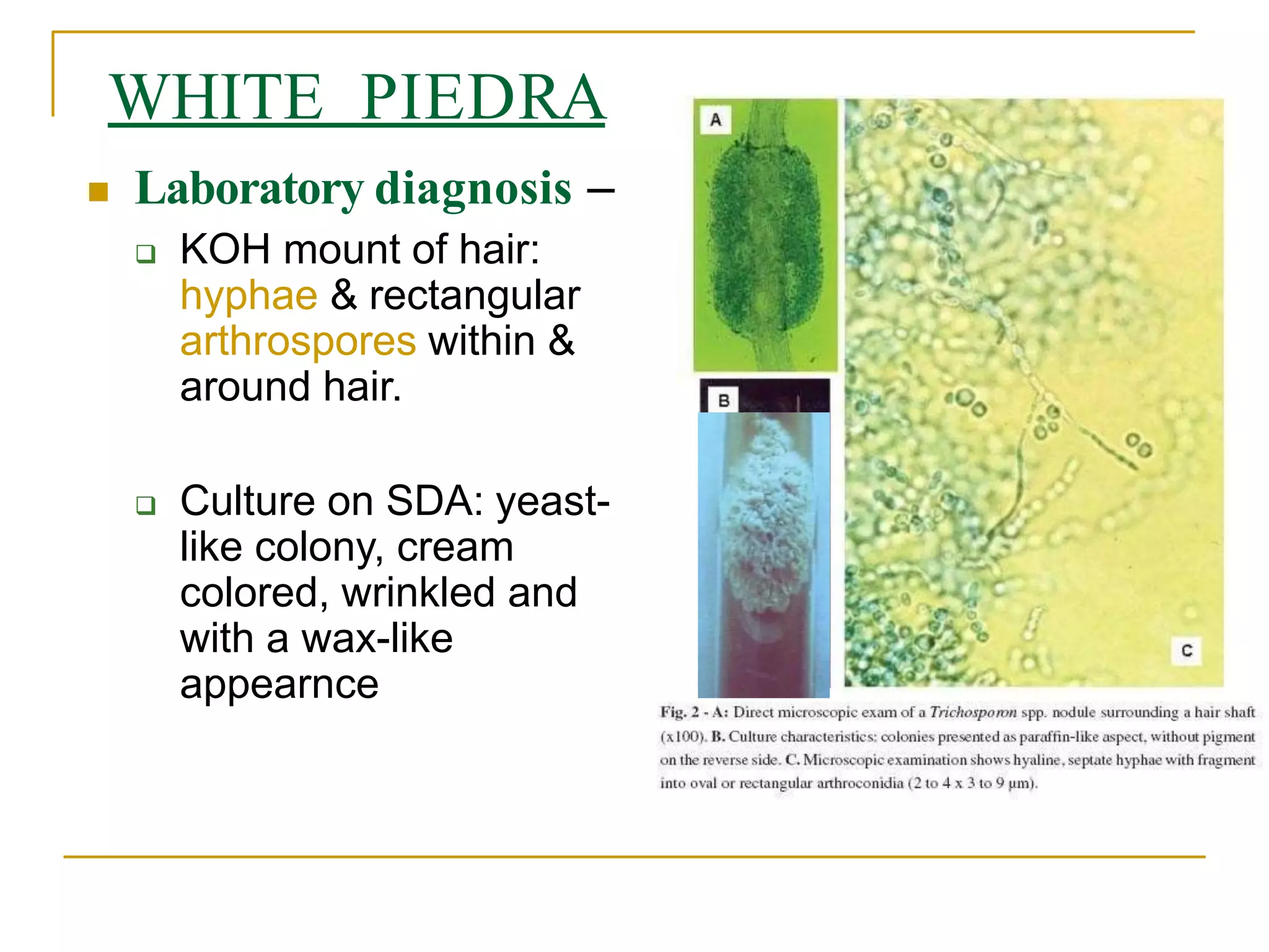
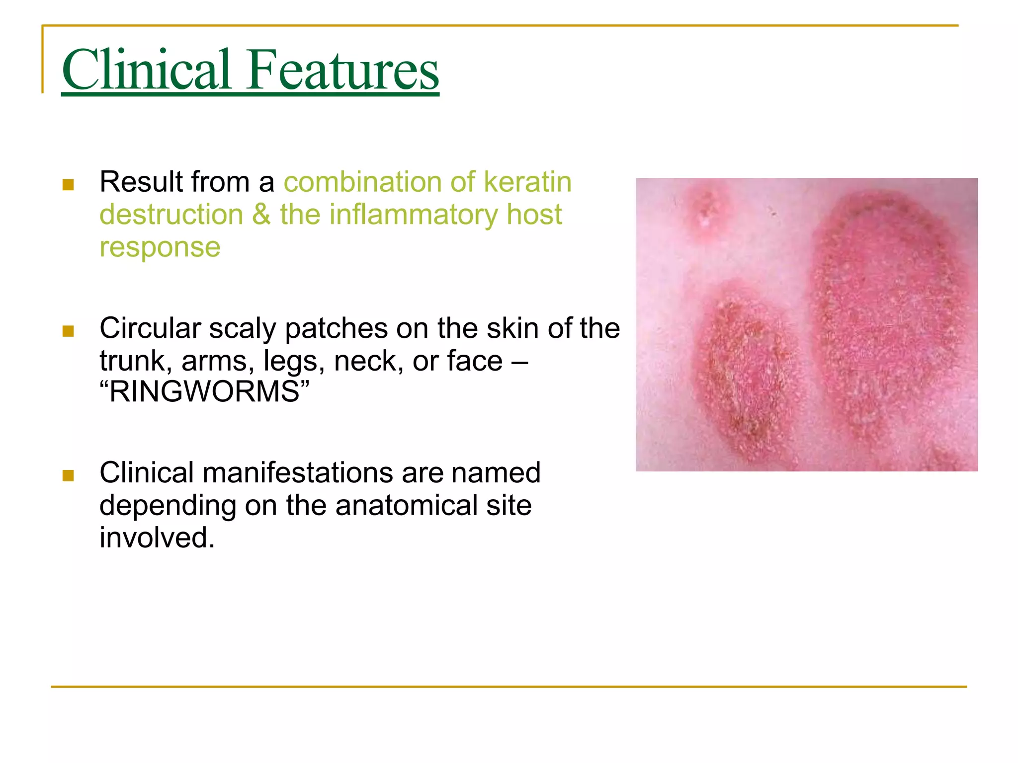
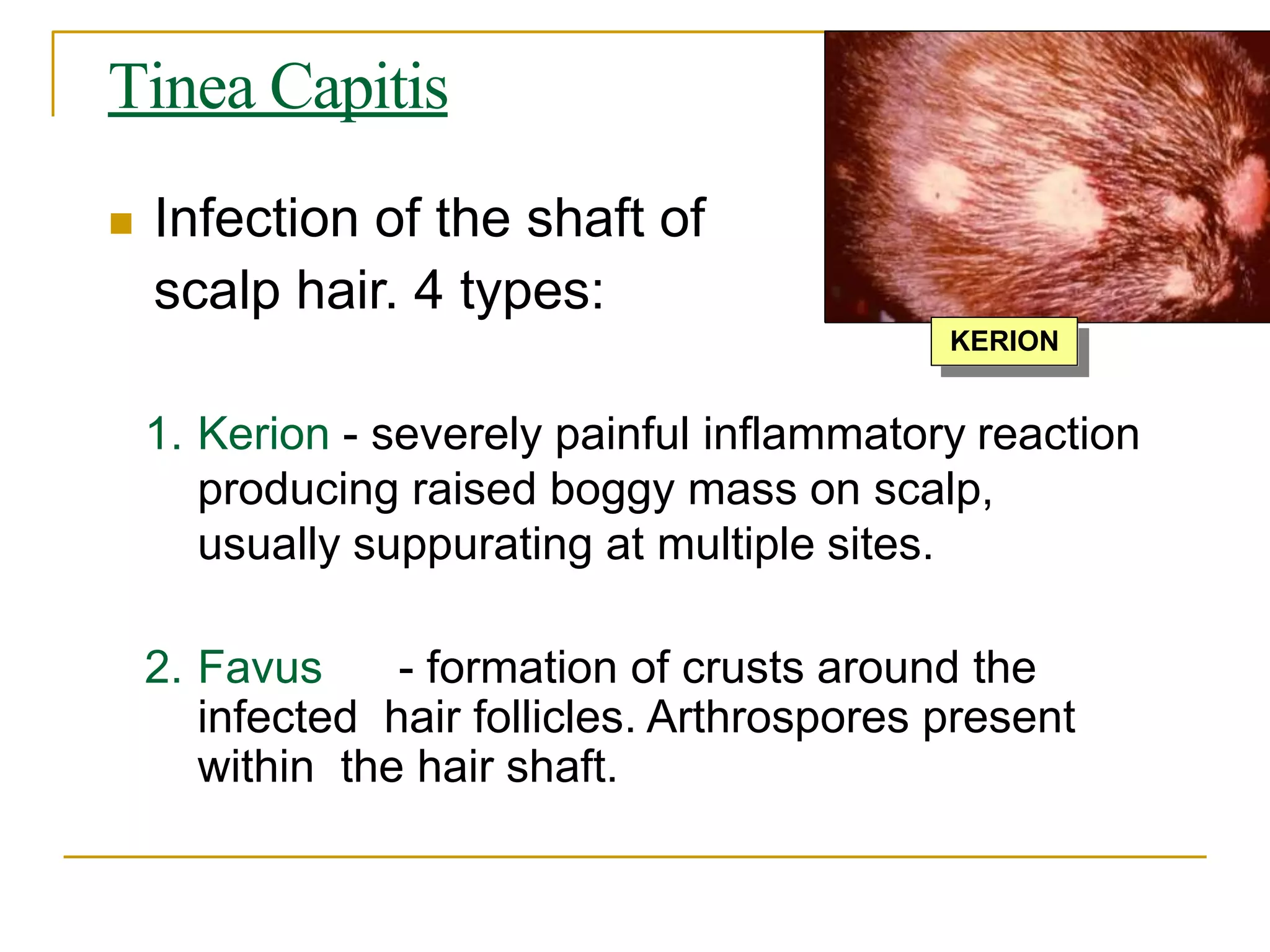
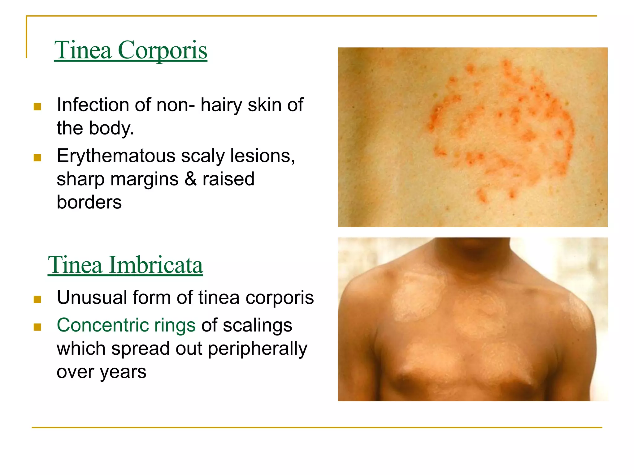
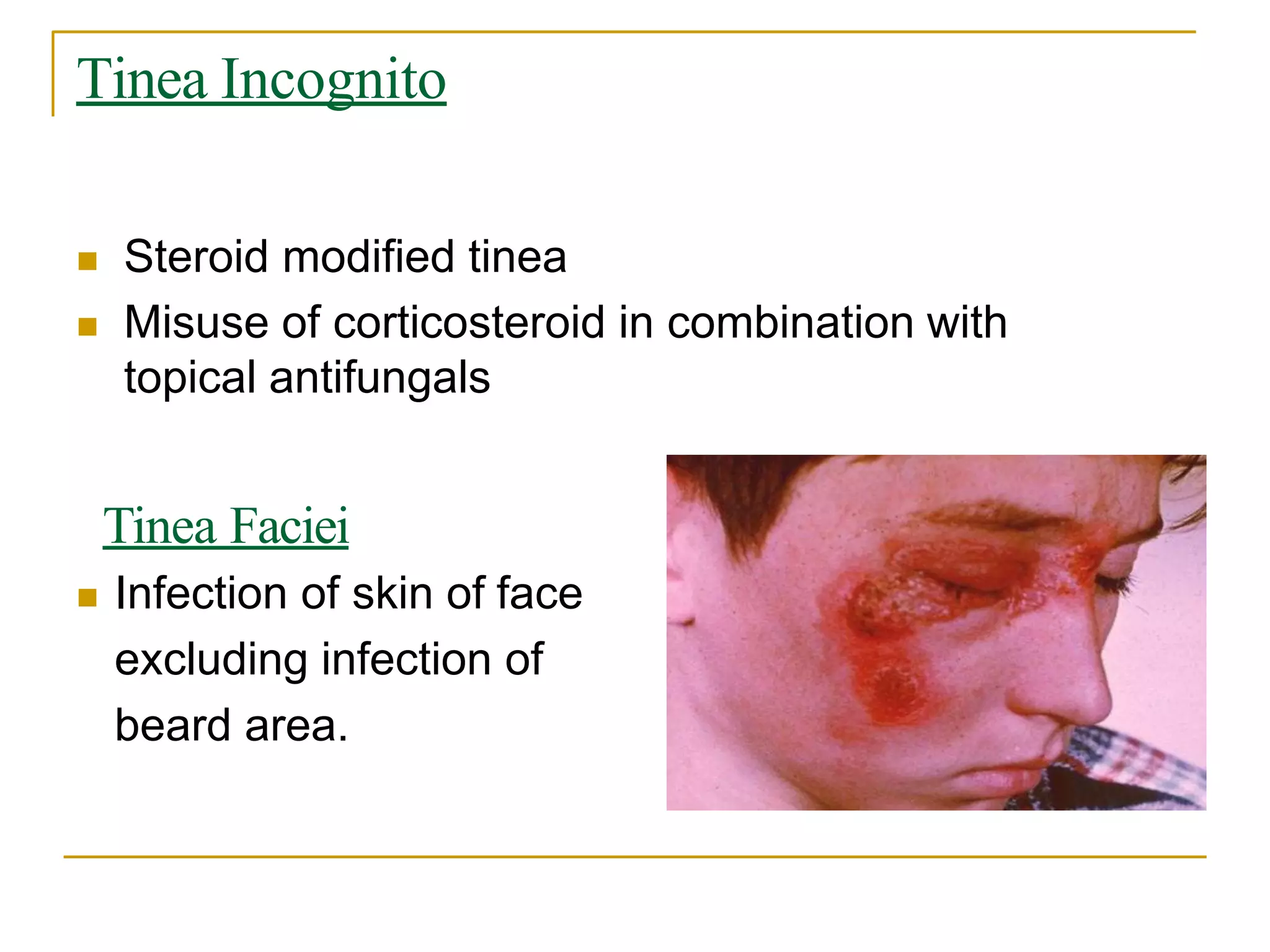
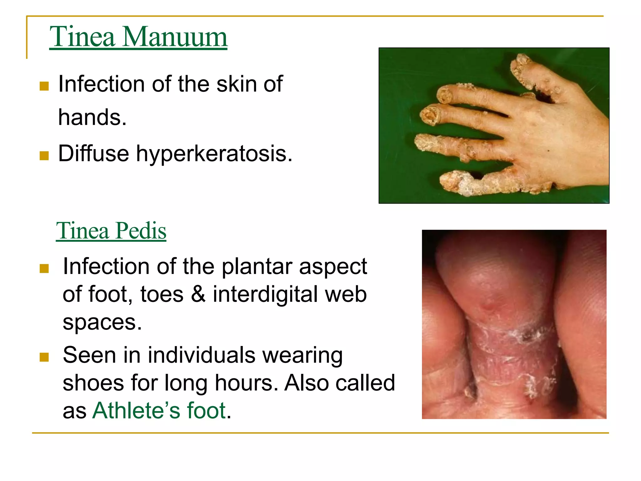
Superficial Mycoses
Dr. Dinesh Jain
Assistant Professor
Deptt. of Microbiology
SMS MC Jaipur
Superficial Mycoses
1. Surface Mycoses - includes




 Malassezia Infections
Pityriasis versicolor
Seborrheic dermatitis
Atopic dermatitis Folliculitis
 Tinea nigra
 Piedra
 Black piedra
 White piedra
2. Cutaneous mycoses - dermatophytoses
Surface Mycoses
the skin & its Involves dead layers of
appendages like hair & nails
 Only cosmetic effects:
 changes in the skin pigmentation or
 formation of nodules along the hair shaft.
 Infections included are :
Malassezia infections, Tinea nigra & Piedra.
Predisposing factors
 high temperature
 high humidity
 greasy skin
 use of corticosteroids
 underlying immunodeficiency
1. MALASSEZIA INFECTIONS
 Clinical presentations
1. Pityriasis versicolor - the commonest presenting disease
2. Seborrheic dermatitis
3. Atopic dermatitis - Chronic, intensely pruritic dermatoses;
involves scalp, face & neck of adults
4. Folliculitis – in females
 Site of lesions - areas of the skin rich in sebaceous
glands.
Pityriasis versicolor
 Caused by Malassezia furfur
 Asymptomatic, chronic recurrent
infection of stratum corneum
 Patchy discoloration ranging from
hypo- to hyperpigmentation:
interferes with melanin production
 Chest, abdomen, upper limbs & back
mainly involved.
Seborrheic dermatitis
 Whitish, dry, loose flakes on scalp. Generally
called as Dandruff.
 Sites rich in sebaceous glands including
head, neck & intertrigenous areas.
 Has been recognised as an early sign of
AIDS.
Laboratory Diagnosis
 Specimen- skin, hair
 Direct Examination
KOH mount : clusters of round yeast
cells, 2 to 7 with occasional buddings.
Hyphae are blunt, short, stout that may
be curved or branched - called “banana
& grapes” or “spaghetti & meat balls”
appearance.
 Culture : SDA covered with a layer of
olive oil (lipophilic fungus)
Treatment
 Topical : 10% sulfur ointment, 1-2%
imidazole creams, selenium sulfide shampoo
 Systemic : Itraconazole, Fluconazole
 Ketoconazole with zinc pyrithione in a
shampoo base is very effective for dandruff.
2. TINEA NIGRA
 Black or brownish, flat macular/
patchy lesions affecting skin of
the palms & occasionally the
soles. Solitary lesions showing
peripheral extensions.
 May also affect neck or trunk
 Caused by Hortaea werneckii, a
Halophilic fungi - lives in rotting
wood, soil, compost, or sewage.
Laboratory Diagnosis
1. Direct examination
 Specimen: skin scrapings from
active border of the lesion
 20% KOH mount: Shows brown,
septate, branching hyphae (5-6) &
budding yeast cells (2-8).
1. Fungal Culture
 on SDA with 15% salt at 25 to 30C.
 colonies are brown to greenish-black
with black pigmentation on the reverse
Treatment
 Keratolytic agent : Whitfield’s ointment, 5-
10% salicylic acid
 Topical antifungals : treatment of choice
1-2% clotrimazole or miconazole
 Systemic antifungals : Itraconazole orally
3. PIEDRA
 Superficial infection of the hair shaft
 Two types :
 Black piedra caused by Piedraia hortae.
 White piedra caused by Trichosporon sps.
 Treatment & Prevention
 Shaving off the hair
 Good personal hygiene
BLACK PIEDRA
 Discrete gritty hard, brown black nodules of about
1mm diameter firmly attached to hair shaft. Makes
hair brittle.
 Mainly involves scalp hair.
Moustache, beard & pubic
hairs may also be affected.
 Transmitted by sharing comb,
pillows, bed sheets & towel.
 In populations where hair care is usually done with
oily substances.
Laboratory Diagnosis
 Direct examination
- Crushed brittle hair in 10% KOH : dark
colored septate hyphae around hair surface
with round to oval asci containing 2-8 ascospores.
 Fungal culture
- On SDA with glycerin
WHITE PIEDRA
 Presence of softer nodule which is white to
light brown
 Occurs mainly on facial, axillary, beard,
moustache & pubic hairs.
 Pruritis or pain may occur at the site.
WHITE PIEDRA
 Laboratory diagnosis –
 KOH mount of hair:
hyphae & rectangular
arthrospores within &
around hair.
 Culture on SDA: yeast-
like colony, cream
colored, wrinkled and
with a wax-like
appearnce
DERMATOPHYTOSES
 Popularly called as Tinea or Ringworm.
 Most common fungal infection of skin, hair & nails.
 3 genera : Trichophyton (skin, hair & nails)
Microsporum (skin & hair)
Epidermophyton (skin & nails)
 It is the only true contagious fungal infection –
 transmitted through fomites i.e. contaminated clothing,
linens, towels, shoes, furniture, athletic equipment, or
personal care items
 also possible to catch ringworm from animals such as cats
or dogs or from fungus in the soil
Clinical Features
 Result from a combination of keratin
destruction & the inflammatory host
response
 Circular scaly patches on the skin of the
trunk, arms, legs, neck, or face –
“RINGWORMS”
 Clinical manifestations are named
depending on the anatomical site
involved.
Clinical Types &Causative Agents
 Tinea capitis - Trichophyton sps,
Microsporum sps.
 Tinea barbae - T. schoenleinii, M. gypseum
 Tinea imbricata - T. concentricum
 Tinea corporis - T. rubrum, other dermatophytes
 Tinea cruris - E. floccosum, T. rubrum
 Tinea pedis - T. rubrum, E. floccosum
 Tinea unguium
 Tinea manuum
Tinea Capitis
 Infection of the shaft of
scalp hair. 4 types:
1. Kerion - severely painful inflammatory reaction
producing raised boggy mass on scalp,
usually suppurating at multiple sites.
2. Favus - formation of crusts around the
infected hair follicles. Arthrospores present
within the hair shaft.
KERION
3. ECTOTHRIX OF HAIR - arthrospores
form a sheath or appear as chains on
the surface of hair shaft.
4. ENDOTHRIX OF HAIR -
arthrospores present within the hair
shaft.
Tinea Corporis
 Infection of non- hairy skin of
the body.
 Erythematous scaly lesions,
sharp margins & raised
borders
Tinea Imbricata
 Unusual form of tinea corporis
 Concentric rings of scalings
which spread out peripherally
over years
Tinea Gladiatorum
 Infection of wrestlers & athletes.
 Direct skin to skin contact
 Lesions on the arms, trunk or head & neck
(corresponding to the areas of greatest contact)
Tinea Incognito
 Steroid modified tinea
 Misuse of corticosteroid in combination with
topical antifungals
Tinea Faciei
 Infection of skin of face
excluding infection of
beard area.
Tinea Barbae
 Infection of the beard &
moustache area of the face
including the hair.
Tinea Cruris
 Most prevalent in tropical
countries.
 Infection of the groin, mostly seen
in men wearing tight fitting
garments.
 Involves perineum, scrotum &
peri-anal areas, may also involve
intertrigenous areas
Tinea Manuum
 Infection of the skin of
hands.
 Diffuse hyperkeratosis.
Tinea Pedis
 Infection of the plantar aspect
of foot, toes & interdigital web
spaces.
 Seen in individuals wearing
shoes for long hours. Also called
as Athlete’s foot.
Tinea Unguium
 Infection of the nail plates
 Commonly seen in adults
Onychomycosis
 Infection of nail.
Laboratory Diagnosis
 Woodlamp’ s examination – of infected hair
 Direct examination
KOH mount : skin scales, nail clippings & hair stubs
- shows branching hyaline mycelia with arthrospore
production.
 Fungal culture
3 genera are differentiated on the basis of shape,
arrangement & number of macroconidia &
microconidia and other associated features like spiral
hypha, racquet hypha, nodular organ & favic
chandelier
Fungal Culture
 Microconidia are unicellular whereas macroconidia are
multicellular.
Genera Macroconidia Microconidia
Trichophyton Scanty, cylindrical Abundant, in clusters
Microsporum Large, fusiform Scanty
Epidermophyton Pear shaped, in clusters Absent
Cylindrical
macroconidia
Fusiform
macroconidia
Club/ pear shaped
macroconidia
Fungal Culture
 SDA with antibiotics & actidione - Slow growth,
takes 10 days to 3 wks.
 Dermatophyte Test Medium (DTM) at 25C -to
isolate & distinguish dermatophytes from fungal
or bacterial contaminants
 Dermatophytes turn the medium red by raising the pH.
 Dermatophyte Identification Medium (DIM) can
be used for presumptive identification.
Other tests for identification
 Hair perforation test
 Urease test
 Hairbrush sampling technique.
 Skin tests using dermatophytin
 PCR
Treatment & Prophylaxis
 Topical antifungals : Azole derivatives,
Whitfield’s ointment
 Oral : Griseofulvin is the drug of choice
 Itraconazole is preferred for onychomycosis

Superficial mycosis

  • 1.
    Superficial Mycoses Dr. DineshJain Assistant Professor Deptt. of Microbiology SMS MC Jaipur
  • 2.
    Superficial Mycoses 1. SurfaceMycoses - includes      Malassezia Infections Pityriasis versicolor Seborrheic dermatitis Atopic dermatitis Folliculitis  Tinea nigra  Piedra  Black piedra  White piedra 2. Cutaneous mycoses - dermatophytoses
  • 3.
    Surface Mycoses the skin& its Involves dead layers of appendages like hair & nails  Only cosmetic effects:  changes in the skin pigmentation or  formation of nodules along the hair shaft.  Infections included are : Malassezia infections, Tinea nigra & Piedra.
  • 4.
    Predisposing factors  hightemperature  high humidity  greasy skin  use of corticosteroids  underlying immunodeficiency
  • 5.
    1. MALASSEZIA INFECTIONS Clinical presentations 1. Pityriasis versicolor - the commonest presenting disease 2. Seborrheic dermatitis 3. Atopic dermatitis - Chronic, intensely pruritic dermatoses; involves scalp, face & neck of adults 4. Folliculitis – in females  Site of lesions - areas of the skin rich in sebaceous glands.
  • 6.
    Pityriasis versicolor  Causedby Malassezia furfur  Asymptomatic, chronic recurrent infection of stratum corneum  Patchy discoloration ranging from hypo- to hyperpigmentation: interferes with melanin production  Chest, abdomen, upper limbs & back mainly involved.
  • 7.
    Seborrheic dermatitis  Whitish,dry, loose flakes on scalp. Generally called as Dandruff.  Sites rich in sebaceous glands including head, neck & intertrigenous areas.  Has been recognised as an early sign of AIDS.
  • 8.
    Laboratory Diagnosis  Specimen-skin, hair  Direct Examination KOH mount : clusters of round yeast cells, 2 to 7 with occasional buddings. Hyphae are blunt, short, stout that may be curved or branched - called “banana & grapes” or “spaghetti & meat balls” appearance.  Culture : SDA covered with a layer of olive oil (lipophilic fungus)
  • 9.
    Treatment  Topical :10% sulfur ointment, 1-2% imidazole creams, selenium sulfide shampoo  Systemic : Itraconazole, Fluconazole  Ketoconazole with zinc pyrithione in a shampoo base is very effective for dandruff.
  • 10.
    2. TINEA NIGRA Black or brownish, flat macular/ patchy lesions affecting skin of the palms & occasionally the soles. Solitary lesions showing peripheral extensions.  May also affect neck or trunk  Caused by Hortaea werneckii, a Halophilic fungi - lives in rotting wood, soil, compost, or sewage.
  • 11.
    Laboratory Diagnosis 1. Directexamination  Specimen: skin scrapings from active border of the lesion  20% KOH mount: Shows brown, septate, branching hyphae (5-6) & budding yeast cells (2-8). 1. Fungal Culture  on SDA with 15% salt at 25 to 30C.  colonies are brown to greenish-black with black pigmentation on the reverse
  • 12.
    Treatment  Keratolytic agent: Whitfield’s ointment, 5- 10% salicylic acid  Topical antifungals : treatment of choice 1-2% clotrimazole or miconazole  Systemic antifungals : Itraconazole orally
  • 13.
    3. PIEDRA  Superficialinfection of the hair shaft  Two types :  Black piedra caused by Piedraia hortae.  White piedra caused by Trichosporon sps.  Treatment & Prevention  Shaving off the hair  Good personal hygiene
  • 14.
    BLACK PIEDRA  Discretegritty hard, brown black nodules of about 1mm diameter firmly attached to hair shaft. Makes hair brittle.  Mainly involves scalp hair. Moustache, beard & pubic hairs may also be affected.  Transmitted by sharing comb, pillows, bed sheets & towel.  In populations where hair care is usually done with oily substances.
  • 15.
    Laboratory Diagnosis  Directexamination - Crushed brittle hair in 10% KOH : dark colored septate hyphae around hair surface with round to oval asci containing 2-8 ascospores.  Fungal culture - On SDA with glycerin
  • 16.
    WHITE PIEDRA  Presenceof softer nodule which is white to light brown  Occurs mainly on facial, axillary, beard, moustache & pubic hairs.  Pruritis or pain may occur at the site.
  • 17.
    WHITE PIEDRA  Laboratorydiagnosis –  KOH mount of hair: hyphae & rectangular arthrospores within & around hair.  Culture on SDA: yeast- like colony, cream colored, wrinkled and with a wax-like appearnce
  • 18.
    DERMATOPHYTOSES  Popularly calledas Tinea or Ringworm.  Most common fungal infection of skin, hair & nails.  3 genera : Trichophyton (skin, hair & nails) Microsporum (skin & hair) Epidermophyton (skin & nails)  It is the only true contagious fungal infection –  transmitted through fomites i.e. contaminated clothing, linens, towels, shoes, furniture, athletic equipment, or personal care items  also possible to catch ringworm from animals such as cats or dogs or from fungus in the soil
  • 19.
    Clinical Features  Resultfrom a combination of keratin destruction & the inflammatory host response  Circular scaly patches on the skin of the trunk, arms, legs, neck, or face – “RINGWORMS”  Clinical manifestations are named depending on the anatomical site involved.
  • 20.
    Clinical Types &CausativeAgents  Tinea capitis - Trichophyton sps, Microsporum sps.  Tinea barbae - T. schoenleinii, M. gypseum  Tinea imbricata - T. concentricum  Tinea corporis - T. rubrum, other dermatophytes  Tinea cruris - E. floccosum, T. rubrum  Tinea pedis - T. rubrum, E. floccosum  Tinea unguium  Tinea manuum
  • 21.
    Tinea Capitis  Infectionof the shaft of scalp hair. 4 types: 1. Kerion - severely painful inflammatory reaction producing raised boggy mass on scalp, usually suppurating at multiple sites. 2. Favus - formation of crusts around the infected hair follicles. Arthrospores present within the hair shaft. KERION
  • 22.
    3. ECTOTHRIX OFHAIR - arthrospores form a sheath or appear as chains on the surface of hair shaft. 4. ENDOTHRIX OF HAIR - arthrospores present within the hair shaft.
  • 23.
    Tinea Corporis  Infectionof non- hairy skin of the body.  Erythematous scaly lesions, sharp margins & raised borders Tinea Imbricata  Unusual form of tinea corporis  Concentric rings of scalings which spread out peripherally over years
  • 24.
    Tinea Gladiatorum  Infectionof wrestlers & athletes.  Direct skin to skin contact  Lesions on the arms, trunk or head & neck (corresponding to the areas of greatest contact)
  • 25.
    Tinea Incognito  Steroidmodified tinea  Misuse of corticosteroid in combination with topical antifungals Tinea Faciei  Infection of skin of face excluding infection of beard area.
  • 26.
    Tinea Barbae  Infectionof the beard & moustache area of the face including the hair. Tinea Cruris  Most prevalent in tropical countries.  Infection of the groin, mostly seen in men wearing tight fitting garments.  Involves perineum, scrotum & peri-anal areas, may also involve intertrigenous areas
  • 27.
    Tinea Manuum  Infectionof the skin of hands.  Diffuse hyperkeratosis. Tinea Pedis  Infection of the plantar aspect of foot, toes & interdigital web spaces.  Seen in individuals wearing shoes for long hours. Also called as Athlete’s foot.
  • 28.
    Tinea Unguium  Infectionof the nail plates  Commonly seen in adults Onychomycosis  Infection of nail.
  • 29.
    Laboratory Diagnosis  Woodlamp’s examination – of infected hair  Direct examination KOH mount : skin scales, nail clippings & hair stubs - shows branching hyaline mycelia with arthrospore production.  Fungal culture 3 genera are differentiated on the basis of shape, arrangement & number of macroconidia & microconidia and other associated features like spiral hypha, racquet hypha, nodular organ & favic chandelier
  • 30.
    Fungal Culture  Microconidiaare unicellular whereas macroconidia are multicellular. Genera Macroconidia Microconidia Trichophyton Scanty, cylindrical Abundant, in clusters Microsporum Large, fusiform Scanty Epidermophyton Pear shaped, in clusters Absent
  • 31.
  • 32.
    Fungal Culture  SDAwith antibiotics & actidione - Slow growth, takes 10 days to 3 wks.  Dermatophyte Test Medium (DTM) at 25C -to isolate & distinguish dermatophytes from fungal or bacterial contaminants  Dermatophytes turn the medium red by raising the pH.  Dermatophyte Identification Medium (DIM) can be used for presumptive identification.
  • 33.
    Other tests foridentification  Hair perforation test  Urease test  Hairbrush sampling technique.  Skin tests using dermatophytin  PCR
  • 34.
    Treatment & Prophylaxis Topical antifungals : Azole derivatives, Whitfield’s ointment  Oral : Griseofulvin is the drug of choice  Itraconazole is preferred for onychomycosis