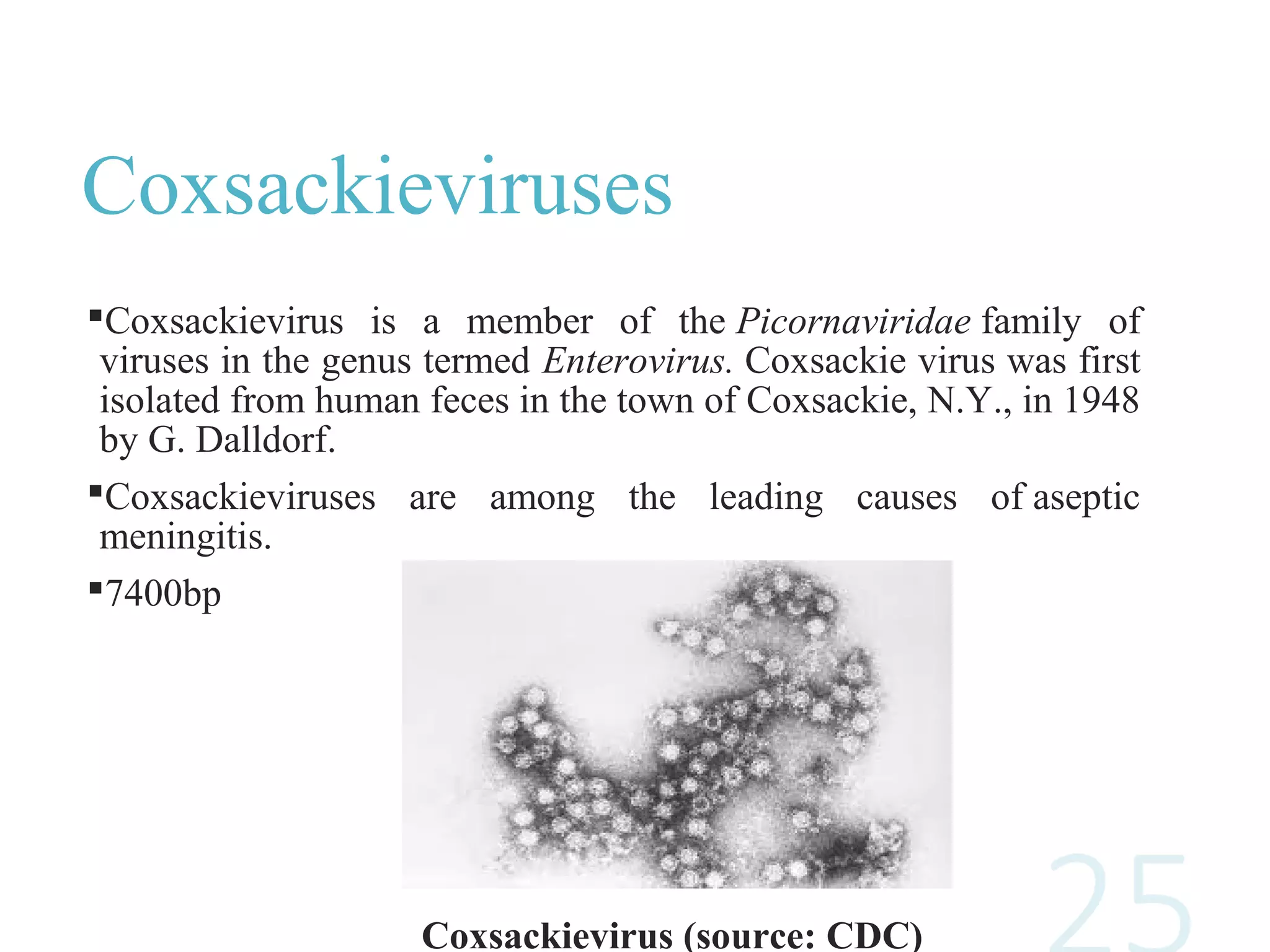
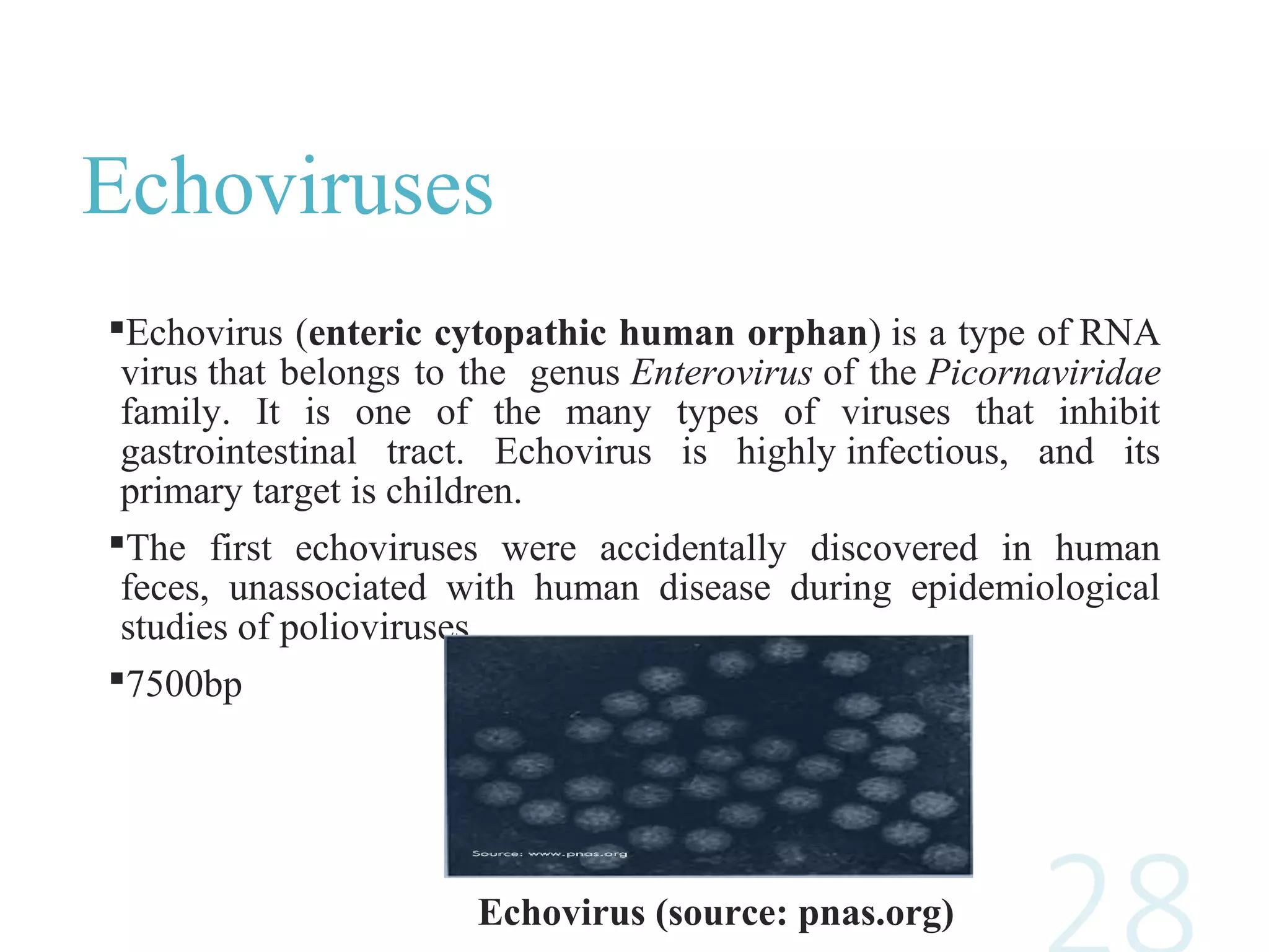
Enteroviruses
BY GROUP NO 4
Introduction
Enteroviruses
Enterovirus, a genus in the family Picornaviridae, consists of 10
virus species, including four human enterovirus species and three
human rhinovirus species.
However, the Picornavirus Study Group of ICTV proposes the
addition of two new species and the renaming of several species
within the enterovirus genus.
 The enteroviruses include the polioviruses, echoviruses,
coxsackieviruses and the human enteroviruses.
Enterovirus Particles
Courtesy of Linda M. Stannard, University of Cape Town,
S.A.h
History
Much of what is known about enteroviruses, has been learned from
the study of poliovirus.
The designation of Coxsackievirus are based on the geographic site
of the initial discovery of the first member of the group (Coxsackie,
NY).
The echoviruses were designated “orphans” because their role as a
potential cause of disease was unknown at the time they were first
identified almost 50 years ago.
Structure
Enteroviruses are small, approximately 25-30 nm in diameter, and
icosahedral in shape.
The viruses are non-enveloped, and the virions are relatively simple,
consisting of a protein capsid surrounding a single-stranded, positive
sense RNA genome.
The genome has approximately 7500 nucleotides, and contains a single
open reading frame that encodes a polyprotein that is then processed to
yield the structural (i.e., capsid) proteins VP1, VP2, VP3, and VP4 and
the non-structural proteins.
Fig:01 Enterovirus
structure
Clinical features
The majority of enterovirus infections are asymptomatic.
However, there are many different types of diseases that can result,
depending somewhat on the specific virus causing the infection.
The incubation period from exposure to onset of symptoms may be
between 2 and 30 days for different strains but is most commonly 5–
14 days.
Virus is shed in both asymptomatic and symptomatic infections in
faeces and in oral secretions.
Classification
The enterovirus genus includes 12 serotypes, which are named as
Enterovirus A to Enterovirus L and RHINOVIRUSES. These serotypes
include the following mentioned species.
1.On the basis of pathogenesis in humans and animals
a)Poliovirus
b)Coxsackie A
c)Coxsackie B
d)Echoviruses
2. Due to overlap in biological properties. Isolated more recently named
with the system of consecutive numbers (numbered enteroviruses)
a)EV-68
b)EV-69
c)EV-70
d)EV-71
Genome and
Replication cycle
Genome
Single stranded RNA virus
Genome size- 7500 nucleotides
Compose of untranslated regions-UTRs
Coding regions – P1, P2, P3
Structural proteins
Non-structural proteins
FIGURE: Structure of the enterovirus RNA genome
http:// www.medcraveonline.com
Replication cycle
Attachment
Attachment of virus to cellular receptor
Viruses become adapted to get entry into the cell
Uncoating
Interaction with cellular receptors
Leads to release of internal VP4 polypeptide and subsequently
the RNA genome
Replication
RNA dependant RNA polymerase
Negative strand is synthesized initially
Genomic RNA is packed inside the capsid
Polypeptide encoding region is proceeded by 5'UTR and 3'UTR in
poly-A tract.
http://www.researchgate.net
Assembly
Assembly of new virions is initiated by the release of P1
capsid.
A small polypeptide (VPg) participate the assembly process
Associated with 5'end of genome
Release
Maturation of virion particle
Virus particles are released upon cell lysis of the host cell.
FIGURE: Replication cycle of Enterovirus
http://www.researchgate.net
Polio virus
• Polio virus is a human entrovirus and belong to family picornaviridae.
• It is a non-enveloped virus, composed of protein capsid and RNA
genome.
• RNA is single stranded, 7500 nucleotides long positive sense that acts as
a mRNA
• The diameter of virus is 30nm and the symmetry is icosahedral.
• There are three serotypes of poliovirus PV1, PV2 and PV3
• It is a human specific virus
Pathogenesis
The incubation period is usually 7-14 days
After ingestion, the virus multiplies in oropharyngeal and intestinal
mucosa.
The lymphatic system, in particular the toncils and the peyer’s
patches of ileum are invaded and the virus enters the blood resulting
in transient viraemia.
In a minority of cases, the virus may involve the CNS following
dissemination. The entry into the CNS is not well understood and
this causes paralytic poliomyletis.
Clinical Manifestations
There are 3 possible outcomes of infection:
 Subclinical infection (90 - 95%) - inapparent subclinical infection
account for the vast majority of poliovirus infections.
 Abortive infection (4 - 8%) - a minor influenza-like illness occurs,
recovery occurs within a few days and the diagnosis can only be
made by the laboratory. The minor illness may be accompanied
by aseptic meningitis
 Major illness (1 - 2%) - the major illness may present 2 - 3 days
following the minor illness or without any preceding minor
illness. Signs of aseptic meningitis are common. Involvement of
the anterior horn cells lead to flaccid paralysis. involvement of the
medulla may lead to respiratory paralysis and death.
Laboratory diagnosis
Virus isolation:
oliovirus can be readily isolated from throat swabs, faeces, and
rectal swabs. It is rarely isolated from the CSF
equires molecular techniques to differentiate between the wild type
and the vaccine type.
RT-PCR : a rapid diagnosis of poliovirus infection may be made by
the use of RT-PCR.
Serology :Very rarely used for diagnosis since cell culture is
efficient. Occasionally used for immune status screening for
immunocompromised individuals
Preventions
 No specific antiviral therapy is available. However the disease may be
prevented through vaccination. There are two vaccines available.
Intramuscular Poliovirus Vaccine (IPV)
 consists of formalin inactivated virus of all 3 poliovirus serotypes.
 Produces serum antibodies only: does not induce local immunity
and thus will not prevent local infection of the gut.
 However, it will prevent paralytic poliomyelitis since viraemia is
essential for the pathogenesis of the disease.
Oral Poliovirus Vaccine (OPV)
 Consists of live attenuated virus of all 3 serotypes.
 Rarely causes paralytic poliomyelitis, around 1 in 3 million doses.
 Most countries use OPV because of its ability to induce local
immunity and also it is much cheaper to produce than IPV.
 The normal response rate to OPV is close to 100%.
 OPV is used for the WHO poliovirus eradication campaign.
 Poliovirus was targeted for eradication by the WHO by the end of
year 2000. To this end, an extensive monitoring network had been
set up.
 Poliovirus has been eradicated from most regions of the world
except the Indian subcontinent and sub-Saharan Africa. It is
possible that the WHO target may be achieved.
Polio in Pakistan
Polio virus remain endemic in three countries Pakistan, Nigeria and
Afghanistan.
The polio eradication campaign started in 1994 but still in 2018, 8
cases have been reported. Some of the factors are:
Misinformation campaign and terrorism.
Poor health conditions
Environment
Perception of Pakistani parents
Malnutrition
Coxsackieviruses
and Echoviruses
Coxsackieviruses
Coxsackievirus is a member of the Picornaviridae family of
viruses in the genus termed Enterovirus. Coxsackie virus was first
isolated from human feces in the town of Coxsackie, N.Y., in 1948
by G. Dalldorf.
Coxsackieviruses are among the leading causes of aseptic
meningitis.
7400bp
Coxsackievirus (source: CDC)
Groups
Coxsackieviruses are distinguished from other enteroviruses based on
their pathogenicity in neonatal mice. They are divided into 2 groups on
the basis of the lesions observed in suckling mice.
Group A viruses were noted to cause flaccid paralysis (which is caused
by diffuse myositis) with acute inflammation and necrosis of fibers of
voluntary muscles.
roup B viruses were noted to cause spastic paralysis (due to focal
muscle injury and degeneration of neuronal tissue), and inflammatory
changes in the dorsal fat pads, the pancreas and occasionally the
myocardium.
At least 23 serotypes (1–22, 24) of group A and six serotypes (1–6) of
group B are recognized.
Coxsackie A viruses:
In general, group A coxsackieviruses tend to infect the skin and mucous
membranes, causing mouth blisters, acute hemorrhagic conjunctivitis,
and hand, foot, and mouth (HFM) diseases.
Coxsackie B Viruses:
Group B coxsackieviruses tend to infect the heart, pleura, pancreas, and
liver causing myocarditis, pericarditis, and hepatitis. Coxsackie B
infection of the heart can lead to pericardial effusion.
The development of insulin-dependent diabetes (IDDM) has recently
been associated with recent enteroviral infection.
Sjogren's syndrome is also being studied in connection with
coxsackievirus.
Echoviruses
Echovirus (enteric cytopathic human orphan) is a type of RNA
virus that belongs to the genus Enterovirus of the Picornaviridae
family. It is one of the many types of viruses that inhibit
gastrointestinal tract. Echovirus is highly infectious, and its
primary target is children.
The first echoviruses were accidentally discovered in human
feces, unassociated with human disease during epidemiological
studies of polioviruses.
7500bp
Echovirus (source: pnas.org)
Symptoms include typically mild upper-respiratory symptoms, such as,
cough, sore throat, flu, rash and viral meningitis.
Echovirus can be confirmed by following laboratory tests: a rectal
culture, a stool culture, a throat culture and a spinal fluid culture.
Echovirus typically go away without treatment. There’s no antiviral
treatment available for this virus infection.
New Enteroviruses
New Enteroviruses
5 types of Enteroviruses (68-72)
EV-68
EV-69
EV-70
EV-71
EV-72
EV-68
First isolated in California in 1962
Once considered rare, it has been on worldwide upswing in
21st century
Unlike all enteroviruses, EV68 displays
➡acid lability
➡lower optimum temperature
Characteristic features of human rhinoviruses
Epidemiology
 In 2012/13, EV68 was found in 2 out of 5 children,
cluster of polio-like disease in California
 In 2016, 29 cases were reported in Europe (5 in France
and Scotland. 3 each in Sweden, Norway and Spain)
 occur late in the enterovirus season (August and
September)
2014 North American, the virus caused clusters of
respiratory diseases in the United States.
691 people in 46 states and the District of Columbia had
come down with a respiratory illness caused by EV-D68.
Five children died.
Sign And
Symptoms
➡Initial symptoms are similar
to those for the common
cold
‣ runny nose
‣ sore throat
‣ cough, and fever.
➡As the disease progresses,
more serious symptoms may
occur
‣ Difficulty in breathing as in
pneumonia
‣ reduced alertness
‣ reduction in urine production
‣ dehydration
‣ may lead to respiratory failure.
Acute Flaccid Myelitis
 A rare muscle weakness, usually due to polio.
 Two California children who tested positive for the virus had
paralysis of one or more limbs reaching peak severity within 48
hours of onset. Recovery of motor function was poor at 6-
month follow-up.
 As of October 2014, the Centers for Disease Control (CDC)
was investigating 10 cases of paralysis and/or cranial
dysfunction in Colorado and other reports around the country,
coinciding with the increase in enterovirus D68 activity.
 As of 2018 the link of EV-D68 and the paralysis is strong,
meeting six Bradford Hill criteria fully and two partially.
The CDC recently issued a statement
on October 17, 2018 claiming:
"Right now, we know that poliovirus
is not the cause of these AFM cases.
CDC has tested every stool specimen
from the AFM patients, none of the
specimens have tested positive for
the poliovirus. https://www.pulseheadlines.com
EV-70
Most recently discovered
EV70 infection is confined to conjunctiva of human, but in
some cases involvement of central nervous system is also
observed.
Epidemiology
Pakistan is reported to have been swept by pandemic of 1971
but no epidemiology records are available
It can give rise to neurological diseases but no such cases has
been reported in Pakistan. except for one paralysis case —low
antibodies for EV70
During the epidemic of 1981, AHC appeared in Karachi.
Conjunctivitis
 Inflammation or swelling of Conjunctiva
 A thin transparent layer of tissue that lines the inner
surface of eyelid and covers the white part of eye
 often called “pink eye”
Symptoms
 itching or burning
 swollen eyelids
 discharge
 excessive tearing
 increased sensitivity
https://www.aoa.org/Images/public/Conjunctivitis_Child.jpeg
EV-71
Epidemiology
 In 1965, with the 1st known strain isolated, the virus seems to
evolve recently.
 This virus has spread to various countries in Asia and most
recently Cambodia(2012)
 Large outbreaks — U.S, Australia, Asia and Europe
Diseases
EV71 infections manifest self-
limited and mild illness
characterized by
lesions on hands, mouth,
oropharyngeal mucosa and feet
small portion of acute
infections are associated with
fetal neurological symptoms
https://www.nejm.org/doi/full/10.1056/NEJMicm1713548
Diseases
associated with
Enteroviruses
Clinical manifestations of
infection
Initial ingestion of virus leads to replication in the pharynx
and small intestine.
 ‘minor’ viremia which allows spread of virus to lymphoid
tissue throughout the body.
 <10% of individuals, ‘major’ viremia occurs with fever,
malaise, fatigue, nausea, vomiting and CNS symptoms 8-12
days following exposure.
Diseases associated with
Enteroviruses
Diseases poliovirus Cox A Cox B Echovirus
Paralytic disease + + + +
Carditis + + + +
Neonatal disease - - + +
Pleurodynia - - + -
Herpangina - + - -
Rash disease - + + +
Haemorr.
conjunctivitis
- + - -
Respiratory infections + + + +
Undifferentiated fever + + + +
Diabetes/pancreatitis - - + -
Undifferentiated febrile illness:
• May be seen with all groups of Enteroviruses symptom include
sweating chills, muscle aches, loss of appetite and dehydration.
Respiratory Infections:
• Several Enteroviruses are associated with the common cold
Symptoms congestion, runny nose, cough, sore throat etc.
Myocarditis:
•Majorly caused by coxsackie B viruses but also caused by
other Enteroviruses.
• Present in neonates as part of neonatal infection and is often
fatal.
Herpangina
•Cox A virus, cause a painful
ulcer on a palate with tongue
leading to problems like
swallowing and vomiting
•Symptoms persist for several
weeks.
Figure: Herpangina- Wikipedia
Epidemic Pleurodynia
•Cox B virus, bornholdm
disease, cause fever,
headache , severe pain in lower
abdomen.
•Symptoms subside for 2 to 4
days.
Figure: bornholdm disease
Rubelliform rashes
•A rash disease consist of erythematous macules. may be seen
with several coxsackie A, B, and echoviruses.
Neonatal Infection
• Some coxsackie B viruses and echoviruses may cause infection
in newborn infants.
•Transmitted perinatally during the birth process and symptoms
vary from a mild febrile illness to a severe fulminating
multisystem disease and death.
General properties of
Enterovisuses
Properties of Enteroviruses
Family :Picronaviridae
Genus :Enterovirus (10 specie)
Single Strand RNA viruses
Enteroviruses are small viruses
Non-enveloped viruses
 Capsid is Icosahedral
Size is from 25-30nm
Laboratory Diagnosis
Virus Isolation
oxsackie B and Echoviruses can be readily grown in cell culture
from throat swabs, feces, and rectal swabs.
RT-PCR - PCR assays are becoming increasingly used for the
detection and identification of enteroviruses. They are especially
useful in the case of Coxsackie A viruses and also for the
diagnosis of menigitis.
Serology
eutralization tests or EIAs are used but are very cumbersome and
thus not offered by most diagnostic laboratories.
General prevention Of
Enteroviruses
Wash your hands with soap regularly.
Don’t touch your hands mouth and nose if your hands are
unwashed.
Don’t hug or share food with infected person.
Stay home when your sick .
Don’t share your towel and cloths.
General prevention Of
Enteroviruses
Cover Sneeze or cough with tissue.
Clean and disinfect frequently touches surfaces such as door
knobs and toys, especially if someone is sick
Disinfect patients care equipments.
Conclusions

Enteroviruses life cycle structure genome organization classification

  • 1.
  • 2.
  • 3.
    Enteroviruses Enterovirus, a genusin the family Picornaviridae, consists of 10 virus species, including four human enterovirus species and three human rhinovirus species. However, the Picornavirus Study Group of ICTV proposes the addition of two new species and the renaming of several species within the enterovirus genus.  The enteroviruses include the polioviruses, echoviruses, coxsackieviruses and the human enteroviruses.
  • 4.
    Enterovirus Particles Courtesy ofLinda M. Stannard, University of Cape Town, S.A.h
  • 5.
    History Much of whatis known about enteroviruses, has been learned from the study of poliovirus. The designation of Coxsackievirus are based on the geographic site of the initial discovery of the first member of the group (Coxsackie, NY). The echoviruses were designated “orphans” because their role as a potential cause of disease was unknown at the time they were first identified almost 50 years ago.
  • 6.
    Structure Enteroviruses are small,approximately 25-30 nm in diameter, and icosahedral in shape. The viruses are non-enveloped, and the virions are relatively simple, consisting of a protein capsid surrounding a single-stranded, positive sense RNA genome. The genome has approximately 7500 nucleotides, and contains a single open reading frame that encodes a polyprotein that is then processed to yield the structural (i.e., capsid) proteins VP1, VP2, VP3, and VP4 and the non-structural proteins.
  • 7.
  • 8.
    Clinical features The majorityof enterovirus infections are asymptomatic. However, there are many different types of diseases that can result, depending somewhat on the specific virus causing the infection. The incubation period from exposure to onset of symptoms may be between 2 and 30 days for different strains but is most commonly 5– 14 days. Virus is shed in both asymptomatic and symptomatic infections in faeces and in oral secretions.
  • 9.
    Classification The enterovirus genusincludes 12 serotypes, which are named as Enterovirus A to Enterovirus L and RHINOVIRUSES. These serotypes include the following mentioned species. 1.On the basis of pathogenesis in humans and animals a)Poliovirus b)Coxsackie A c)Coxsackie B d)Echoviruses 2. Due to overlap in biological properties. Isolated more recently named with the system of consecutive numbers (numbered enteroviruses) a)EV-68 b)EV-69 c)EV-70 d)EV-71
  • 10.
  • 11.
    Genome Single stranded RNAvirus Genome size- 7500 nucleotides Compose of untranslated regions-UTRs Coding regions – P1, P2, P3 Structural proteins Non-structural proteins FIGURE: Structure of the enterovirus RNA genome http:// www.medcraveonline.com
  • 12.
    Replication cycle Attachment Attachment ofvirus to cellular receptor Viruses become adapted to get entry into the cell Uncoating Interaction with cellular receptors Leads to release of internal VP4 polypeptide and subsequently the RNA genome
  • 13.
    Replication RNA dependant RNApolymerase Negative strand is synthesized initially Genomic RNA is packed inside the capsid Polypeptide encoding region is proceeded by 5'UTR and 3'UTR in poly-A tract. http://www.researchgate.net
  • 14.
    Assembly Assembly of newvirions is initiated by the release of P1 capsid. A small polypeptide (VPg) participate the assembly process Associated with 5'end of genome Release Maturation of virion particle Virus particles are released upon cell lysis of the host cell.
  • 15.
    FIGURE: Replication cycleof Enterovirus http://www.researchgate.net
  • 16.
  • 17.
    • Polio virusis a human entrovirus and belong to family picornaviridae. • It is a non-enveloped virus, composed of protein capsid and RNA genome. • RNA is single stranded, 7500 nucleotides long positive sense that acts as a mRNA • The diameter of virus is 30nm and the symmetry is icosahedral. • There are three serotypes of poliovirus PV1, PV2 and PV3 • It is a human specific virus
  • 18.
    Pathogenesis The incubation periodis usually 7-14 days After ingestion, the virus multiplies in oropharyngeal and intestinal mucosa. The lymphatic system, in particular the toncils and the peyer’s patches of ileum are invaded and the virus enters the blood resulting in transient viraemia. In a minority of cases, the virus may involve the CNS following dissemination. The entry into the CNS is not well understood and this causes paralytic poliomyletis.
  • 19.
    Clinical Manifestations There are3 possible outcomes of infection:  Subclinical infection (90 - 95%) - inapparent subclinical infection account for the vast majority of poliovirus infections.  Abortive infection (4 - 8%) - a minor influenza-like illness occurs, recovery occurs within a few days and the diagnosis can only be made by the laboratory. The minor illness may be accompanied by aseptic meningitis  Major illness (1 - 2%) - the major illness may present 2 - 3 days following the minor illness or without any preceding minor illness. Signs of aseptic meningitis are common. Involvement of the anterior horn cells lead to flaccid paralysis. involvement of the medulla may lead to respiratory paralysis and death.
  • 20.
    Laboratory diagnosis Virus isolation: olioviruscan be readily isolated from throat swabs, faeces, and rectal swabs. It is rarely isolated from the CSF equires molecular techniques to differentiate between the wild type and the vaccine type. RT-PCR : a rapid diagnosis of poliovirus infection may be made by the use of RT-PCR. Serology :Very rarely used for diagnosis since cell culture is efficient. Occasionally used for immune status screening for immunocompromised individuals
  • 21.
    Preventions  No specificantiviral therapy is available. However the disease may be prevented through vaccination. There are two vaccines available. Intramuscular Poliovirus Vaccine (IPV)  consists of formalin inactivated virus of all 3 poliovirus serotypes.  Produces serum antibodies only: does not induce local immunity and thus will not prevent local infection of the gut.  However, it will prevent paralytic poliomyelitis since viraemia is essential for the pathogenesis of the disease. Oral Poliovirus Vaccine (OPV)  Consists of live attenuated virus of all 3 serotypes.  Rarely causes paralytic poliomyelitis, around 1 in 3 million doses.
  • 22.
     Most countriesuse OPV because of its ability to induce local immunity and also it is much cheaper to produce than IPV.  The normal response rate to OPV is close to 100%.  OPV is used for the WHO poliovirus eradication campaign.  Poliovirus was targeted for eradication by the WHO by the end of year 2000. To this end, an extensive monitoring network had been set up.  Poliovirus has been eradicated from most regions of the world except the Indian subcontinent and sub-Saharan Africa. It is possible that the WHO target may be achieved.
  • 23.
    Polio in Pakistan Poliovirus remain endemic in three countries Pakistan, Nigeria and Afghanistan. The polio eradication campaign started in 1994 but still in 2018, 8 cases have been reported. Some of the factors are: Misinformation campaign and terrorism. Poor health conditions Environment Perception of Pakistani parents Malnutrition
  • 24.
  • 25.
    Coxsackieviruses Coxsackievirus is amember of the Picornaviridae family of viruses in the genus termed Enterovirus. Coxsackie virus was first isolated from human feces in the town of Coxsackie, N.Y., in 1948 by G. Dalldorf. Coxsackieviruses are among the leading causes of aseptic meningitis. 7400bp Coxsackievirus (source: CDC)
  • 26.
    Groups Coxsackieviruses are distinguishedfrom other enteroviruses based on their pathogenicity in neonatal mice. They are divided into 2 groups on the basis of the lesions observed in suckling mice. Group A viruses were noted to cause flaccid paralysis (which is caused by diffuse myositis) with acute inflammation and necrosis of fibers of voluntary muscles. roup B viruses were noted to cause spastic paralysis (due to focal muscle injury and degeneration of neuronal tissue), and inflammatory changes in the dorsal fat pads, the pancreas and occasionally the myocardium. At least 23 serotypes (1–22, 24) of group A and six serotypes (1–6) of group B are recognized.
  • 27.
    Coxsackie A viruses: Ingeneral, group A coxsackieviruses tend to infect the skin and mucous membranes, causing mouth blisters, acute hemorrhagic conjunctivitis, and hand, foot, and mouth (HFM) diseases. Coxsackie B Viruses: Group B coxsackieviruses tend to infect the heart, pleura, pancreas, and liver causing myocarditis, pericarditis, and hepatitis. Coxsackie B infection of the heart can lead to pericardial effusion. The development of insulin-dependent diabetes (IDDM) has recently been associated with recent enteroviral infection. Sjogren's syndrome is also being studied in connection with coxsackievirus.
  • 28.
    Echoviruses Echovirus (enteric cytopathichuman orphan) is a type of RNA virus that belongs to the genus Enterovirus of the Picornaviridae family. It is one of the many types of viruses that inhibit gastrointestinal tract. Echovirus is highly infectious, and its primary target is children. The first echoviruses were accidentally discovered in human feces, unassociated with human disease during epidemiological studies of polioviruses. 7500bp Echovirus (source: pnas.org)
  • 29.
    Symptoms include typicallymild upper-respiratory symptoms, such as, cough, sore throat, flu, rash and viral meningitis. Echovirus can be confirmed by following laboratory tests: a rectal culture, a stool culture, a throat culture and a spinal fluid culture. Echovirus typically go away without treatment. There’s no antiviral treatment available for this virus infection.
  • 30.
  • 31.
    New Enteroviruses 5 typesof Enteroviruses (68-72) EV-68 EV-69 EV-70 EV-71 EV-72
  • 32.
    EV-68 First isolated inCalifornia in 1962 Once considered rare, it has been on worldwide upswing in 21st century Unlike all enteroviruses, EV68 displays ➡acid lability ➡lower optimum temperature Characteristic features of human rhinoviruses
  • 33.
    Epidemiology  In 2012/13,EV68 was found in 2 out of 5 children, cluster of polio-like disease in California  In 2016, 29 cases were reported in Europe (5 in France and Scotland. 3 each in Sweden, Norway and Spain)  occur late in the enterovirus season (August and September) 2014 North American, the virus caused clusters of respiratory diseases in the United States. 691 people in 46 states and the District of Columbia had come down with a respiratory illness caused by EV-D68. Five children died.
  • 34.
    Sign And Symptoms ➡Initial symptomsare similar to those for the common cold ‣ runny nose ‣ sore throat ‣ cough, and fever. ➡As the disease progresses, more serious symptoms may occur ‣ Difficulty in breathing as in pneumonia ‣ reduced alertness ‣ reduction in urine production ‣ dehydration ‣ may lead to respiratory failure.
  • 35.
    Acute Flaccid Myelitis A rare muscle weakness, usually due to polio.  Two California children who tested positive for the virus had paralysis of one or more limbs reaching peak severity within 48 hours of onset. Recovery of motor function was poor at 6- month follow-up.  As of October 2014, the Centers for Disease Control (CDC) was investigating 10 cases of paralysis and/or cranial dysfunction in Colorado and other reports around the country, coinciding with the increase in enterovirus D68 activity.  As of 2018 the link of EV-D68 and the paralysis is strong, meeting six Bradford Hill criteria fully and two partially.
  • 36.
    The CDC recentlyissued a statement on October 17, 2018 claiming: "Right now, we know that poliovirus is not the cause of these AFM cases. CDC has tested every stool specimen from the AFM patients, none of the specimens have tested positive for the poliovirus. https://www.pulseheadlines.com
  • 37.
    EV-70 Most recently discovered EV70infection is confined to conjunctiva of human, but in some cases involvement of central nervous system is also observed. Epidemiology Pakistan is reported to have been swept by pandemic of 1971 but no epidemiology records are available It can give rise to neurological diseases but no such cases has been reported in Pakistan. except for one paralysis case —low antibodies for EV70 During the epidemic of 1981, AHC appeared in Karachi.
  • 38.
    Conjunctivitis  Inflammation orswelling of Conjunctiva  A thin transparent layer of tissue that lines the inner surface of eyelid and covers the white part of eye  often called “pink eye” Symptoms  itching or burning  swollen eyelids  discharge  excessive tearing  increased sensitivity https://www.aoa.org/Images/public/Conjunctivitis_Child.jpeg
  • 39.
    EV-71 Epidemiology  In 1965,with the 1st known strain isolated, the virus seems to evolve recently.  This virus has spread to various countries in Asia and most recently Cambodia(2012)  Large outbreaks — U.S, Australia, Asia and Europe
  • 40.
    Diseases EV71 infections manifestself- limited and mild illness characterized by lesions on hands, mouth, oropharyngeal mucosa and feet small portion of acute infections are associated with fetal neurological symptoms https://www.nejm.org/doi/full/10.1056/NEJMicm1713548
  • 41.
  • 42.
    Clinical manifestations of infection Initialingestion of virus leads to replication in the pharynx and small intestine.  ‘minor’ viremia which allows spread of virus to lymphoid tissue throughout the body.  <10% of individuals, ‘major’ viremia occurs with fever, malaise, fatigue, nausea, vomiting and CNS symptoms 8-12 days following exposure.
  • 43.
    Diseases associated with Enteroviruses Diseasespoliovirus Cox A Cox B Echovirus Paralytic disease + + + + Carditis + + + + Neonatal disease - - + + Pleurodynia - - + - Herpangina - + - - Rash disease - + + + Haemorr. conjunctivitis - + - - Respiratory infections + + + + Undifferentiated fever + + + + Diabetes/pancreatitis - - + -
  • 44.
    Undifferentiated febrile illness: •May be seen with all groups of Enteroviruses symptom include sweating chills, muscle aches, loss of appetite and dehydration. Respiratory Infections: • Several Enteroviruses are associated with the common cold Symptoms congestion, runny nose, cough, sore throat etc. Myocarditis: •Majorly caused by coxsackie B viruses but also caused by other Enteroviruses. • Present in neonates as part of neonatal infection and is often fatal.
  • 45.
    Herpangina •Cox A virus,cause a painful ulcer on a palate with tongue leading to problems like swallowing and vomiting •Symptoms persist for several weeks. Figure: Herpangina- Wikipedia Epidemic Pleurodynia •Cox B virus, bornholdm disease, cause fever, headache , severe pain in lower abdomen. •Symptoms subside for 2 to 4 days. Figure: bornholdm disease
  • 46.
    Rubelliform rashes •A rashdisease consist of erythematous macules. may be seen with several coxsackie A, B, and echoviruses. Neonatal Infection • Some coxsackie B viruses and echoviruses may cause infection in newborn infants. •Transmitted perinatally during the birth process and symptoms vary from a mild febrile illness to a severe fulminating multisystem disease and death.
  • 47.
  • 48.
    Properties of Enteroviruses Family:Picronaviridae Genus :Enterovirus (10 specie) Single Strand RNA viruses Enteroviruses are small viruses Non-enveloped viruses  Capsid is Icosahedral Size is from 25-30nm
  • 49.
    Laboratory Diagnosis Virus Isolation oxsackieB and Echoviruses can be readily grown in cell culture from throat swabs, feces, and rectal swabs. RT-PCR - PCR assays are becoming increasingly used for the detection and identification of enteroviruses. They are especially useful in the case of Coxsackie A viruses and also for the diagnosis of menigitis. Serology eutralization tests or EIAs are used but are very cumbersome and thus not offered by most diagnostic laboratories.
  • 50.
    General prevention Of Enteroviruses Washyour hands with soap regularly. Don’t touch your hands mouth and nose if your hands are unwashed. Don’t hug or share food with infected person. Stay home when your sick . Don’t share your towel and cloths.
  • 51.
    General prevention Of Enteroviruses CoverSneeze or cough with tissue. Clean and disinfect frequently touches surfaces such as door knobs and toys, especially if someone is sick Disinfect patients care equipments.
  • 52.