Campylobacter
Made By : Niteesh Kumar
Introduction
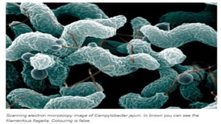
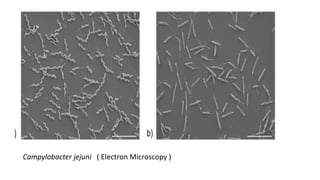
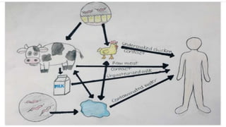
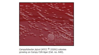
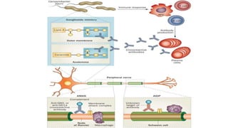
Campylobacter species are microaerophilic
curved gram-negative bacilli. They are zoonotic,
cause both diarrheal ( C. jejuni ) and systemic
diseases ( C. fetus ).
Morphology
1. Gram negative rods
2. 0.2-0.5μm – 1.5-5μm
3. Curved or spiral shaped , sometimes “Gull’s wing” appearance
4. Darting Motility
5. Single polar flagellum
6. Non Sporing
Campylobacter jejuni ( Electron Microscopy )
Epidemiology
• Mode of transmission:
(i) By ingestion of raw or undercooked food-
poultry
(most common), raw (unpasteurized) milk or
water,
(ii) Through direct contact with animals,
(iii) Oral-Faecal contact
• MC Age affected: C.jejuni (children) and C.fetus (extremes of
age)
• Developing versus developed countries:
 In developing countries : It is hyperendemic, mostly
asymptomatic
except < 2 years (symptomatic).
 In developed countries, Campylobacter is the leading bacterial
cause
of diarrheal disease.
• Seasonality: Incidence peaks during summer and early autumn.
Cultural Properties
1. Microaerophilic.
(5-10% CO2, 2-5% O2 and high humidity).
2. Non Fermenter
3. Oxidase +ve
4. Catalase +ve
5. Nitrate Reducer
6. Thermophilic Grow at 42°C except C. fetus ( non thermophilic )
 Fastidious organism requires enriched &
selective growth media as:
- Blood agar.
- Campy BAP selective media.
- Butzler’s selective media.
- Modified Skirrow’s blood agar (selective
medium, blood agar with antibiotics) containing
vancomycin, polymyxin & trimethoprim.
 Transport medium : Cary-Blair medium can be used.
It can survive for 1-2 weeks at 4°C.
 Incubation for 2-5 days.
 Non-proteolytic and unable to attack carbohydrates.
 Colony morphology is :
1: Watery and spreading
2 : Round and convex
Pathogenesis
• Motility of the strain (possesses single polar flagellum and exhibits
darting motility)
• Capacity to adhere to host tissues
• Toxins play a minor role by inflammation :
○ Enterotoxin (Heat-labile, similar to cholera toxin)
○ Cytotoxins (Cytolethal distending toxin, or CDT)
• Proteinaceous capsule-like structure (S-layer) expressed by C.fetus.
It prevents the bacteria from complement mediated killing and
opsonisation and may contribute to die chronicity and high rate of
recurrence of
C. fetus infections in immunocompromised hosts.
Clinical Manifestations
The clinical manifestations seen in campylobacteriosis are as follows :
• Intestinal infection: It is characterized by inflammatory diarrhoea,
abdominal pain and fever. Degree of diarrhoea varies from several
loose stools to grossly bloody stools. It is self-limiting; however, relapse
is seen in 5- 10% of untreated cases.
• Extraintestinal infection:
It is mainly due to C. fetus developing mostly in immunocompromised
hosts and at the extremes of age. Common manifestations include
bacteremia, sepsis, meningitis, vascular infections (endocarditis,
aneurysm, and thrombophlebitis).
 ln persons with the HLA-B27 phenotype: Reactive arthritis and
other rheumatologic manifestations may develop several
weeks after infection with Campylobacter.
 Campylobacter triggers the pathogenesis of various
other diseases such as:
• Guillain -Barre syndrome (mainly by C. Jejuni serotype O19).
• Alpha chain disease, a form of lymphoma that originates in small
intestinal mucosa-associated lymphoid tissue.
Guillain-Barré syndrome
An acute inflammatory demyelinating polyneuropathy characterized
by progressive muscle weakness and areflexia.
Results in progressive loss of nerve impulse conduction leads to
- Loss of sensation.
- Symmetrical Muscle paralysis in limb
- Can lead to respiratory failure.
Laboratory Diagnosis of Campylobacter
 Specimen : Diarrhoeal stool
 Smear Preparation :- Gram Staining is negative with Gull’s wing shape.
 Dark ground microscopy demonstrates the darting motility of the
bacilli.
 Culture Preparation.
 Serologic detection of antibodies by ELISA is useful in patients with
Reactive Arthritis or Guillain-Barre syndrome.
 Antibiotic Sensitivity Testing
Gram Staining : Negative
•Biochemical Reaction Testing :
1. Urease Negative.
2. Oxidase Positive.
3. Catalase positive.
4. Hippurate Positive.
5. Nitrate Reducer
6. H₂S Production : Negative
Urease Negative
Oxidase Negative
Catalase Negative
Hippurate Positive
H₂S Production : Negative
Thank You

Campylobacter

  • 1.
  • 2.
    Introduction Campylobacter species aremicroaerophilic curved gram-negative bacilli. They are zoonotic, cause both diarrheal ( C. jejuni ) and systemic diseases ( C. fetus ).
  • 4.
    Morphology 1. Gram negativerods 2. 0.2-0.5μm – 1.5-5μm 3. Curved or spiral shaped , sometimes “Gull’s wing” appearance 4. Darting Motility 5. Single polar flagellum 6. Non Sporing
  • 5.
    Campylobacter jejuni (Electron Microscopy )
  • 6.
    Epidemiology • Mode oftransmission: (i) By ingestion of raw or undercooked food- poultry (most common), raw (unpasteurized) milk or water, (ii) Through direct contact with animals, (iii) Oral-Faecal contact
  • 8.
    • MC Ageaffected: C.jejuni (children) and C.fetus (extremes of age) • Developing versus developed countries:  In developing countries : It is hyperendemic, mostly asymptomatic except < 2 years (symptomatic).  In developed countries, Campylobacter is the leading bacterial cause of diarrheal disease. • Seasonality: Incidence peaks during summer and early autumn.
  • 9.
    Cultural Properties 1. Microaerophilic. (5-10%CO2, 2-5% O2 and high humidity). 2. Non Fermenter 3. Oxidase +ve 4. Catalase +ve 5. Nitrate Reducer 6. Thermophilic Grow at 42°C except C. fetus ( non thermophilic )
  • 11.
     Fastidious organismrequires enriched & selective growth media as: - Blood agar. - Campy BAP selective media. - Butzler’s selective media. - Modified Skirrow’s blood agar (selective medium, blood agar with antibiotics) containing vancomycin, polymyxin & trimethoprim.
  • 13.
     Transport medium: Cary-Blair medium can be used. It can survive for 1-2 weeks at 4°C.  Incubation for 2-5 days.  Non-proteolytic and unable to attack carbohydrates.  Colony morphology is : 1: Watery and spreading 2 : Round and convex
  • 15.
    Pathogenesis • Motility ofthe strain (possesses single polar flagellum and exhibits darting motility) • Capacity to adhere to host tissues • Toxins play a minor role by inflammation : ○ Enterotoxin (Heat-labile, similar to cholera toxin) ○ Cytotoxins (Cytolethal distending toxin, or CDT) • Proteinaceous capsule-like structure (S-layer) expressed by C.fetus. It prevents the bacteria from complement mediated killing and opsonisation and may contribute to die chronicity and high rate of recurrence of C. fetus infections in immunocompromised hosts.
  • 17.
    Clinical Manifestations The clinicalmanifestations seen in campylobacteriosis are as follows : • Intestinal infection: It is characterized by inflammatory diarrhoea, abdominal pain and fever. Degree of diarrhoea varies from several loose stools to grossly bloody stools. It is self-limiting; however, relapse is seen in 5- 10% of untreated cases.
  • 18.
    • Extraintestinal infection: Itis mainly due to C. fetus developing mostly in immunocompromised hosts and at the extremes of age. Common manifestations include bacteremia, sepsis, meningitis, vascular infections (endocarditis, aneurysm, and thrombophlebitis).
  • 19.
     ln personswith the HLA-B27 phenotype: Reactive arthritis and other rheumatologic manifestations may develop several weeks after infection with Campylobacter.  Campylobacter triggers the pathogenesis of various other diseases such as: • Guillain -Barre syndrome (mainly by C. Jejuni serotype O19). • Alpha chain disease, a form of lymphoma that originates in small intestinal mucosa-associated lymphoid tissue.
  • 20.
    Guillain-Barré syndrome An acuteinflammatory demyelinating polyneuropathy characterized by progressive muscle weakness and areflexia. Results in progressive loss of nerve impulse conduction leads to - Loss of sensation. - Symmetrical Muscle paralysis in limb - Can lead to respiratory failure.
  • 26.
    Laboratory Diagnosis ofCampylobacter  Specimen : Diarrhoeal stool  Smear Preparation :- Gram Staining is negative with Gull’s wing shape.  Dark ground microscopy demonstrates the darting motility of the bacilli.  Culture Preparation.  Serologic detection of antibodies by ELISA is useful in patients with Reactive Arthritis or Guillain-Barre syndrome.  Antibiotic Sensitivity Testing
  • 27.
  • 28.
    •Biochemical Reaction Testing: 1. Urease Negative. 2. Oxidase Positive. 3. Catalase positive. 4. Hippurate Positive. 5. Nitrate Reducer 6. H₂S Production : Negative
  • 29.
  • 30.
  • 31.
  • 32.
  • 33.
  • 34.