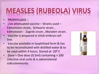
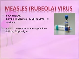
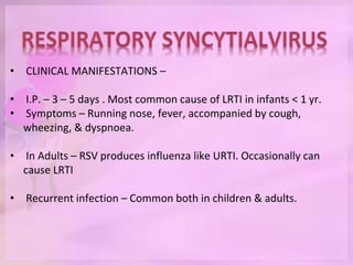
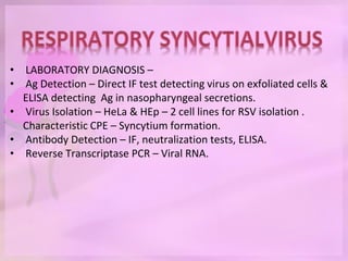
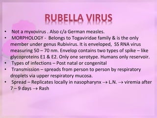
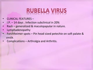
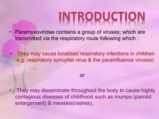
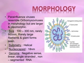
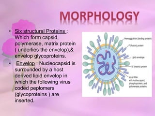
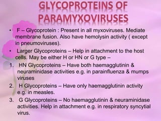
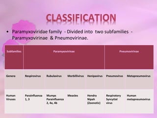
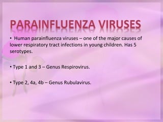
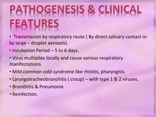
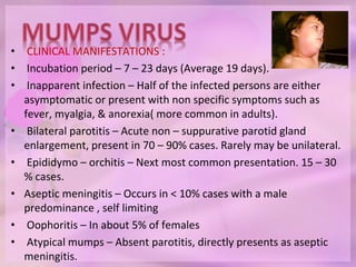
• Paramyxoviridae contains a group of viruses; which are
transmitted via the respiratory route following which :
• They may cause localized respiratory infections in children
e.g. respiratory syncytial virus & the parainfluenza viruses)
or
• They may disseminate throughout the body to cause highly
contagious diseases of childhood such as mumps (parotid
enlargement) & measles(rashes).
• Parainfluenza viruses
resemble Orthomyxoviruses
in morphology but are larger
& pleomorphic.
• Size : 100 – 300 nm, rarely
800nm, Rarely large
filaments & giant forms
seen.
• Symmetry : Helical
• Nucleocapsid : 18nm
• Genome : Negative sense,
linear, single stranded , non
– segmented RNA
• Six structural Proteins :
Which form capsid,
polymerase, matrix protein
( underlies the envelop),&
envelop glycoproteins.
• Envelop : Nucleocapsid is
surrounded by a host
derived lipid envelop in
which the following virus
coded peplomers
(glycoproteins ) are
inserted.
• F – Glycoprotein : Present in all myxoviruses. Mediate
membrane fusion. Also have hemolysin activity ( except
in pneumoviruses).
• Larger Glycoproteins – Help in attachment to the host
cells. May be either H or HN or G type –
1. HN Glycoproteins – Have both haemagglutinin &
neuraminidase activities e.g. in parainfluenza & mumps
viruses
2. H Glycoproteins – Have only haemagglutinin activity
e.g. in measles.
3. G Glycoproteins – No haemagglutinin & neuraminidase
activities. Help in attachment e.g. in respiratory syncytial
virus.
• Paramyxoviridae family - Divided into two subfamilies -
Paramyxovirinae & Pneumovirinae.
Subfamilies Paramyxovirinae Pneumovirinae
Genera Respirovirus Rubulavirus Morbillivirus Henipavirus Pneumovirus Metapneumovirus
Human
Viruses
Parainfluenza
1, 3
Mumps
Parainfluenza
2, 4a, 4b
Measles Hendra
Nipah
(Zoonotic)
Respiratory
Syncytial
virus
Human
metapneumovirus
• Human parainfluenza viruses – one of the major causes of
lower respiratory tract infections in young children. Has 5
serotypes.
• Type 1 and 3 – Genus Respirovirus.
• Type 2, 4a, 4b – Genus Rubulavirus.
• Transmission by respiratory route ( By direct salivary contact or
by large – droplet aerosols).
• Incubation Period – 5 to 6 days.
• Virus multiplies locally and cause various respiratory
manifestations.
• Mild common cold syndrome like rhinitis, pharyngitis.
• Laryngotracheobronchitis ( croup) – with type 1 & 2 viruses.
• Bronchitis & Pneumonia
• Reinfection.
• Worldwide in distribution
• Type 3 – Most prevalent serotype & exists as endemic
throughout the year with annual epidemics during spring.
• Type 1 & Type 2 – Less common. Tend to cause epidemics
during rainfall or winter.
• Type 4a & 4b cause milder illness and are most difficult to
isolate.
• Parainfluenza viruses – Important cause of outbreaks in
pediatric wards, day care centers & in schools.
• Antigen detection : Viral antigens in the infected exfoliated
epithelial cells of nasopharynx – Detected by direct
immunofluorescence by using specific monoclonal antibodies. It
is rapid but less sensitive.
• Viral isolation :
1. Specimens – Nasal washes, bronchoalveolar lavage fluid, lung
tissue. Specimens to be inoculated as early as possible.
2. Tissue culture – Primary monkey kidney cell line – most
sensitive. Other cell lines used – LLC – MK2.
3. Produce little or no cytopathic effects
4. Viral growth detected by performing haemadsorption using
guinea pig erythrocytes or Ag detection by direct IF.
•Serum antibodies : Can be measured by neutralization test,
haemagglutination inhibition test or ELISA. Presence of IgM or
fourfold rise of IgG titer – Indicative of active infection.
• Reverse transcriptase PCR : Highly specific & sensitive. But
available only in limited settings.
• Most common cause of parotid gland enlargement in children. In
severe cases, it can also cause orchitis & aseptic meningitis.
• PATHOGENESIS :
 Transmission – Through respiratory route via droplets, saliva, &
fomites.
 Primary replication – Occurs in the nasal mucosa or upper
respiratory mucosa → infects mononuclear cells & regional lymph
nodes→ spills over to blood stream resulting in viremia →
dissemination.
 Target sites – Mumps virus has special affinity for glandular
epithelium. Classic sites – salivary glands, testes, pancreas,
ovaries, mammary glands, & central nervous system.
• CLINICAL MANIFESTATIONS :
• Incubation period – 7 – 23 days (Average 19 days).
• Inapparent infection – Half of the infected persons are either
asymptomatic or present with non specific symptoms such as
fever, myalgia, & anorexia( more common in adults).
• Bilateral parotitis – Acute non – suppurative parotid gland
enlargement, present in 70 – 90% cases. Rarely may be unilateral.
• Epididymo – orchitis – Next most common presentation. 15 – 30
% cases.
• Aseptic meningitis – Occurs in < 10% cases with a male
predominance , self limiting
• Oophoritis – In about 5% of females
• Atypical mumps – Absent parotitis, directly presents as aseptic
meningitis.
• EPIDEMIOLOGY –
• Endemic worldwide. Peak in winter & spring.
• Period of communicability – Pts are infectious from 1 wk. before
to 1 wk. after the onset of symptoms. It is shed in saliva,
respiratory droplets, & urine.
• Source – Clinical & subclinical cases. No carrier state.
• Reservoir – Humans are the only reservoir of infection.
• Incidence – 100 – 1000 cases per 10000.
• Age – 5 – 9 years. No age spared.
• Immunity – One attack – lifelong immunity.
• LABORATORY DIAGNOSIS –
• Specimens – Buccal or oral swab, CSF, Saliva, urine
• Antigen detection – By direct IF test
• Viral isolation – 1. Primary monkey kidney cells, 2. Shell viral
technique. Cytopathic effect – cell rounding & giant cell formation.
• Serum Abs – By ELISA, neutralization test, haemagglutination
inhibition test.
• RT – PCR – Detects viral RNA.
• TREATMENT –
• No specific antiviral drug. T/t – mostly symptomatic
• PROPHYLAXIS –
• LIVE ATTENUATED VACCINE – From Jeryl Lynn or RIT 4385, or
Urabe strain. It is prepared in chick embryo cell line.
• MUMPS VACCINE IS AVAILABLE AS – 1. Trivalent MMR vaccine
(Live attenuated Measles – Mumps – Rubella vaccine) or 2.
Quadrivalent MMR – V vaccine ( contains additional live
attenuated varicella vaccine) or 3. Monovalent mumps vaccine (
not commonly used)
• Schedule – 2 doses of MMR is given by IM route at 1 yr. & 4 – 6
yr.(before starting school)
• Efficacy – 90% after second dose.
• Measles is an acute, highly contagious childhood disease,
characterized by fever, respiratory symptoms, followed by typical
maculopapular rash.
• PATHOGENESIS –
 Transmission – Via respiratory route through droplet inhalation &
aerosols.
 Spread – Virus multiplies locally in the respiratory tract →Then
spreads to regional lymph nodes → enters the blood stream in
infected monocytes (primary viremia)→ further multiplies in
reticuloendothelial system → spill over into blood ( secondary
viremia)→ disseminates to various sites.
 Target sites – Predominantly the virus is seeded in the epithelial
surfaces of the body, including skin, respiratory tract &
conjunctiva.
• CLINICAL MANIFESTATIONS –
• Incubation period – 10 days to 3 wks.
1. PRODROMAL STAGE – lasts for 4 days.
Characterized by – Fever – On day 1
i.e. 10th day of inf.
• Koplik’s spots - are pathognomic of
measles, appear on 12th day of inf. – 1
mm white to bluish spot surrounded by
an erythema on buccal mucosa near 2nd
lower molar. Rapidly spread to entire
buccal mucosa & fades away on onset of
rash.
• Non specific symptoms – Cough, coryza,
nasal discharge, redness of eye, diarrhea
or vomiting.
• CLINICAL MANIFESTATIONS –
2. Eruptive stage –
• Maculopapular dusky red rashes after 4
days of fever ( i.e. 14th day of infection).
Typically appear first behind the ears→
spread to face, arm, trunk & legs→ then
fade in the same order after 4 days of
onset.
3. Post Measles Stage –
• Characterized by weight loss, weakness
,disorientation , chronic illness.
• COMPLICATIONS –
• Secondary bacterial infections – Otitis media,
Bronchopneumonia.
• Recurrence of fever or failure of fever to subside with rash.
• Giant cell pneumonitis ( Hecht’s pneumonia) in
Immunocompromised pts. Acute Laryngotracheobronchitis &
diarrhoea.
• CNS complications –
1. Post measles encephalomyelitis
2. Measles inclusion body encephalitis
3. Subacute sclerosing panencephelitis
• LABORATORY DIAGNOSIS :
• Specimen – Nasopharyngeal swab
• Antigen detection by using anti –
nucleoprotein antibodies.
• Virus isolation –
1. Monkey or human kidney cells or Vero /
hSLAM cell line – produces CPE as
multinucleated giant cells ( Warthin – Finkeledy
cells.
2. Shell viral culture
• Antibody detection – Against nucleoprotein Ag
by ELISA or neutralization tests.
• Reverse – transcriptase PCR – detects viral
RNA.
• PROPHYLAXIS –
• Live attenuated vaccine – Strains used –
Edmonston strain, Schwartz strain ,
Edmonston - Zagreb strain , Moraten strain.
• Vaccine is prepared in chick embryo cell
line.
• Vaccine available in lyophilized form & has
to be reconstituted with distilled water & to
be used within 4 hours. Stored at -200 C
• Dose – One dose (0.5ml) containing > 100
infective viral units & is administered
subcutaneously.
• PROPHYLAXIS –
• Combined vaccines – MMR or MMR – V
vaccines
• Contacts – Measles immunoglobulin –
0.25 mg / kg/body wt.
• EPIDEMIOLOGY –
• Source – Cases are only source of infection.
• Reservoir – Humans only
• Infective material – Virus shed in secretions of nose, throat, &
respiratory tract of cases of measles.
• Period of communicability - Pts. Are infectious from 4 days
before to 4 days after the onset of rash.
• Secondary attack rate – high
• Age – Children 6 months to 3 years in developing countries &
older children > 5 yrs. In developed countries.
• RSV is a major respiratory pathogen of young children & is the
most common cause of LRTI ( Bronchiolitis & pneumonia) in
infants.
• PATHOGENESIS –
• Transmission – Direct contact( contaminated fingers, fomites, self
inoculation onto conjunctiva or anterior nares or by large
droplets.
• Spread – It replicates locally in the epithelial cells of nasopharynx
 spread to LRT  cause bronchiolitis & pneumonia
• Pathology – Peribronchiolar infiltration by lymphocytes.
Submucosal edema, Necrosis of bronchiolar epithelium &
formation of plugs consisting of mucus, cellular debris & fibrin
which occlude the smaller bronchioles.
• CLINICAL MANIFESTATIONS –
• I.P. – 3 – 5 days . Most common cause of LRTI in infants < 1 yr.
• Symptoms – Running nose, fever, accompanied by cough,
wheezing, & dyspnoea.
• In Adults – RSV produces influenza like URTI. Occasionally can
cause LRTI
• Recurrent infection – Common both in children & adults.
• LABORATORY DIAGNOSIS –
• Ag Detection – Direct IF test detecting virus on exfoliated cells &
ELISA detecting Ag in nasopharyngeal secretions.
• Virus Isolation – HeLa & HEp – 2 cell lines for RSV isolation .
Characteristic CPE – Syncytium formation.
• Antibody Detection – IF, neutralization tests, ELISA.
• Reverse Transcriptase PCR – Viral RNA.
• Not a myxovirus . Also c/a German measles.
• MORPHOLOGY - Belongs to Togaviridae family & is the only
member under genus Rubivirus. It is enveloped, SS RNA virus
measuring 50 – 70 nm. Envelop contains two types of spike – like
glycoproteins E1 & E2. Only one serotype. Humans only reservoir.
• Types of infections – Post natal or congenital
• Transmission – spreads from person to person by respiratory
droplets via upper respiratory mucosa.
• Spread – Replicates locally in nasopharynx  L.N.  viremia after
7 – 9 days  Rash
• CLINICAL FEATURES –
• I.P. – 14 days . Infection subclinical in 20%
• Rash – generalized & maculopapular in nature.
• Lymphadenopathy
• Forchheimer spots – Pin head sized petechie on soft palate &
uvula.
• Complications – Arthralgia and Arthritis.
• LABORATORY DIAGNOSIS –
• Specimens – Nasopharyngeal & Throat swab.
• Virus Isolation – In monkey or rabbit origin cell lines & then
growth detected by viral interference. Shell viral technique.
• Antibody Detection – By HAI or ELISA
• CONGENITAL RUBELLA SYNDROME – Has teratogenic effect.
Transmission to fetus if mother is infected during first trimester .
• Causes ear defect, ocular defect, cardiac defect & CNS defects.
• VACCINATION –
• RA 27/3 is live attenuated vaccine for rubella prepared from
human diploid fibroblast cell line. Available singly or in
combination of mumps & measles – MMR.
• Schedule – Single dose (0.5 ml) of vaccine is administered
subcutaneously.
Paramyxoviruses lecture dwd

Paramyxoviruses lecture dwd

  • 2.
    • Paramyxoviridae containsa group of viruses; which are transmitted via the respiratory route following which : • They may cause localized respiratory infections in children e.g. respiratory syncytial virus & the parainfluenza viruses) or • They may disseminate throughout the body to cause highly contagious diseases of childhood such as mumps (parotid enlargement) & measles(rashes).
  • 3.
    • Parainfluenza viruses resembleOrthomyxoviruses in morphology but are larger & pleomorphic. • Size : 100 – 300 nm, rarely 800nm, Rarely large filaments & giant forms seen. • Symmetry : Helical • Nucleocapsid : 18nm • Genome : Negative sense, linear, single stranded , non – segmented RNA
  • 4.
    • Six structuralProteins : Which form capsid, polymerase, matrix protein ( underlies the envelop),& envelop glycoproteins. • Envelop : Nucleocapsid is surrounded by a host derived lipid envelop in which the following virus coded peplomers (glycoproteins ) are inserted.
  • 5.
    • F –Glycoprotein : Present in all myxoviruses. Mediate membrane fusion. Also have hemolysin activity ( except in pneumoviruses). • Larger Glycoproteins – Help in attachment to the host cells. May be either H or HN or G type – 1. HN Glycoproteins – Have both haemagglutinin & neuraminidase activities e.g. in parainfluenza & mumps viruses 2. H Glycoproteins – Have only haemagglutinin activity e.g. in measles. 3. G Glycoproteins – No haemagglutinin & neuraminidase activities. Help in attachment e.g. in respiratory syncytial virus.
  • 6.
    • Paramyxoviridae family- Divided into two subfamilies - Paramyxovirinae & Pneumovirinae. Subfamilies Paramyxovirinae Pneumovirinae Genera Respirovirus Rubulavirus Morbillivirus Henipavirus Pneumovirus Metapneumovirus Human Viruses Parainfluenza 1, 3 Mumps Parainfluenza 2, 4a, 4b Measles Hendra Nipah (Zoonotic) Respiratory Syncytial virus Human metapneumovirus
  • 7.
    • Human parainfluenzaviruses – one of the major causes of lower respiratory tract infections in young children. Has 5 serotypes. • Type 1 and 3 – Genus Respirovirus. • Type 2, 4a, 4b – Genus Rubulavirus.
  • 8.
    • Transmission byrespiratory route ( By direct salivary contact or by large – droplet aerosols). • Incubation Period – 5 to 6 days. • Virus multiplies locally and cause various respiratory manifestations. • Mild common cold syndrome like rhinitis, pharyngitis. • Laryngotracheobronchitis ( croup) – with type 1 & 2 viruses. • Bronchitis & Pneumonia • Reinfection.
  • 9.
    • Worldwide indistribution • Type 3 – Most prevalent serotype & exists as endemic throughout the year with annual epidemics during spring. • Type 1 & Type 2 – Less common. Tend to cause epidemics during rainfall or winter. • Type 4a & 4b cause milder illness and are most difficult to isolate. • Parainfluenza viruses – Important cause of outbreaks in pediatric wards, day care centers & in schools.
  • 10.
    • Antigen detection: Viral antigens in the infected exfoliated epithelial cells of nasopharynx – Detected by direct immunofluorescence by using specific monoclonal antibodies. It is rapid but less sensitive. • Viral isolation : 1. Specimens – Nasal washes, bronchoalveolar lavage fluid, lung tissue. Specimens to be inoculated as early as possible. 2. Tissue culture – Primary monkey kidney cell line – most sensitive. Other cell lines used – LLC – MK2. 3. Produce little or no cytopathic effects 4. Viral growth detected by performing haemadsorption using guinea pig erythrocytes or Ag detection by direct IF.
  • 11.
    •Serum antibodies :Can be measured by neutralization test, haemagglutination inhibition test or ELISA. Presence of IgM or fourfold rise of IgG titer – Indicative of active infection. • Reverse transcriptase PCR : Highly specific & sensitive. But available only in limited settings.
  • 12.
    • Most commoncause of parotid gland enlargement in children. In severe cases, it can also cause orchitis & aseptic meningitis. • PATHOGENESIS :  Transmission – Through respiratory route via droplets, saliva, & fomites.  Primary replication – Occurs in the nasal mucosa or upper respiratory mucosa → infects mononuclear cells & regional lymph nodes→ spills over to blood stream resulting in viremia → dissemination.  Target sites – Mumps virus has special affinity for glandular epithelium. Classic sites – salivary glands, testes, pancreas, ovaries, mammary glands, & central nervous system.
  • 13.
    • CLINICAL MANIFESTATIONS: • Incubation period – 7 – 23 days (Average 19 days). • Inapparent infection – Half of the infected persons are either asymptomatic or present with non specific symptoms such as fever, myalgia, & anorexia( more common in adults). • Bilateral parotitis – Acute non – suppurative parotid gland enlargement, present in 70 – 90% cases. Rarely may be unilateral. • Epididymo – orchitis – Next most common presentation. 15 – 30 % cases. • Aseptic meningitis – Occurs in < 10% cases with a male predominance , self limiting • Oophoritis – In about 5% of females • Atypical mumps – Absent parotitis, directly presents as aseptic meningitis.
  • 14.
    • EPIDEMIOLOGY – •Endemic worldwide. Peak in winter & spring. • Period of communicability – Pts are infectious from 1 wk. before to 1 wk. after the onset of symptoms. It is shed in saliva, respiratory droplets, & urine. • Source – Clinical & subclinical cases. No carrier state. • Reservoir – Humans are the only reservoir of infection. • Incidence – 100 – 1000 cases per 10000. • Age – 5 – 9 years. No age spared. • Immunity – One attack – lifelong immunity.
  • 15.
    • LABORATORY DIAGNOSIS– • Specimens – Buccal or oral swab, CSF, Saliva, urine • Antigen detection – By direct IF test • Viral isolation – 1. Primary monkey kidney cells, 2. Shell viral technique. Cytopathic effect – cell rounding & giant cell formation. • Serum Abs – By ELISA, neutralization test, haemagglutination inhibition test. • RT – PCR – Detects viral RNA. • TREATMENT – • No specific antiviral drug. T/t – mostly symptomatic
  • 16.
    • PROPHYLAXIS – •LIVE ATTENUATED VACCINE – From Jeryl Lynn or RIT 4385, or Urabe strain. It is prepared in chick embryo cell line. • MUMPS VACCINE IS AVAILABLE AS – 1. Trivalent MMR vaccine (Live attenuated Measles – Mumps – Rubella vaccine) or 2. Quadrivalent MMR – V vaccine ( contains additional live attenuated varicella vaccine) or 3. Monovalent mumps vaccine ( not commonly used) • Schedule – 2 doses of MMR is given by IM route at 1 yr. & 4 – 6 yr.(before starting school) • Efficacy – 90% after second dose.
  • 17.
    • Measles isan acute, highly contagious childhood disease, characterized by fever, respiratory symptoms, followed by typical maculopapular rash. • PATHOGENESIS –  Transmission – Via respiratory route through droplet inhalation & aerosols.  Spread – Virus multiplies locally in the respiratory tract →Then spreads to regional lymph nodes → enters the blood stream in infected monocytes (primary viremia)→ further multiplies in reticuloendothelial system → spill over into blood ( secondary viremia)→ disseminates to various sites.  Target sites – Predominantly the virus is seeded in the epithelial surfaces of the body, including skin, respiratory tract & conjunctiva.
  • 18.
    • CLINICAL MANIFESTATIONS– • Incubation period – 10 days to 3 wks. 1. PRODROMAL STAGE – lasts for 4 days. Characterized by – Fever – On day 1 i.e. 10th day of inf. • Koplik’s spots - are pathognomic of measles, appear on 12th day of inf. – 1 mm white to bluish spot surrounded by an erythema on buccal mucosa near 2nd lower molar. Rapidly spread to entire buccal mucosa & fades away on onset of rash. • Non specific symptoms – Cough, coryza, nasal discharge, redness of eye, diarrhea or vomiting.
  • 19.
    • CLINICAL MANIFESTATIONS– 2. Eruptive stage – • Maculopapular dusky red rashes after 4 days of fever ( i.e. 14th day of infection). Typically appear first behind the ears→ spread to face, arm, trunk & legs→ then fade in the same order after 4 days of onset. 3. Post Measles Stage – • Characterized by weight loss, weakness ,disorientation , chronic illness.
  • 20.
    • COMPLICATIONS – •Secondary bacterial infections – Otitis media, Bronchopneumonia. • Recurrence of fever or failure of fever to subside with rash. • Giant cell pneumonitis ( Hecht’s pneumonia) in Immunocompromised pts. Acute Laryngotracheobronchitis & diarrhoea. • CNS complications – 1. Post measles encephalomyelitis 2. Measles inclusion body encephalitis 3. Subacute sclerosing panencephelitis
  • 21.
    • LABORATORY DIAGNOSIS: • Specimen – Nasopharyngeal swab • Antigen detection by using anti – nucleoprotein antibodies. • Virus isolation – 1. Monkey or human kidney cells or Vero / hSLAM cell line – produces CPE as multinucleated giant cells ( Warthin – Finkeledy cells. 2. Shell viral culture • Antibody detection – Against nucleoprotein Ag by ELISA or neutralization tests. • Reverse – transcriptase PCR – detects viral RNA.
  • 22.
    • PROPHYLAXIS – •Live attenuated vaccine – Strains used – Edmonston strain, Schwartz strain , Edmonston - Zagreb strain , Moraten strain. • Vaccine is prepared in chick embryo cell line. • Vaccine available in lyophilized form & has to be reconstituted with distilled water & to be used within 4 hours. Stored at -200 C • Dose – One dose (0.5ml) containing > 100 infective viral units & is administered subcutaneously.
  • 23.
    • PROPHYLAXIS – •Combined vaccines – MMR or MMR – V vaccines • Contacts – Measles immunoglobulin – 0.25 mg / kg/body wt.
  • 24.
    • EPIDEMIOLOGY – •Source – Cases are only source of infection. • Reservoir – Humans only • Infective material – Virus shed in secretions of nose, throat, & respiratory tract of cases of measles. • Period of communicability - Pts. Are infectious from 4 days before to 4 days after the onset of rash. • Secondary attack rate – high • Age – Children 6 months to 3 years in developing countries & older children > 5 yrs. In developed countries.
  • 25.
    • RSV isa major respiratory pathogen of young children & is the most common cause of LRTI ( Bronchiolitis & pneumonia) in infants. • PATHOGENESIS – • Transmission – Direct contact( contaminated fingers, fomites, self inoculation onto conjunctiva or anterior nares or by large droplets. • Spread – It replicates locally in the epithelial cells of nasopharynx  spread to LRT  cause bronchiolitis & pneumonia • Pathology – Peribronchiolar infiltration by lymphocytes. Submucosal edema, Necrosis of bronchiolar epithelium & formation of plugs consisting of mucus, cellular debris & fibrin which occlude the smaller bronchioles.
  • 26.
    • CLINICAL MANIFESTATIONS– • I.P. – 3 – 5 days . Most common cause of LRTI in infants < 1 yr. • Symptoms – Running nose, fever, accompanied by cough, wheezing, & dyspnoea. • In Adults – RSV produces influenza like URTI. Occasionally can cause LRTI • Recurrent infection – Common both in children & adults.
  • 27.
    • LABORATORY DIAGNOSIS– • Ag Detection – Direct IF test detecting virus on exfoliated cells & ELISA detecting Ag in nasopharyngeal secretions. • Virus Isolation – HeLa & HEp – 2 cell lines for RSV isolation . Characteristic CPE – Syncytium formation. • Antibody Detection – IF, neutralization tests, ELISA. • Reverse Transcriptase PCR – Viral RNA.
  • 28.
    • Not amyxovirus . Also c/a German measles. • MORPHOLOGY - Belongs to Togaviridae family & is the only member under genus Rubivirus. It is enveloped, SS RNA virus measuring 50 – 70 nm. Envelop contains two types of spike – like glycoproteins E1 & E2. Only one serotype. Humans only reservoir. • Types of infections – Post natal or congenital • Transmission – spreads from person to person by respiratory droplets via upper respiratory mucosa. • Spread – Replicates locally in nasopharynx  L.N.  viremia after 7 – 9 days  Rash
  • 29.
    • CLINICAL FEATURES– • I.P. – 14 days . Infection subclinical in 20% • Rash – generalized & maculopapular in nature. • Lymphadenopathy • Forchheimer spots – Pin head sized petechie on soft palate & uvula. • Complications – Arthralgia and Arthritis.
  • 30.
    • LABORATORY DIAGNOSIS– • Specimens – Nasopharyngeal & Throat swab. • Virus Isolation – In monkey or rabbit origin cell lines & then growth detected by viral interference. Shell viral technique. • Antibody Detection – By HAI or ELISA • CONGENITAL RUBELLA SYNDROME – Has teratogenic effect. Transmission to fetus if mother is infected during first trimester . • Causes ear defect, ocular defect, cardiac defect & CNS defects.
  • 31.
    • VACCINATION – •RA 27/3 is live attenuated vaccine for rubella prepared from human diploid fibroblast cell line. Available singly or in combination of mumps & measles – MMR. • Schedule – Single dose (0.5 ml) of vaccine is administered subcutaneously.