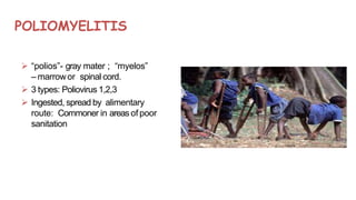
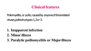
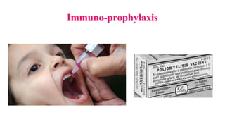
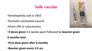
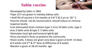
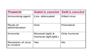
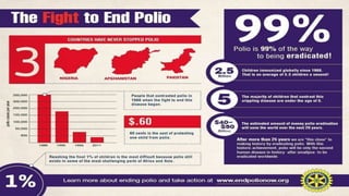
PICORNAVIRIDAE
Introduction to Picornaviruses
Family Picornaviridae
Consists of a large number of very small RNAviruses,
27-30 nm in size
Resistant to lipid solvents like Ether, Chloroform, Bile salts
Morphology of Enterovirus
Classification
• 4 Genera pathogenic tohumans:
1. Enterovirus – Infects intestinal tract
2. Rhinovirus – infects nasal mucosa
3. Hepatovirus- Hepatitis A
4. Paraechovirus- Echo viruses
• 2 more Genera pathogenic foranimals
1. Aphthovirus – Foot and Mouth disease of catlle
2. Cardiovirus – Encephalomyocarditis virus that infects mice
ENTEROVIRUSES–Classification
Group Serotype
Poliovirus (3) Type 1, Type 2, Type3
Coxsackie virus A(24) 1-22, 24
Coxsackie virus (6) 1-6
Echovirus (34) 1-9, 11-27, 29-31
Numbered enterovirus
(EVsince 1969 – Numbered 68 and so)
68-71
Poliovirus - Introduction
• FamilyPicornaviridae; Genus Enterovirus; Species
Poliovirus
• The virus is composed of an RNA genome and a protein capsule. The
genome is single-stranded positive-sense RNA genome that is about
7500 nucleotides long.
• Often called the simplest significant virus - First isolated in 1909 by
Karl Landsteiner and Erwin Popper
• Egyptian paintings depicted the
effects of polio shows a priest
with a deformity of his leg
characteristic of the flaccid
paralysis typical of poliomyelitis.
• and the ‘oldest’ known viruses
(temple record from Egypt 1400
B.C.)
Morphology of Poliovirus
Electronmicroscopy(EM)ofPoliovirus
POLIOMYELITIS
 “polios”- gray mater ; “myelos”
– marrowor spinal cord.
 3 types: Poliovirus 1,2,3
 Ingested, spread by alimentary
route: Commoner in areas of poor
sanitation
Morphology
• Spherical Virion – Icosahedral symmetry
• 27 nm in diameter in size
• Composed of 60 subunits
• Consists of 4 Viral Proteins (VP1, VP2,VP3 &VP4)
• VP1 faces outside – Major antigenic site for combination
• VP1 has type-specific neutralizingantibodies
• Viral Genome: Single stranded positive strand (ss RNA+vesense)
• Virus can be crystallized – seen in cytoplasm of infected cells
Poliomyelitis is an acute infectious disease which affects
the CNS with destruction of motor neurons of the
anterior horns in the spinal cord resulting in flaccid
paralysis (less than 0.1%). However, most poliovirus
infections are subclinical.
During epidemic outbreaks, type I is most frequently
isolated (in 65-95% of cases) while types II and III account for
the remaining5-35% of cases.
Resistance
• Resistant to lipid solvents – ether, chloroform, bile, proteolytic
enzymes of intestinal contents and detergents
• Stable at a pH of 3
• In feces, it can survive for 4 months at 4ºC and for years at-20ºC
• Room temperature survival of virus in feces vary (one day to several
weeks) and It depends on temperature, moisture, pH and amount of
virus
• Readily inactivated by heat (55ºC X30 minutes)
• Molar MgCl2 , Milk or Icecream protects virus against heat
inactivation
Resistance
• Formaldehyde and Oxidising disinfectants destroy the virus
• Chlorination destroy the virus in water
• Organic matter present delays inactivation of virus
• Phenolic disinfectants not effective
• Does not survive lyophilisation well
Host range&Cultivation
• Natural infection occurs only in humans
• Experimental transmission in monkeys by intracerebral or
intraspinal inoculation
• Chimpanzees and Cynomolgus monkeys – can be infected orally
• Established non-fresh strains can be grown in rodents, chick
embryos
• Virus grows readily in Tissue cultures of primate origin
• Primary Monkey Kidney cultures are used for Diagnostic purpose
and for Vaccination
Pathogenicity
1. Virus transmitted by Fecal-oral route through ingestion. Other
possible modes in close contacts in patients of early stages:
Inhalation or Entry through conjunctiva of droplets of respiratory
secretions.
2. Virus multiplies initially in the epithelial cells of the alimentary
canal and in the lymphatic tissues, from the tonsils to peyer’s
patches
3. Spreads to regional lymph nodes and enters blood stream (Primary
viremia)
4. Further multiplication takes place in reticulo-endothelial system
Pathogenicity - Continues
5. Virus enters the Blood stream again (Secondary viremia)
6. Virus is now carried to Central Nervous System (CNS) Spinal cord
and Brain.
7. In CNS,Virus multiplies in selective neurons and destroys them
8. Earliest change: Degeneration of Nissl’s bodies.
9. Lesions are mostly in the anterior horns of the spinal cord causing
Flaccid paralysis, but posterior and intermediate horns can also be
involved.
Clinical features
Poliomyelitis,orpolio,causedby anyoneofthreerelated
viruses,poliovirustypes1,2or3.
1. Inapparent infection
2. Minor illness
3. Paralytic poiliomyelitis or Majorillness
Clinical features
Inapparent infection:
90-95% susceptible individuals develop only inapparent polio infection
with Seroconversion alone.
Only 5-10% among them develop clinical infection. Incubationperiod:
About 10 days on average (Range:4 days – 4weeks)
Minorillness:
Early manifestation is fever, headache, sore throat and malaise (Phase of
primary viremia) lasting 1-5 days called Minor illness or Abortive
poliomyelitis
Paralytic poliomyelitis or majorillness:
Progression of infection 3-4 days after minor illness results in major
illness. Fever returns (Biphasic fever), along with headache, stiff neck and
other features of meningitis due to viral invasion of CNS (Poliocase).
Poliomyelitis types andComplication
• Non- Paralytic polio: Disease does not progress beyond stage of
aseptic meningitis
• So, Typesof polio: Nonparalytic polio &Paralyticpolio.
• Paralytic polio can further be divided into: Spinal polio, bulbar polio,
and bulbospinal polio based on distribution of paralysis.
• Complication: Post-polio syndrome may also occur in which
symptoms ranging form breathing and swallowing problems to joint
pain, start many decades after the initial sickness.
Laboratorydiagnosis
• Samples to be collected: Blood, CSF,Throatswab,Feces
• Transport: Immediately to lab in viral transport media.
• Storage: 4⁰C(Days),-20⁰C(monthsto years)
• LaboratoryDiagnostic testsavailable:
Viral isolation
Serodiagnosis
Molecular diagnosis
1. Viralisolation
• In tissue culture – during primary Viremia 3-5 days afterinfection
(from blood)
• Early stages – isolation from throat swabs
• First week of infection: 80-85% viral isolation from feces
• Second week of infection: 50% viral isolation from feces
• Third week of infection: 25% viral isolation from feces
• Fecal excretion – intermittent (So two samples needed to be tested)
• Prolonged fecal excretion in immunocompromised, but no
permanent carriers,
1. Viral isolation - continues
• poliovirus isolated from CSF,but it can be isolated from spinal cord
and brain, post-mortem diagnosis. (Unlike enteroviruses)
• Primary monkey kidney cells are employed commonly
• Human or Simian kidney cells can also be used.
• Inference:Viralgrowth indicated by Cytopathic effect (CPE)in 2-3
days.
• Mere isolation does not confirm the diagnosis
2. Serodiagnosis
• Antibody rise appears after the onset of paralysis – demonstrated
by Neutralisation tests or Complement Fixation Tests (CFT).
•CFTis useful to identify the exposure to Poliovirus but
not for type-specific diagnosis
3. Molecular diagnosis
•Reverse Transcriptase PCR:
Viral RNAin CSFdemonstrated
•RNASequencing: In circulation 3 types of strainscirculate
1. Wild virus
2. OPVstrain (Oral polio Virus)
3. VDPV– Vaccine Derived PolioVirus
Immuno-prophylaxis
Salk vaccine
•Developed by salk in 1953
•Formalin inactivated vaccine
•Given I/M or subcutanous
•3 doses given 4-6 weeks apart followed by booster given
6 months later
•First dose given after 6 months
•Booster given every 3-5 yrs
Sabin vaccine
•Developed by Sabin in 1962
•Type 127r are grown in monkey kidney cells.
• shelf life of vaccine is 4-6 months at 4-8 °C & 2 yrs at -20 ° C
•Vaccine should not be neurovirulent, should induce an immune
response locally.
•Live attenuated strain contains type 1 virus 10 lakhs units, type 2
virus 2 lakhs units & type 3 3 lakhs units
•Stimulates local IgA and humoral IgM & IgG
•Virus excreted in feces so protects the community
•Given orally 3 doses are given zero dose is given at birth Ist dose
at 6 weeks and 2nd & 3rd dose at difference of 6 weeks
•Booster is given at 18-24 months age
Differentiating features of killed & live vaccine
Virus Killed Polio Virus Live attenuated Virus
Safety in immunodeficient persons Safe Not safe
Administration Injectable (subcutaneous or
Intramuscular
Orally
Economy Costlier Cheaper
Nature of immunity Only systemic antibody response but
no local (intestinal) immunity
Both local immunity & systemic
antibody response
Duration of immunity To be maintained by booster doses
periodically
Life-long
Community protection No Yes
Storage Doesn’t require stringent storage
conditions. Has a longer self life.
To be transported in cold conditions
Usefulness in epidemics Not useful Useful
Eradication of Poliomyelitis
• TheWHOin 1988 proposed global eradication of poliomyelitis by the year
2000
• Byglobal immunization with OPV
• Poor progress in immunization in many countries hasbeen asetback to this objective
• In 2015 – only 74 casesin entire World
• In INDIA– 2009- 741cases
2010- 42cases
2011- 1 case(Howrah)
2012- NOCASE– INDIA’sname wastaken off the list of World’s Endemic
countries
2014- INDIAwasdeclared POLIOERADICA
TED
COUNTRY
• Present – Pakistan,Afganistan, Nigeria
PulsePolio Immunisation Programme
• Massadministration of OPVon asingle day toall
children aged0-5 years in the community
• Thestrategy is to give rounds of dosesat
an interval of 4-6 weeks during the low
polio transmission period in winter(Nov-
Feb)
Treatment
• Currently there is no treatment to cure Polio.Treatment is focused on
supportive care. Moderate exercise - Anutritious diet
• Medication and rest to lower the fever and to reduce the pain and
improve the strength. Breathing assistance with a ventilator
• To Prevent Poliovirus: The most effective and most commonly used is
the Polio vaccine. This vaccine is given to young children in specific
increments.
• Vaccine works by strengthening and preparing the immune system
to a future encounter with the Poliovirus.
Coxsackie virus
Isolated in 1949 from coxsackie village of New York
Resemble polio virus
2 types
Coxsackie A causes myositis & flaccid paralysis
Coxsackie B produces patchy myositis, spastic paralysis ,
pancreatitis , hepatitis, and myocarditis
Clinical features
Infection transmitted by faeco-oral route , IP 2-9 days
Group A virus causes aseptic meningitis, Vesicular
pharyngitis, headache, fever, pharyngitis, pain abdomen
and hand foot mouth disease (vesicles in mouth, hands &
feet)
Group B virus Bornholm disease(epidemic myalgia ),
myocarditis, pericarditis, aseptic meningitis, juvenile
diabetes, neonatal infections, post viral fatigue syndrome
Hand foot mouth disease
Lab Diagnosis
•Specimen feces,
Inoculated in suckling mouse and histopathology done
followed by neutralization test
Tissue culture done show cytopathic effect
Serology not effective
Vaccination not effective
Echo virus
•Entero cytopathic human orphan viruses (Echo) infect only
human
•Resemble polio virus
•Spread by feco oral route and they remain in GIT
•Clinical features aseptic meningitis , paralysis, rash & fever ,
diarrhoea ,respiratory disease, pericarditis and myocarditis
Enterovirus
•Similar to polio virus
•Cause pneumonia, acute hemarragic
conjunctivitis , meningitis & encephalitis
Acute hemmorrghic conjunctivitis
•IP 24 hrs
•causes swelling, congestion, watering &
pain in the eyes, subconjunctival
haemrrohage recovery occurs in 3-7 days
Rhinoviruses
•Causes common cold,
•isolated from nose and throat
•Acid labile so no GIT symptoms
•optimal temp for growth is 33°C
•Virus resemble other picrona virus except that it
is destroyed in acid pH virus causes common cold
infection transmitted by droplet infection.
•IP is 2-4 days
• patient have watery nose, sneezing, cough ,
headache , sore throat and malaise symptoms
resolve in 7 days
Rhino virus
Rhinoviruses contd…
Lab diagnosis:
•Specimen: nose & pharyngeal swab
•Specimen inoculated on human & monkey cell lines and
incubated at 33oC.
•Virus Ag seen by ELISA, PCR
•Serology not useful
Summary
• Polio virus – Belong to Entero virus – ss RNAvirus +sense
• Polio virus: 3 types – 1, 2&3
• Feco-oral transmission
• Paralytic or Non –paralytic polio
• CSF,Blood, Throat swab&feces
• Neutralisation tests, ELISA,CFT,RT-PCR
• OPV
,IPV–Salk and Sabin
• Pulse polio immunization programme.
Poliovirus
• Clinical case scenarion:
• A12 years old boy from Kovilpalayam presented to the emergency
department witha h/o mild fever and sore throat X8 days,
condition worsened and became severe accompanied by neck
rigidity and vomiting for last 2 days after a brief asymptomatic
period of 2 days.
• On morning, boy experienced pain in lower limbs which
increased and progressed as weakness at the time of presentation.
• Mother’s history: Childhood (<5 years) vaccination not remembered
Poliovirus
• Diagnostic &Management strategy of Case scenarion:
• Throat swab, Stool, CSFsent to Diagnostic Microbiology Dept. for
viral studies
• Throat swab and stool specimen tested positive for Cytopathic effect
(CPE)in tissueculture.
• Virus was confirmed as Poliovirus type 1 by Neutralizationtest
• ReverseTranscriptase PCRwas positive for Poliovirus in CSF
• Patient improved on supportive treatment.
THANKYOU

picornaviruses ppt

  • 1.
  • 2.
    Introduction to Picornaviruses FamilyPicornaviridae Consists of a large number of very small RNAviruses, 27-30 nm in size Resistant to lipid solvents like Ether, Chloroform, Bile salts
  • 3.
  • 4.
    Classification • 4 Generapathogenic tohumans: 1. Enterovirus – Infects intestinal tract 2. Rhinovirus – infects nasal mucosa 3. Hepatovirus- Hepatitis A 4. Paraechovirus- Echo viruses • 2 more Genera pathogenic foranimals 1. Aphthovirus – Foot and Mouth disease of catlle 2. Cardiovirus – Encephalomyocarditis virus that infects mice
  • 5.
    ENTEROVIRUSES–Classification Group Serotype Poliovirus (3)Type 1, Type 2, Type3 Coxsackie virus A(24) 1-22, 24 Coxsackie virus (6) 1-6 Echovirus (34) 1-9, 11-27, 29-31 Numbered enterovirus (EVsince 1969 – Numbered 68 and so) 68-71
  • 6.
    Poliovirus - Introduction •FamilyPicornaviridae; Genus Enterovirus; Species Poliovirus • The virus is composed of an RNA genome and a protein capsule. The genome is single-stranded positive-sense RNA genome that is about 7500 nucleotides long. • Often called the simplest significant virus - First isolated in 1909 by Karl Landsteiner and Erwin Popper
  • 7.
    • Egyptian paintingsdepicted the effects of polio shows a priest with a deformity of his leg characteristic of the flaccid paralysis typical of poliomyelitis. • and the ‘oldest’ known viruses (temple record from Egypt 1400 B.C.)
  • 8.
  • 9.
    POLIOMYELITIS  “polios”- graymater ; “myelos” – marrowor spinal cord.  3 types: Poliovirus 1,2,3  Ingested, spread by alimentary route: Commoner in areas of poor sanitation
  • 10.
    Morphology • Spherical Virion– Icosahedral symmetry • 27 nm in diameter in size • Composed of 60 subunits • Consists of 4 Viral Proteins (VP1, VP2,VP3 &VP4) • VP1 faces outside – Major antigenic site for combination • VP1 has type-specific neutralizingantibodies • Viral Genome: Single stranded positive strand (ss RNA+vesense) • Virus can be crystallized – seen in cytoplasm of infected cells
  • 11.
    Poliomyelitis is anacute infectious disease which affects the CNS with destruction of motor neurons of the anterior horns in the spinal cord resulting in flaccid paralysis (less than 0.1%). However, most poliovirus infections are subclinical. During epidemic outbreaks, type I is most frequently isolated (in 65-95% of cases) while types II and III account for the remaining5-35% of cases.
  • 12.
    Resistance • Resistant tolipid solvents – ether, chloroform, bile, proteolytic enzymes of intestinal contents and detergents • Stable at a pH of 3 • In feces, it can survive for 4 months at 4ºC and for years at-20ºC • Room temperature survival of virus in feces vary (one day to several weeks) and It depends on temperature, moisture, pH and amount of virus • Readily inactivated by heat (55ºC X30 minutes) • Molar MgCl2 , Milk or Icecream protects virus against heat inactivation
  • 13.
    Resistance • Formaldehyde andOxidising disinfectants destroy the virus • Chlorination destroy the virus in water • Organic matter present delays inactivation of virus • Phenolic disinfectants not effective • Does not survive lyophilisation well
  • 14.
    Host range&Cultivation • Naturalinfection occurs only in humans • Experimental transmission in monkeys by intracerebral or intraspinal inoculation • Chimpanzees and Cynomolgus monkeys – can be infected orally • Established non-fresh strains can be grown in rodents, chick embryos • Virus grows readily in Tissue cultures of primate origin • Primary Monkey Kidney cultures are used for Diagnostic purpose and for Vaccination
  • 15.
    Pathogenicity 1. Virus transmittedby Fecal-oral route through ingestion. Other possible modes in close contacts in patients of early stages: Inhalation or Entry through conjunctiva of droplets of respiratory secretions. 2. Virus multiplies initially in the epithelial cells of the alimentary canal and in the lymphatic tissues, from the tonsils to peyer’s patches 3. Spreads to regional lymph nodes and enters blood stream (Primary viremia) 4. Further multiplication takes place in reticulo-endothelial system
  • 16.
    Pathogenicity - Continues 5.Virus enters the Blood stream again (Secondary viremia) 6. Virus is now carried to Central Nervous System (CNS) Spinal cord and Brain. 7. In CNS,Virus multiplies in selective neurons and destroys them 8. Earliest change: Degeneration of Nissl’s bodies. 9. Lesions are mostly in the anterior horns of the spinal cord causing Flaccid paralysis, but posterior and intermediate horns can also be involved.
  • 17.
    Clinical features Poliomyelitis,orpolio,causedby anyoneofthreerelated viruses,poliovirustypes1,2or3. 1.Inapparent infection 2. Minor illness 3. Paralytic poiliomyelitis or Majorillness
  • 18.
    Clinical features Inapparent infection: 90-95%susceptible individuals develop only inapparent polio infection with Seroconversion alone. Only 5-10% among them develop clinical infection. Incubationperiod: About 10 days on average (Range:4 days – 4weeks) Minorillness: Early manifestation is fever, headache, sore throat and malaise (Phase of primary viremia) lasting 1-5 days called Minor illness or Abortive poliomyelitis Paralytic poliomyelitis or majorillness: Progression of infection 3-4 days after minor illness results in major illness. Fever returns (Biphasic fever), along with headache, stiff neck and other features of meningitis due to viral invasion of CNS (Poliocase).
  • 19.
    Poliomyelitis types andComplication •Non- Paralytic polio: Disease does not progress beyond stage of aseptic meningitis • So, Typesof polio: Nonparalytic polio &Paralyticpolio. • Paralytic polio can further be divided into: Spinal polio, bulbar polio, and bulbospinal polio based on distribution of paralysis. • Complication: Post-polio syndrome may also occur in which symptoms ranging form breathing and swallowing problems to joint pain, start many decades after the initial sickness.
  • 21.
    Laboratorydiagnosis • Samples tobe collected: Blood, CSF,Throatswab,Feces • Transport: Immediately to lab in viral transport media. • Storage: 4⁰C(Days),-20⁰C(monthsto years) • LaboratoryDiagnostic testsavailable: Viral isolation Serodiagnosis Molecular diagnosis
  • 22.
    1. Viralisolation • Intissue culture – during primary Viremia 3-5 days afterinfection (from blood) • Early stages – isolation from throat swabs • First week of infection: 80-85% viral isolation from feces • Second week of infection: 50% viral isolation from feces • Third week of infection: 25% viral isolation from feces • Fecal excretion – intermittent (So two samples needed to be tested) • Prolonged fecal excretion in immunocompromised, but no permanent carriers,
  • 23.
    1. Viral isolation- continues • poliovirus isolated from CSF,but it can be isolated from spinal cord and brain, post-mortem diagnosis. (Unlike enteroviruses) • Primary monkey kidney cells are employed commonly • Human or Simian kidney cells can also be used. • Inference:Viralgrowth indicated by Cytopathic effect (CPE)in 2-3 days. • Mere isolation does not confirm the diagnosis
  • 24.
    2. Serodiagnosis • Antibodyrise appears after the onset of paralysis – demonstrated by Neutralisation tests or Complement Fixation Tests (CFT). •CFTis useful to identify the exposure to Poliovirus but not for type-specific diagnosis
  • 25.
    3. Molecular diagnosis •ReverseTranscriptase PCR: Viral RNAin CSFdemonstrated •RNASequencing: In circulation 3 types of strainscirculate 1. Wild virus 2. OPVstrain (Oral polio Virus) 3. VDPV– Vaccine Derived PolioVirus
  • 26.
  • 27.
    Salk vaccine •Developed bysalk in 1953 •Formalin inactivated vaccine •Given I/M or subcutanous •3 doses given 4-6 weeks apart followed by booster given 6 months later •First dose given after 6 months •Booster given every 3-5 yrs
  • 28.
    Sabin vaccine •Developed bySabin in 1962 •Type 127r are grown in monkey kidney cells. • shelf life of vaccine is 4-6 months at 4-8 °C & 2 yrs at -20 ° C •Vaccine should not be neurovirulent, should induce an immune response locally. •Live attenuated strain contains type 1 virus 10 lakhs units, type 2 virus 2 lakhs units & type 3 3 lakhs units •Stimulates local IgA and humoral IgM & IgG •Virus excreted in feces so protects the community •Given orally 3 doses are given zero dose is given at birth Ist dose at 6 weeks and 2nd & 3rd dose at difference of 6 weeks •Booster is given at 18-24 months age
  • 30.
    Differentiating features ofkilled & live vaccine Virus Killed Polio Virus Live attenuated Virus Safety in immunodeficient persons Safe Not safe Administration Injectable (subcutaneous or Intramuscular Orally Economy Costlier Cheaper Nature of immunity Only systemic antibody response but no local (intestinal) immunity Both local immunity & systemic antibody response Duration of immunity To be maintained by booster doses periodically Life-long Community protection No Yes Storage Doesn’t require stringent storage conditions. Has a longer self life. To be transported in cold conditions Usefulness in epidemics Not useful Useful
  • 31.
    Eradication of Poliomyelitis •TheWHOin 1988 proposed global eradication of poliomyelitis by the year 2000 • Byglobal immunization with OPV • Poor progress in immunization in many countries hasbeen asetback to this objective • In 2015 – only 74 casesin entire World • In INDIA– 2009- 741cases 2010- 42cases 2011- 1 case(Howrah) 2012- NOCASE– INDIA’sname wastaken off the list of World’s Endemic countries 2014- INDIAwasdeclared POLIOERADICA TED COUNTRY • Present – Pakistan,Afganistan, Nigeria
  • 32.
    PulsePolio Immunisation Programme •Massadministration of OPVon asingle day toall children aged0-5 years in the community • Thestrategy is to give rounds of dosesat an interval of 4-6 weeks during the low polio transmission period in winter(Nov- Feb)
  • 34.
    Treatment • Currently thereis no treatment to cure Polio.Treatment is focused on supportive care. Moderate exercise - Anutritious diet • Medication and rest to lower the fever and to reduce the pain and improve the strength. Breathing assistance with a ventilator • To Prevent Poliovirus: The most effective and most commonly used is the Polio vaccine. This vaccine is given to young children in specific increments. • Vaccine works by strengthening and preparing the immune system to a future encounter with the Poliovirus.
  • 35.
    Coxsackie virus Isolated in1949 from coxsackie village of New York Resemble polio virus 2 types Coxsackie A causes myositis & flaccid paralysis Coxsackie B produces patchy myositis, spastic paralysis , pancreatitis , hepatitis, and myocarditis
  • 36.
    Clinical features Infection transmittedby faeco-oral route , IP 2-9 days Group A virus causes aseptic meningitis, Vesicular pharyngitis, headache, fever, pharyngitis, pain abdomen and hand foot mouth disease (vesicles in mouth, hands & feet) Group B virus Bornholm disease(epidemic myalgia ), myocarditis, pericarditis, aseptic meningitis, juvenile diabetes, neonatal infections, post viral fatigue syndrome
  • 37.
  • 38.
  • 39.
  • 40.
    Lab Diagnosis •Specimen feces, Inoculatedin suckling mouse and histopathology done followed by neutralization test Tissue culture done show cytopathic effect Serology not effective Vaccination not effective
  • 41.
    Echo virus •Entero cytopathichuman orphan viruses (Echo) infect only human •Resemble polio virus •Spread by feco oral route and they remain in GIT •Clinical features aseptic meningitis , paralysis, rash & fever , diarrhoea ,respiratory disease, pericarditis and myocarditis
  • 42.
    Enterovirus •Similar to poliovirus •Cause pneumonia, acute hemarragic conjunctivitis , meningitis & encephalitis Acute hemmorrghic conjunctivitis •IP 24 hrs •causes swelling, congestion, watering & pain in the eyes, subconjunctival haemrrohage recovery occurs in 3-7 days
  • 43.
    Rhinoviruses •Causes common cold, •isolatedfrom nose and throat •Acid labile so no GIT symptoms •optimal temp for growth is 33°C •Virus resemble other picrona virus except that it is destroyed in acid pH virus causes common cold infection transmitted by droplet infection. •IP is 2-4 days • patient have watery nose, sneezing, cough , headache , sore throat and malaise symptoms resolve in 7 days
  • 44.
  • 45.
    Rhinoviruses contd… Lab diagnosis: •Specimen:nose & pharyngeal swab •Specimen inoculated on human & monkey cell lines and incubated at 33oC. •Virus Ag seen by ELISA, PCR •Serology not useful
  • 46.
    Summary • Polio virus– Belong to Entero virus – ss RNAvirus +sense • Polio virus: 3 types – 1, 2&3 • Feco-oral transmission • Paralytic or Non –paralytic polio • CSF,Blood, Throat swab&feces • Neutralisation tests, ELISA,CFT,RT-PCR • OPV ,IPV–Salk and Sabin • Pulse polio immunization programme.
  • 47.
    Poliovirus • Clinical casescenarion: • A12 years old boy from Kovilpalayam presented to the emergency department witha h/o mild fever and sore throat X8 days, condition worsened and became severe accompanied by neck rigidity and vomiting for last 2 days after a brief asymptomatic period of 2 days. • On morning, boy experienced pain in lower limbs which increased and progressed as weakness at the time of presentation. • Mother’s history: Childhood (<5 years) vaccination not remembered
  • 48.
    Poliovirus • Diagnostic &Managementstrategy of Case scenarion: • Throat swab, Stool, CSFsent to Diagnostic Microbiology Dept. for viral studies • Throat swab and stool specimen tested positive for Cytopathic effect (CPE)in tissueculture. • Virus was confirmed as Poliovirus type 1 by Neutralizationtest • ReverseTranscriptase PCRwas positive for Poliovirus in CSF • Patient improved on supportive treatment.
  • 50.