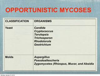
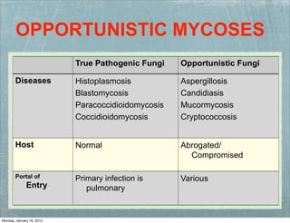
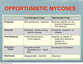
OPPORTUNISTIC MYCOSES
Dr. Dinesh Jain
Assistant Professor
Deptt. of Microbiology
SMS MC Jaipur
OPPORTUNISTIC MYCOSES
CLASSIFICATION ORGANISMS
Yeast Candida
Cryptococcus
Torulopsis
Trichosporon
Rhodotorula
Geotrichium
Molds Aspergillus
Pseudoallescheria
Zygomycetes (Rhizopus, Mucor, and Absidia
Monday, January 16, 2012
OPPORTUNISTIC MYCOSES
True Pathogenic Fungi Opportunistic Fungi
Diseases Histoplasmosis
Blastomycosis
Paracoccidioidomycosis
Coccidioidomycosis
Aspergillosis
Candidiasis
Mucormycosis
Cryptococcosis
Host Normal Abrogated/
Compromised
Portal of
Entry
Primary infection is
pulmonary
Various
Monday, January 16, 2012
OPPORTUNISTIC MYCOSES
True Pathogenic Fungi Opportunistic Fungi
Prognosis 99% spontaneous resolution Recovery depends on the
severity of impairment of
host defenses
Immunity Resolution results to strong
specific immunity
No specific resistance
infection
to
Host Response Tuberculoid granuloma,
mixed pyogenic
Depends on degree of
impairment necrosis to
pyogenic to
granulomatous
Morphology in
Tissue
All agents showed
dimorphism to a
form
tissue
No change in morphology
Distribution Geographically restricted Ubiquitous
Monday, January 16, 2012
CANDIDIASIS
C. albicans is the most common (4-6 um;
budding)
Multiplication: blastospore formation
producing either pseudohyphae or
septate hyphae
Identification: assimilation and
fermentation of CHOs; physiologic and
morphologic responses they exhibit
when grown under controlled
nutritional conditions “germ tubes”
Monday, January 16, 2012
CANDIDIASIS
“chlamydoconidia”
Monday, January 16, 2012
FACTORS THAT AFFECT CANDIDA
NORMAL POPULATION
poor oral hygiene
use of antibiotics
use of oral contraceptives
diet
presence of antagonistic inhibitory bacteria
Monday, January 16, 2012
Candida albicans is a resident flora of the
skin, mouth, vagina and stool!
Imbalance will lead to infection....HOW?
Changes in the Physiology: e.g.
pregnancy, use of steroids and diabetes
Prolonged administration of antibiotics
Immunocompromised patients
Monday, January 16, 2012
MUCOCUTANEOUS CANDIDIASIS
(MC)
a condition caused by a fungus from
the candida family (lives on the
surface of skin) that develops a
diffuse and persistent type of
infection of the mouth, nails, skin, and
at times other organs
affects infants (starts before age 3) and
young adults, is rarely seen in adults
with other diseases
including chronic mucocutaneous
candidaisis or CMCC
Monday, January 16, 2012
SYMPTOMS: ORAL
“thrush” “glossitis” “stomatitis”
“cheilitis” “perleche”
Monday, January 16, 2012
SYMPTOMS: VAGINITIS &
BALANITIS
“VAGINITIS = female”
“BALANITIS = male”
Monday, January 16, 2012
“Esophageal growth”
OTHERS: gastritis, peritonitis, enteric and perianal disease
SYMPTOMS: ALIMENTARY
Monday, January 16, 2012
CANDIDIASIS IN NAILS
Monday, January 16, 2012
CANDIDIASIS IN DIAPER RASH
“Candida may come from fecal origin”
Monday, January 16, 2012
SYSTEMIC INVOLVEMENT
Urinary tract
Endocarditis
Meningitis
Septicemia
Latrogenic candidemia
Dissemination to other organ systems
Monday, January 16, 2012
DISSEMINATED CANDIDIASIS
originate at a gastrointestinal site
CA enters epithelial microvilli through persorption of yeast cells
or by germination (a,c)
In both cases, organisms enter the vasculature (b,d) for
dissemination into tissues such as the kidney (e)
localizes in the cortex (f) where it grows as hyphae/
pseudohyphae
A vigorous host response occurs at this site consisting of both
mononuclear and polymorphonuclear leukocytes
Virulence factors (adhesins, morphogenesis, switch phenotypes,
antioxidant proteins and invasive enzymes) promote the invasion
of the organism
Monday, January 16, 2012
ALLERGIC CANDIDIASIS
Eczema
Asthma
Gastritis
Monday, January 16, 2012
LABORATORY DIAGNOSIS:
CADIDIASIS
Direct microscopic
examination
Specimen for examination can
be sputum, skin scrapings,
vaginal swabs, biopsy material,
from any types of organs or
even in blood.
The specimen is treated with
1-2 drops of 10-20% KOH.
Monday, January 16, 2012
LABORATORY DIAGNOSIS:
CADIDIASIS
The presence of the capsule
and budding yeast cells are
considered as the positive
results.
Aside from KOH, other
stains can be used such as
India ink and Papanicolaou
stain.
Monday, January 16, 2012
GERM TUBE TEST
Most isolates of
C. albicans produce a
hyphal growth from
blastospores when
they are suspended in
serum at 37°C for 2-3
hours.
Monday, January 16, 2012
IN CULTURE...
SDA at either room temperature or at 37°C
Colonies: usually develop in 2-3 days as
white, typical yeast colonies
In vitro: monomorphic, growing as non
encapsulated yeast cells at any temperature
Monday, January 16, 2012
IN CULTURE...
Monday, January 16, 2012
FROM CORN MEAL AGAR
Monday, January 16, 2012
TREATMENT OF CANDIDIASIS
Most localized, cutaneous, candidiasis infections may
be treated with any number of topical antifungal
agents (eg, clotrimazole, econazole, ciclopirox,
miconazole, ketoconazole, nystatin).
For Candida onychomycosis, oral itraconazole
(Sporanox)
For Genitourinary tract candidiasis, VVC can be
managed with either topical antifungal agents or
Monday, January 16, 2012
TREATMENT OF CANDIDIASIS
Caspofungin acetate (Cancidas) as a 70-mg
loading dose is followed by 50 mg/d IV for a
minimum of 2 weeks after improvement or
after blood cultures have cleared.
Chronic mucocutaneous candidiasis is treated
with oral azoles, either fluconazole (Diflucan)
Monday, January 16, 2012
ASPERGILLOSIS
One of the largest of the fungal genera
Hundred of species have been recorded
The most important species:
A. fumigatus
A. flavus
A. niger
Monday, January 16, 2012
ASPERGILLUS FUMIGATUS
Aspergillus fumigatus
identified according to
the pattern of
conidiophore
development,
morphologic features
and color of the
conidia
Monday, January 16, 2012
IMPORTANT PARTS
Monday, January 16, 2012
SPECTRA OF ASPERGILLOSIS
Toxicity due to ingestion of contaminated foods
Allergy and sequelae to the presence of conidia or transient
growth of the organism in body orifices
Colonization without extension in preformed cavities and
debilitated tissues
Invasive, inflammatory, granulomatous, necrotizing disease of
lungs and other organs
Systemic and fatal disseminated disease
Monday, January 16, 2012
ALLERGIC ASPERILLOSIS
Allergic aspergillosis maybe benign early on and
severe as the patient grows older
In secondary colonization, a chronic clinical
situation may exist with little distress except
occasional bout of hemoptysis and some
pathological changes in the lungs that may lead to
the formation of fungus ball.
Monday, January 16, 2012
ALLERGIC ASPERILLOSIS
SKIN FUNGAL SPECIMEN
IN THE TISSUE
Monday, January 16, 2012
SYSTEMIC ASPERGILLOSIS
An extreme serious disorder that is usually
rapidly fatal unless diagnosed early and treated
aggressively
The status of the host’s immune system
contributes to the prognosis of the patient
Monday, January 16, 2012
SYSTEMIC ASPERGILLOSIS
FUNGUS BALL/
ASPERGILLOMA
Monday, January 16, 2012
Disease Etiologic Factors
Mycotoxicoses Ingestion of contaminated food
products
Hypersensitivity
peumonitis
Allergic bronchopulmonary
disease
Secondary
colonization
Colonization of preexisting
cavity (pulmonary abscess)
without invasion into
contiguous tissue
Systemic disease Invasive disease involving
multiple organs
Monday, January 16, 2012
DISSEMINATED ASPERGILLOSIS
Aerosols of Aspergillus fumigatus conidia are inhaled and
travel to the alveoli
In the healthy host, alveolar macrophages (AM) phagocytose
and kill the organism after swelling of the conidium, an
essential pre-germination stage
The production of reactive oxygen intermediates by AM is
required to eliminate the organism, but
polymorphonuclear cells (PMNs) also contribute
In the immunosuppressed patient, reduced numbers of PMNs
and inefficient AM allow growth of the fungus
Consequently, the conidia germinate and escape from the AM
Monday, January 16, 2012
LABORATORY DIAGNOSIS
Aspergillosis is easy to isolate and identify....BUT!
also important to distinguish a true pathogen
from a contaminant
If sputum sample is to be collected, it is expected
to be thick and gelatinous
In invasive sampling, lung aspirates or tissue
biopsy is used
Monday, January 16, 2012
LABORATORY DIAGNOSIS
Direct microscopic examination will show
hyaline, dichotomously branched and septate
hyphae
Occasionally in sputum, in cases of pulmonary
aspergillosis, one may also sees very small, rough
walled spores (3-4 um in diameter).
Monday, January 16, 2012
PULMONARY ASPERGILLOSIS
Monday, January 16, 2012
TREATMENT
Amphotericin B was used
for many years BUT!!! with
disappointing results
In 1990 itraconazole was
introduced as a new broad
spectrum anti-fungal agent.
Monday, January 16, 2012
ZYGOMYCOSIS/PHYCOMYSIS
Class Phycomycetes
Rhizopus
Absidia
Mucor
They formed coenocytic hyphae and reproduce
asexually by producing sporangiosphores within
which develops sporangiospores
Monday, January 16, 2012
ZYGOMYCOSIS/PHYCOMYSIS
Repeated isolation of the
organisms from consecutive
specimens provides strong
evidence that the organisms
may be relevant, even though
coenocytic hyphal elements
are not seen in
histopathologic examination
of tissue.
Monday, January 16, 2012
MUCORMYCOSIS (ORAL CAVITY)
Monday, January 16, 2012
CATEGORIES COMMENTS
Rhinocerebral It is the most frequent presentation
ketoacidosis.
overall and classically affects diabetics with
Usually presents with facial and/or eye pain, proptosis and progressive signs of
involvement of orbital structures (muscles, nerves and vessels).
Common complications include cavernous sinus and internal carotid artery
thrombosis.
Pulmonary It occurs most frequently among neutropenic patients.
It presents with nonspecific symptoms such as fever, cough and dyspnea;
hemoptysis may occur with vascular invasion.
Radiological presentation includes segmental consolidation that progresses to
contiguous areas of the lung, with occasional cavitation.
Gastrointestinal Usually affects patients with severe malnutrition
May involve the stomach, ileum, and colon
Clinical picture mimics intra-abdominal abscess. The diagnosis is often made at
autopsy.
Cutaneous It has been
burns.
reported with minor trauma, insect bites, no sterile dressing, wounds, and
The necrotic lesions progressively evolve from the epidermis into dermis and even
muscle.
Others Heart, bone, kidneys, bladder, trachea, and mediastinum
Monday, January 16, 2012
DIRECT EXAMINATION: ZYGOMYCOSIS
A rapid diagnosis is critical
Fungal elements are usually not numerous in discharges
Scrapings from the upper turbinates, aspirated material
from sinuses, sputum in pulmonary disease, and biopsy
material mounted in 10% KOH typically contain thick-
walled, refractile hyphae 6-15 um in diameter
Swollen cells (up to 50 um) and distorted hyphae may be
present
Monday, January 16, 2012
IN CULTURE...
Sabouraud dextrose agar:
Incubate at 30°C
DON’T: cycloheximide =
sensitive
Sterile bread:
for recovery of Zygomycetes
when other media fail
WHY bread???
Monday, January 16, 2012
TREATMENT
Control of the diabetes
Aggressive surgical
debridement of involved tissue
High doses of amphotericin B
are recommended
Monday, January 16, 2012

Opportunisticmycoses

  • 1.
    OPPORTUNISTIC MYCOSES Dr. DineshJain Assistant Professor Deptt. of Microbiology SMS MC Jaipur
  • 2.
    OPPORTUNISTIC MYCOSES CLASSIFICATION ORGANISMS YeastCandida Cryptococcus Torulopsis Trichosporon Rhodotorula Geotrichium Molds Aspergillus Pseudoallescheria Zygomycetes (Rhizopus, Mucor, and Absidia Monday, January 16, 2012
  • 3.
    OPPORTUNISTIC MYCOSES True PathogenicFungi Opportunistic Fungi Diseases Histoplasmosis Blastomycosis Paracoccidioidomycosis Coccidioidomycosis Aspergillosis Candidiasis Mucormycosis Cryptococcosis Host Normal Abrogated/ Compromised Portal of Entry Primary infection is pulmonary Various Monday, January 16, 2012
  • 4.
    OPPORTUNISTIC MYCOSES True PathogenicFungi Opportunistic Fungi Prognosis 99% spontaneous resolution Recovery depends on the severity of impairment of host defenses Immunity Resolution results to strong specific immunity No specific resistance infection to Host Response Tuberculoid granuloma, mixed pyogenic Depends on degree of impairment necrosis to pyogenic to granulomatous Morphology in Tissue All agents showed dimorphism to a form tissue No change in morphology Distribution Geographically restricted Ubiquitous Monday, January 16, 2012
  • 5.
    CANDIDIASIS C. albicans isthe most common (4-6 um; budding) Multiplication: blastospore formation producing either pseudohyphae or septate hyphae Identification: assimilation and fermentation of CHOs; physiologic and morphologic responses they exhibit when grown under controlled nutritional conditions “germ tubes” Monday, January 16, 2012
  • 6.
  • 7.
    FACTORS THAT AFFECTCANDIDA NORMAL POPULATION poor oral hygiene use of antibiotics use of oral contraceptives diet presence of antagonistic inhibitory bacteria Monday, January 16, 2012
  • 8.
    Candida albicans isa resident flora of the skin, mouth, vagina and stool! Imbalance will lead to infection....HOW? Changes in the Physiology: e.g. pregnancy, use of steroids and diabetes Prolonged administration of antibiotics Immunocompromised patients Monday, January 16, 2012
  • 9.
    MUCOCUTANEOUS CANDIDIASIS (MC) a conditioncaused by a fungus from the candida family (lives on the surface of skin) that develops a diffuse and persistent type of infection of the mouth, nails, skin, and at times other organs affects infants (starts before age 3) and young adults, is rarely seen in adults with other diseases including chronic mucocutaneous candidaisis or CMCC Monday, January 16, 2012
  • 10.
    SYMPTOMS: ORAL “thrush” “glossitis”“stomatitis” “cheilitis” “perleche” Monday, January 16, 2012
  • 11.
    SYMPTOMS: VAGINITIS & BALANITIS “VAGINITIS= female” “BALANITIS = male” Monday, January 16, 2012
  • 12.
    “Esophageal growth” OTHERS: gastritis,peritonitis, enteric and perianal disease SYMPTOMS: ALIMENTARY Monday, January 16, 2012
  • 13.
  • 14.
    CANDIDIASIS IN DIAPERRASH “Candida may come from fecal origin” Monday, January 16, 2012
  • 15.
    SYSTEMIC INVOLVEMENT Urinary tract Endocarditis Meningitis Septicemia Latrogeniccandidemia Dissemination to other organ systems Monday, January 16, 2012
  • 16.
    DISSEMINATED CANDIDIASIS originate ata gastrointestinal site CA enters epithelial microvilli through persorption of yeast cells or by germination (a,c) In both cases, organisms enter the vasculature (b,d) for dissemination into tissues such as the kidney (e) localizes in the cortex (f) where it grows as hyphae/ pseudohyphae A vigorous host response occurs at this site consisting of both mononuclear and polymorphonuclear leukocytes Virulence factors (adhesins, morphogenesis, switch phenotypes, antioxidant proteins and invasive enzymes) promote the invasion of the organism Monday, January 16, 2012
  • 17.
  • 18.
    LABORATORY DIAGNOSIS: CADIDIASIS Direct microscopic examination Specimenfor examination can be sputum, skin scrapings, vaginal swabs, biopsy material, from any types of organs or even in blood. The specimen is treated with 1-2 drops of 10-20% KOH. Monday, January 16, 2012
  • 19.
    LABORATORY DIAGNOSIS: CADIDIASIS The presenceof the capsule and budding yeast cells are considered as the positive results. Aside from KOH, other stains can be used such as India ink and Papanicolaou stain. Monday, January 16, 2012
  • 20.
    GERM TUBE TEST Mostisolates of C. albicans produce a hyphal growth from blastospores when they are suspended in serum at 37°C for 2-3 hours. Monday, January 16, 2012
  • 21.
    IN CULTURE... SDA ateither room temperature or at 37°C Colonies: usually develop in 2-3 days as white, typical yeast colonies In vitro: monomorphic, growing as non encapsulated yeast cells at any temperature Monday, January 16, 2012
  • 22.
  • 23.
    FROM CORN MEALAGAR Monday, January 16, 2012
  • 24.
    TREATMENT OF CANDIDIASIS Mostlocalized, cutaneous, candidiasis infections may be treated with any number of topical antifungal agents (eg, clotrimazole, econazole, ciclopirox, miconazole, ketoconazole, nystatin). For Candida onychomycosis, oral itraconazole (Sporanox) For Genitourinary tract candidiasis, VVC can be managed with either topical antifungal agents or Monday, January 16, 2012
  • 25.
    TREATMENT OF CANDIDIASIS Caspofunginacetate (Cancidas) as a 70-mg loading dose is followed by 50 mg/d IV for a minimum of 2 weeks after improvement or after blood cultures have cleared. Chronic mucocutaneous candidiasis is treated with oral azoles, either fluconazole (Diflucan) Monday, January 16, 2012
  • 26.
    ASPERGILLOSIS One of thelargest of the fungal genera Hundred of species have been recorded The most important species: A. fumigatus A. flavus A. niger Monday, January 16, 2012
  • 27.
    ASPERGILLUS FUMIGATUS Aspergillus fumigatus identifiedaccording to the pattern of conidiophore development, morphologic features and color of the conidia Monday, January 16, 2012
  • 28.
  • 29.
    SPECTRA OF ASPERGILLOSIS Toxicitydue to ingestion of contaminated foods Allergy and sequelae to the presence of conidia or transient growth of the organism in body orifices Colonization without extension in preformed cavities and debilitated tissues Invasive, inflammatory, granulomatous, necrotizing disease of lungs and other organs Systemic and fatal disseminated disease Monday, January 16, 2012
  • 30.
    ALLERGIC ASPERILLOSIS Allergic aspergillosismaybe benign early on and severe as the patient grows older In secondary colonization, a chronic clinical situation may exist with little distress except occasional bout of hemoptysis and some pathological changes in the lungs that may lead to the formation of fungus ball. Monday, January 16, 2012
  • 31.
    ALLERGIC ASPERILLOSIS SKIN FUNGALSPECIMEN IN THE TISSUE Monday, January 16, 2012
  • 32.
    SYSTEMIC ASPERGILLOSIS An extremeserious disorder that is usually rapidly fatal unless diagnosed early and treated aggressively The status of the host’s immune system contributes to the prognosis of the patient Monday, January 16, 2012
  • 33.
  • 34.
    Disease Etiologic Factors MycotoxicosesIngestion of contaminated food products Hypersensitivity peumonitis Allergic bronchopulmonary disease Secondary colonization Colonization of preexisting cavity (pulmonary abscess) without invasion into contiguous tissue Systemic disease Invasive disease involving multiple organs Monday, January 16, 2012
  • 35.
    DISSEMINATED ASPERGILLOSIS Aerosols ofAspergillus fumigatus conidia are inhaled and travel to the alveoli In the healthy host, alveolar macrophages (AM) phagocytose and kill the organism after swelling of the conidium, an essential pre-germination stage The production of reactive oxygen intermediates by AM is required to eliminate the organism, but polymorphonuclear cells (PMNs) also contribute In the immunosuppressed patient, reduced numbers of PMNs and inefficient AM allow growth of the fungus Consequently, the conidia germinate and escape from the AM Monday, January 16, 2012
  • 36.
    LABORATORY DIAGNOSIS Aspergillosis iseasy to isolate and identify....BUT! also important to distinguish a true pathogen from a contaminant If sputum sample is to be collected, it is expected to be thick and gelatinous In invasive sampling, lung aspirates or tissue biopsy is used Monday, January 16, 2012
  • 37.
    LABORATORY DIAGNOSIS Direct microscopicexamination will show hyaline, dichotomously branched and septate hyphae Occasionally in sputum, in cases of pulmonary aspergillosis, one may also sees very small, rough walled spores (3-4 um in diameter). Monday, January 16, 2012
  • 38.
  • 39.
    TREATMENT Amphotericin B wasused for many years BUT!!! with disappointing results In 1990 itraconazole was introduced as a new broad spectrum anti-fungal agent. Monday, January 16, 2012
  • 40.
    ZYGOMYCOSIS/PHYCOMYSIS Class Phycomycetes Rhizopus Absidia Mucor They formedcoenocytic hyphae and reproduce asexually by producing sporangiosphores within which develops sporangiospores Monday, January 16, 2012
  • 41.
    ZYGOMYCOSIS/PHYCOMYSIS Repeated isolation ofthe organisms from consecutive specimens provides strong evidence that the organisms may be relevant, even though coenocytic hyphal elements are not seen in histopathologic examination of tissue. Monday, January 16, 2012
  • 42.
  • 43.
    CATEGORIES COMMENTS Rhinocerebral Itis the most frequent presentation ketoacidosis. overall and classically affects diabetics with Usually presents with facial and/or eye pain, proptosis and progressive signs of involvement of orbital structures (muscles, nerves and vessels). Common complications include cavernous sinus and internal carotid artery thrombosis. Pulmonary It occurs most frequently among neutropenic patients. It presents with nonspecific symptoms such as fever, cough and dyspnea; hemoptysis may occur with vascular invasion. Radiological presentation includes segmental consolidation that progresses to contiguous areas of the lung, with occasional cavitation. Gastrointestinal Usually affects patients with severe malnutrition May involve the stomach, ileum, and colon Clinical picture mimics intra-abdominal abscess. The diagnosis is often made at autopsy. Cutaneous It has been burns. reported with minor trauma, insect bites, no sterile dressing, wounds, and The necrotic lesions progressively evolve from the epidermis into dermis and even muscle. Others Heart, bone, kidneys, bladder, trachea, and mediastinum Monday, January 16, 2012
  • 44.
    DIRECT EXAMINATION: ZYGOMYCOSIS Arapid diagnosis is critical Fungal elements are usually not numerous in discharges Scrapings from the upper turbinates, aspirated material from sinuses, sputum in pulmonary disease, and biopsy material mounted in 10% KOH typically contain thick- walled, refractile hyphae 6-15 um in diameter Swollen cells (up to 50 um) and distorted hyphae may be present Monday, January 16, 2012
  • 45.
    IN CULTURE... Sabouraud dextroseagar: Incubate at 30°C DON’T: cycloheximide = sensitive Sterile bread: for recovery of Zygomycetes when other media fail WHY bread??? Monday, January 16, 2012
  • 46.
    TREATMENT Control of thediabetes Aggressive surgical debridement of involved tissue High doses of amphotericin B are recommended Monday, January 16, 2012