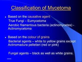
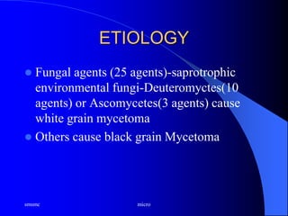
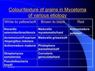
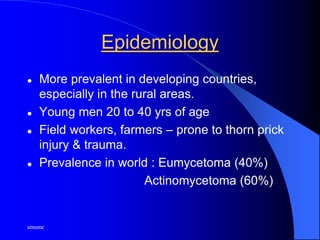
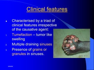
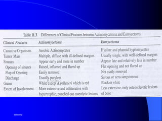
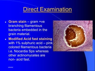
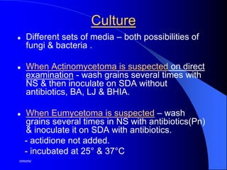
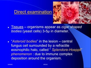
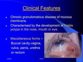
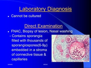
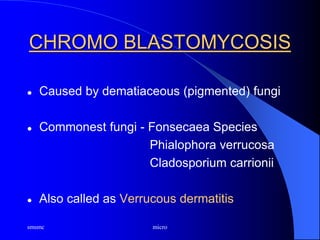
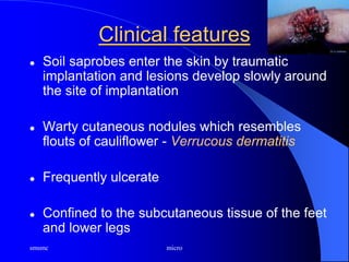
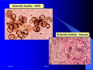
iii sem smsmc
SUBCUTANEOUS
MYCOSIS
Mycetoma
Dr. Dinesh Jain
Assistant Professor
Deptt. of Microbiology
SMS MC Jaipur
INTRODUCTION
 Heterogenous group of fungal infections
which are characterized by development of
clinical lesions in subcutaneous tissues at
the site of inoculation of etiological agents.
Disease process starts following a trivial
trauma, which is the sole source of
infection.
mycology aruna, Microbiology
16/12/07
INTRODUCTION
 Usually follow trauma.
 Lesions develop at the site of implantation of
the etiological agent in the subcutaneous
tissue.
 Includes – Mycetoma
- Sporotrichosis
- Rhinosporidiosis
- Chromoblastomycosis
- Phaeohyphomycosis
- Lobomycosis
16/12/07
MYCETOMA
 Chronic, slowly progressive granulomatous
infection of skin & subcutaneous tissues with
the involvement of underlying fasciae &
bones commonly affecting the extremities.
 Reported by Gill from Madurai, S.India.
 Maduramycosis or Madura foot.
 Tropical & subtropical countries of Asia ,
Africa, Central & S.America.
smsmc
Classification of Mycetoma
 Based on the causative agent
True Fungi – Eumycetoma
Aerobic filamentous bacteria (actinomycetes) -
Actinomycetoma
 Based on the colour of grains
Bacterial agents – white to yellow grains except
Actinomadura pelletieri (red or pink)
Fungal agents – black as well as white grains.
ETIOLOGY
 Fungal agents (25 agents)-saprotrophic
environmental fungi-Deuteromyctes(10
agents) or Ascomycetes(3 agents) cause
white grain mycetoma
 Others cause black grain Mycetoma
smsmc micro
16/12/07
Colour/texture of grains in Mycetoma
of various etiology
White to yellow/soft Brown to black Red
Nocardia
asteroides/brasiliensis
Madurella
mycetomatis/hard
Actinomadura
pelletieri
Acremonium/Fusarium
/Aspergillus nidulans
Madurella grisea/soft
Actinomadura madurai Phialophora
jeanselmei/soft
Streptomyces
somaliensis
Curvularia
geniculata/hard
Pseudollescheria
boydii
smsmc
Epidemiology
 More prevalent in developing countries,
especially in the rural areas.
 Young men 20 to 40 yrs of age
 Field workers, farmers – prone to thorn prick
injury & trauma.
 Prevalence in world : Eumycetoma (40%)
Actinomycetoma (60%)
ssmsmc
Pathogenesis & Pathology
 Causative agent present in soil
 Accidental trauma by thorns or splinter injury
 Minor trauma & skin abrasions
 Mycetoma of ear – use of wicks for removal
of earwax.
 Mycetoma of back – carrying goods like
wood, grain bags, stones, etc
 Mycetoma of the head & neck – bundles of
wood
smsmc
Pathogenesis & Pathology
 Lesion begins as a small subcutaneous
swelling of the foot, which enlarges burrowing
into the deeper tissues & tracking to the
surface as multiple sinuses discharging
viscid, seropurulent fluid containing granules
or grains which are microcolonies of the
causative agent.
smsmc
Clinical features
 Characterised by a triad of
clinical features irrespective
of the causative agent:
1. Tumefaction – tumor like
swelling
2. Multiple draining sinuses
3. Presence of grains or
granules in sinuses.
smsmc
smsmc
Diagnosis
 Radiodiagnosis – Xray, CT, MRI.
 Laboratory diagnosis
- Proper h/o patient
- Gross examination of lesion by a microbiologist
Specimen – grains or granules
- pus / exudates or biopsy
Lesions cleaned with antiseptics & the grains
collected by pressing the sinus from the
periphery.
Gross examination of grains – size, shape,
texture, colour
smsmc
Direct Examination
 KOH mount – grains
Eumycetoma : 2-6µ, wide interwoven hyphae
with large, swollen cells (chlamydospores) at the
margin of the lesion.
Actinomycetoma : filaments with a diameter of
0.5 - 1µ, coccoid to bacillary forms.
If hyphae seen on KOH mount, use special
stains.
smsmc
Direct Examination
 Gram stain – gram +ve
branching filamentous
bacteria embedded in the
grain material.
 Modified Acid fast staining
with 1% sulphuric acid – pink
colored filamentous bacteria
i.e. Nocardia Sps whereas
other actinomycetes are
non- acid fast.
smsmc
Culture
 Different sets of media – both possibilities of
fungi & bacteria .
 When Actinomycetoma is suspected on direct
examination - wash grains several times with
NS & then inoculate on SDA without
antibiotics, BA, LJ & BHIA.
 When Eumycetoma is suspected – wash
grains several times in NS with antibiotics(Pn)
& inoculate it on SDA with antibiotics.
- actidione not added.
- incubated at 25° & 37°C
smsmc micro
Treatment & prophylaxis
 Eumycetoma – Oral Ketoconazole &
Itraconazole
AMB for Madurella & Fusarium species.
 Actinomycetoma – Sulfonamides,
Tetracyclines, Streptomycin, Amoxycillin,
Clavunate & Amikacin
 Protracted case – Surgery (debridement with
skin graft)
smsmc
micro
Botryomycosis
 Similar condition caused by bacteria like
S.aureus, P.aeruginosa, CONS, E.coli.
Pr.vulgaris etc.
 2 types : cutaneous
visceral – ill debilitated pts
smsmc micro
SPOROTRICHOSIS
 Caused by Sporothrix schenckii, a dimorphic
fungus.
 Most common in USA.
 Found on plant, thorns & timber
 Infection is acquired through thorn pricks or
other minor injuries
smsmc micro
Pathogenesis & pathology
 Spreads from primary site
to the regional LNs through
lymphatics
 Mostly involves upper limbs
 Pyogranulomatous reaction
 Clinical features - Nodules
on the skin, subcutaneous
tissue and in the LNs which
later soften & ulcerate.
Lymphocutaneous sporotrichosis
smsmc micro
Laboratory Diagnosis
 Specimens – pus, exudate & aspirate from
nodules.
- curettage or swabs from open lesions.
Direct Examination
 Gram’s stain – gram+ve, irregularly stained
yeast cells.
 CFW – very useful.
smsmc micro
Direct examination
 Tissues – organisms appear as cigar shaped
bodies (yeast cells) 3-5µ in diameter.
 “Asteroid bodies” in the lesion – central
fungus cell surrounded by a refractile
eosinophilic halo, called “ Splendore-Hoeppli”
phenomenon : due to immune complex
deposition around the organism.
smsmc micro
Culture
 Inoculated on 2 sets of SDA, BHIA
 Incubated at 25°& 37°C.
smsmc micro
LPCB mount from Culture
 septate hyphae - very thin & carry flower like
clusters of small conidia on delicate sterigmata.
smsmc micro
Treatment & Prophylaxis
 Saturated solution of KI – drug of choice
 Oral Ketoconazole or Itraconazole
 AMB – disseminated & CNS disease.
smsmc micro
RHINOSPORIDIOSIS
 Caused by a hydrophilic protist,
Rhinosporidium seeberi
 1st identified in Argentina, but majority of cases
occur in India and Sri lanka.
 High incidence among people who frequently
bathe along with domestic animals in ponds,
tanks, lakes
smsmc micro
Clinical Features
 Chronic granulomatous disease of mucous
membrane.
 Characterised by the development of friable
polyps in the nose, mouth or eye.
 Miscellaneous forms –
Buccal cavity,vagina,
vulva, penis, urethra
or rectum
smsmc micro
Laboratory Diagnosis
 Cannot be cultured
Direct Examination
 FNAC, Biopsy of lesion, Nasal washing
- Contains sporangia
filled with thousands of
sporangiospores(6-9µ)
embedded in a stroma
of connective tissue &
capillaries
smsmc micro
Treatment & Prophylaxis
 Radical Surgery:- Excision/ Electrocautery
 Medical therapy :- not useful
DDS (widely used)
 Recurrence common
smsmc micro
CHROMO BLASTOMYCOSIS
 Caused by dematiaceous (pigmented) fungi
 Commonest fungi - Fonsecaea Species
Phialophora verrucosa
Cladosporium carrionii
 Also called as Verrucous dermatitis
smsmc micro
Clinical features
 Soil saprobes enter the skin by traumatic
implantation and lesions develop slowly around
the site of implantation
 Warty cutaneous nodules which resembles
flouts of cauliflower - Verrucous dermatitis
 Frequently ulcerate
 Confined to the subcutaneous tissue of the feet
and lower legs
smsmc micro
Laboratory Diagnosis
Direct Examination
Dry crusty material from the surface of the
lesions
1. KOH w/m –
dark brown, multicellular structures, 5-12μ in
diameter that divide by transverse septation.
-Called sclerotic bodies, medlar bodies, copper-
pennies bodies or muriform cells
smsmc micro
Sclerotic bodies - KOH
Sclerotic bodies - tissues
smsmc micro
Direct examination
Medlar bodies - characteristic tissue form -
facilitates survival of organism in host tissues.
2. Tissue Stains - for Biopsy specimens
HE, Giemsa & Fontana- Masson
- Sclerotic bodies very well seen
Fungal culture - SDA with actidione and
antibiotics
smsmc micro
Treatment & Prophylaxis
 Responds poorly to available therapies.
 Cryotherapy, Thermotherapy, Laser
therapy,Chemotherapy and Surgery.
 Flucytosine (commonly used drug)
 Itraconazole, Fluconazole, Terbinafine
*Relapses are frequently seen
smsmc micro
PHAEOHYPHOMYCOSIS
 Seen in debilitated & immunodeficient hosts.
 Causes subcutaneous & systemic infection.
 Caused by dematiaceous fungi. Commonest
genera involved - Alternaria, Bipolaris,
Curvularia, Exophiala, Phialophora, etc.
smsmc micro
Clinical Features
 Clinical types:
1. Brain abscess caused by Cladosporium
2. Subcutaneous or intramuscular lesions with
abscess or cysts - single circumscribed
lesion with a central cavity filled with pus
and surrounded by a fibrous wall
3. Cutaneous lesions
smsmc micro
Laboratory Diagnosis
Specimen
 Aspirates from cysts
 Curreting from plaques, nodules and drained
abscess
Direct Examination
KOH mount
- Pigmented hyphae 3-4µ in dia.
smsmc micro
Fungal Culture
 SDA with actidione at 25º & 37ºC.
Treatment & Prophylaxis
 Local excision for subcutaneous forms
 Invasive infections – I.V. AMB + Oral
Flucytosine.
smsmc micro
LOBOMYCOSIS
 Caused by Lacazia loboi
(Hydrophilic fungus) : exists
only as yeast cells.
 Involves exposed parts
 Presence of macule, papule,
keloid, verrucous, nodular
lesions or plaques & tumors.
 Lesions are painless with
slight pruritis
smsmc micro
Laboratory Diagnosis
 Direct Examination of curettage / biopsy -
crushed
a. KOH w / m
b. CFW
- spheroid, yeast - like cells, 5 -12µ
- thick - walled & multinucleate.
- form chain with cells joined by bridges.
c. HE – may show ‘asteroid bodies’
• Culture – cannot be cultured
smsmc micro
Fontana Masson stain
smsmc micro
Treatment & prophylaxis
 No effective medical treatment
 Complete excision
 Cryosurgery.

Subcutaneous mycoses

  • 1.
    iii sem smsmc SUBCUTANEOUS MYCOSIS Mycetoma Dr.Dinesh Jain Assistant Professor Deptt. of Microbiology SMS MC Jaipur
  • 2.
    INTRODUCTION  Heterogenous groupof fungal infections which are characterized by development of clinical lesions in subcutaneous tissues at the site of inoculation of etiological agents. Disease process starts following a trivial trauma, which is the sole source of infection. mycology aruna, Microbiology
  • 3.
    16/12/07 INTRODUCTION  Usually followtrauma.  Lesions develop at the site of implantation of the etiological agent in the subcutaneous tissue.  Includes – Mycetoma - Sporotrichosis - Rhinosporidiosis - Chromoblastomycosis - Phaeohyphomycosis - Lobomycosis
  • 4.
    16/12/07 MYCETOMA  Chronic, slowlyprogressive granulomatous infection of skin & subcutaneous tissues with the involvement of underlying fasciae & bones commonly affecting the extremities.  Reported by Gill from Madurai, S.India.  Maduramycosis or Madura foot.  Tropical & subtropical countries of Asia , Africa, Central & S.America.
  • 5.
    smsmc Classification of Mycetoma Based on the causative agent True Fungi – Eumycetoma Aerobic filamentous bacteria (actinomycetes) - Actinomycetoma  Based on the colour of grains Bacterial agents – white to yellow grains except Actinomadura pelletieri (red or pink) Fungal agents – black as well as white grains.
  • 6.
    ETIOLOGY  Fungal agents(25 agents)-saprotrophic environmental fungi-Deuteromyctes(10 agents) or Ascomycetes(3 agents) cause white grain mycetoma  Others cause black grain Mycetoma smsmc micro
  • 7.
    16/12/07 Colour/texture of grainsin Mycetoma of various etiology White to yellow/soft Brown to black Red Nocardia asteroides/brasiliensis Madurella mycetomatis/hard Actinomadura pelletieri Acremonium/Fusarium /Aspergillus nidulans Madurella grisea/soft Actinomadura madurai Phialophora jeanselmei/soft Streptomyces somaliensis Curvularia geniculata/hard Pseudollescheria boydii
  • 8.
    smsmc Epidemiology  More prevalentin developing countries, especially in the rural areas.  Young men 20 to 40 yrs of age  Field workers, farmers – prone to thorn prick injury & trauma.  Prevalence in world : Eumycetoma (40%) Actinomycetoma (60%)
  • 9.
    ssmsmc Pathogenesis & Pathology Causative agent present in soil  Accidental trauma by thorns or splinter injury  Minor trauma & skin abrasions  Mycetoma of ear – use of wicks for removal of earwax.  Mycetoma of back – carrying goods like wood, grain bags, stones, etc  Mycetoma of the head & neck – bundles of wood
  • 10.
    smsmc Pathogenesis & Pathology Lesion begins as a small subcutaneous swelling of the foot, which enlarges burrowing into the deeper tissues & tracking to the surface as multiple sinuses discharging viscid, seropurulent fluid containing granules or grains which are microcolonies of the causative agent.
  • 11.
    smsmc Clinical features  Characterisedby a triad of clinical features irrespective of the causative agent: 1. Tumefaction – tumor like swelling 2. Multiple draining sinuses 3. Presence of grains or granules in sinuses.
  • 12.
  • 13.
    smsmc Diagnosis  Radiodiagnosis –Xray, CT, MRI.  Laboratory diagnosis - Proper h/o patient - Gross examination of lesion by a microbiologist Specimen – grains or granules - pus / exudates or biopsy Lesions cleaned with antiseptics & the grains collected by pressing the sinus from the periphery. Gross examination of grains – size, shape, texture, colour
  • 14.
    smsmc Direct Examination  KOHmount – grains Eumycetoma : 2-6µ, wide interwoven hyphae with large, swollen cells (chlamydospores) at the margin of the lesion. Actinomycetoma : filaments with a diameter of 0.5 - 1µ, coccoid to bacillary forms. If hyphae seen on KOH mount, use special stains.
  • 15.
    smsmc Direct Examination  Gramstain – gram +ve branching filamentous bacteria embedded in the grain material.  Modified Acid fast staining with 1% sulphuric acid – pink colored filamentous bacteria i.e. Nocardia Sps whereas other actinomycetes are non- acid fast.
  • 16.
    smsmc Culture  Different setsof media – both possibilities of fungi & bacteria .  When Actinomycetoma is suspected on direct examination - wash grains several times with NS & then inoculate on SDA without antibiotics, BA, LJ & BHIA.  When Eumycetoma is suspected – wash grains several times in NS with antibiotics(Pn) & inoculate it on SDA with antibiotics. - actidione not added. - incubated at 25° & 37°C
  • 17.
    smsmc micro Treatment &prophylaxis  Eumycetoma – Oral Ketoconazole & Itraconazole AMB for Madurella & Fusarium species.  Actinomycetoma – Sulfonamides, Tetracyclines, Streptomycin, Amoxycillin, Clavunate & Amikacin  Protracted case – Surgery (debridement with skin graft)
  • 18.
    smsmc micro Botryomycosis  Similar conditioncaused by bacteria like S.aureus, P.aeruginosa, CONS, E.coli. Pr.vulgaris etc.  2 types : cutaneous visceral – ill debilitated pts
  • 19.
    smsmc micro SPOROTRICHOSIS  Causedby Sporothrix schenckii, a dimorphic fungus.  Most common in USA.  Found on plant, thorns & timber  Infection is acquired through thorn pricks or other minor injuries
  • 20.
    smsmc micro Pathogenesis &pathology  Spreads from primary site to the regional LNs through lymphatics  Mostly involves upper limbs  Pyogranulomatous reaction  Clinical features - Nodules on the skin, subcutaneous tissue and in the LNs which later soften & ulcerate. Lymphocutaneous sporotrichosis
  • 21.
    smsmc micro Laboratory Diagnosis Specimens – pus, exudate & aspirate from nodules. - curettage or swabs from open lesions. Direct Examination  Gram’s stain – gram+ve, irregularly stained yeast cells.  CFW – very useful.
  • 22.
    smsmc micro Direct examination Tissues – organisms appear as cigar shaped bodies (yeast cells) 3-5µ in diameter.  “Asteroid bodies” in the lesion – central fungus cell surrounded by a refractile eosinophilic halo, called “ Splendore-Hoeppli” phenomenon : due to immune complex deposition around the organism.
  • 23.
    smsmc micro Culture  Inoculatedon 2 sets of SDA, BHIA  Incubated at 25°& 37°C.
  • 24.
    smsmc micro LPCB mountfrom Culture  septate hyphae - very thin & carry flower like clusters of small conidia on delicate sterigmata.
  • 25.
    smsmc micro Treatment &Prophylaxis  Saturated solution of KI – drug of choice  Oral Ketoconazole or Itraconazole  AMB – disseminated & CNS disease.
  • 26.
    smsmc micro RHINOSPORIDIOSIS  Causedby a hydrophilic protist, Rhinosporidium seeberi  1st identified in Argentina, but majority of cases occur in India and Sri lanka.  High incidence among people who frequently bathe along with domestic animals in ponds, tanks, lakes
  • 27.
    smsmc micro Clinical Features Chronic granulomatous disease of mucous membrane.  Characterised by the development of friable polyps in the nose, mouth or eye.  Miscellaneous forms – Buccal cavity,vagina, vulva, penis, urethra or rectum
  • 28.
    smsmc micro Laboratory Diagnosis Cannot be cultured Direct Examination  FNAC, Biopsy of lesion, Nasal washing - Contains sporangia filled with thousands of sporangiospores(6-9µ) embedded in a stroma of connective tissue & capillaries
  • 29.
    smsmc micro Treatment &Prophylaxis  Radical Surgery:- Excision/ Electrocautery  Medical therapy :- not useful DDS (widely used)  Recurrence common
  • 30.
    smsmc micro CHROMO BLASTOMYCOSIS Caused by dematiaceous (pigmented) fungi  Commonest fungi - Fonsecaea Species Phialophora verrucosa Cladosporium carrionii  Also called as Verrucous dermatitis
  • 31.
    smsmc micro Clinical features Soil saprobes enter the skin by traumatic implantation and lesions develop slowly around the site of implantation  Warty cutaneous nodules which resembles flouts of cauliflower - Verrucous dermatitis  Frequently ulcerate  Confined to the subcutaneous tissue of the feet and lower legs
  • 32.
    smsmc micro Laboratory Diagnosis DirectExamination Dry crusty material from the surface of the lesions 1. KOH w/m – dark brown, multicellular structures, 5-12μ in diameter that divide by transverse septation. -Called sclerotic bodies, medlar bodies, copper- pennies bodies or muriform cells
  • 33.
    smsmc micro Sclerotic bodies- KOH Sclerotic bodies - tissues
  • 34.
    smsmc micro Direct examination Medlarbodies - characteristic tissue form - facilitates survival of organism in host tissues. 2. Tissue Stains - for Biopsy specimens HE, Giemsa & Fontana- Masson - Sclerotic bodies very well seen Fungal culture - SDA with actidione and antibiotics
  • 35.
    smsmc micro Treatment &Prophylaxis  Responds poorly to available therapies.  Cryotherapy, Thermotherapy, Laser therapy,Chemotherapy and Surgery.  Flucytosine (commonly used drug)  Itraconazole, Fluconazole, Terbinafine *Relapses are frequently seen
  • 36.
    smsmc micro PHAEOHYPHOMYCOSIS  Seenin debilitated & immunodeficient hosts.  Causes subcutaneous & systemic infection.  Caused by dematiaceous fungi. Commonest genera involved - Alternaria, Bipolaris, Curvularia, Exophiala, Phialophora, etc.
  • 37.
    smsmc micro Clinical Features Clinical types: 1. Brain abscess caused by Cladosporium 2. Subcutaneous or intramuscular lesions with abscess or cysts - single circumscribed lesion with a central cavity filled with pus and surrounded by a fibrous wall 3. Cutaneous lesions
  • 38.
    smsmc micro Laboratory Diagnosis Specimen Aspirates from cysts  Curreting from plaques, nodules and drained abscess Direct Examination KOH mount - Pigmented hyphae 3-4µ in dia.
  • 39.
    smsmc micro Fungal Culture SDA with actidione at 25º & 37ºC. Treatment & Prophylaxis  Local excision for subcutaneous forms  Invasive infections – I.V. AMB + Oral Flucytosine.
  • 40.
    smsmc micro LOBOMYCOSIS  Causedby Lacazia loboi (Hydrophilic fungus) : exists only as yeast cells.  Involves exposed parts  Presence of macule, papule, keloid, verrucous, nodular lesions or plaques & tumors.  Lesions are painless with slight pruritis
  • 41.
    smsmc micro Laboratory Diagnosis Direct Examination of curettage / biopsy - crushed a. KOH w / m b. CFW - spheroid, yeast - like cells, 5 -12µ - thick - walled & multinucleate. - form chain with cells joined by bridges. c. HE – may show ‘asteroid bodies’ • Culture – cannot be cultured
  • 42.
  • 43.
    smsmc micro Treatment &prophylaxis  No effective medical treatment  Complete excision  Cryosurgery.