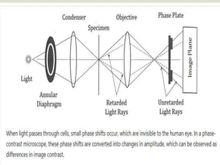
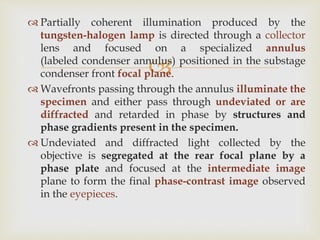
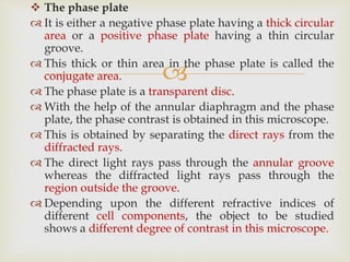
Phase-contrast microscopy is a technique that converts phase shifts in light passing through a transparent specimen to brightness changes in the image, allowing living cells that are otherwise invisible to be seen. It works by separating light rays that pass through a specimen unchanged from those that are diffracted, using an annular diaphragm and phase plate in the light path. Phase-contrast microscopy is widely used in biological research for observing living cells, microorganisms, and other transparent specimens without staining or fixing.
BRIGHT FIELD MICROSCOPY by SIVASANGARI SHANMUGAM
bRIGHT FIELD MICROSCOPY is also called a compound microscope. The name bright - field is derived from the fact that the specimen is dark and contrasted by the surrounding bright viewing field.
DARK FIELD MICROSCOPY by SIVASANGARI SHANMUGAM
Dark-field microscopy is ideally used to illuminate unstained samples causing them to appear brightly lit against a dark background.
This type of microscope contains a special condenser that scatters light and causes it to reflect off the specimen at an angle
5. Microsocope ELECTRON MICROSCOPE (TEM & SEM ) - BasicsNethravathi Siri
Basics only
Electron beam is the source of illumination.
Image is produced by magnetic field.
Contrasting features between light microscope and electron microscope are
construction, working principle, specimen preparation, cost-expenses and designed
room (vacuum chamber).
Microscopy is the technical field of using microscopes to view objects and areas of objects that cannot be seen with the naked eye (objects that are not within the resolution range of the normal eye). There are three well-known branches of microscopy: optical, electron, and scanning probe microscopy, along with the emerging field of X-ray microscopy.
Phase contrast microscopy by sivasangari shanmugam
Phase-contrast microscopy, first described by Dutch physicist Frits Zernike in 1934.
It can be utilized to produce high-contrast images of transparent specimens, such as living cells (usually in culture), microorganisms, thin tissue slices, fibers, latex dispersions, glass fragments, and subcellular particles (including nuclei and other organelles).
Bright field microscopy, Principle and applicationsKAUSHAL SAHU
Introduction
History
Basic Component of Microscope
Light Microscopy
Types of Light Microscopy
What Are Bright Microscopy
Principle of Bright Microscope
Advantage
Disadvantage
Application
Conclusion
Reference
LIGHT MICROSCOPY by SIVASANGARI SHANMUGAM
The optical microscope, The functions of a light microscope is based on its ability to focus a beam of light through, which is very small and transparent, to produce an image.
Electron microscope, principle and applicationKAUSHAL SAHU
Introduction
History
Resolution &Magnification of
Electron microscope
Types of electron microscope
1) Transmission electron microscope (TEM)
- Structural parts of TEM
- Principle & Working of TEM
- Sample preparation for TEM
- Advantages & disadvantages of TEM
Scanning electron microscope (SEM)
- Structural parts of SEM
- Principle & Working of SEM
- Sample preparation for SEM
- Advantages & disadvantages of SEM
3) Scanning transmission electron microscope (STEM)
Applications of electron microscope
Conclusion
References
An isotope is one of two or more atoms having the same atomic number but different mass numbers.
Unstable isotopes are called Radioisotopes.
uses of radioisotopes are many which are discussed in this slide.
To Study Principles of Microscopy: Light Microscope, Phase Contrast Microsco...Om Prakash
To Study Principles of Microscopy: Light Microscope, Phase Contrast Microscope & Electron Microscope
ByOm Prakash
June 13, 2022
Write a Comment
on To Study Principles of Microscopy: Light Microscope, Phase Contrast Microscope & Electron Microscope
Aim: To study principles of Microscopy: Light Microscope, Phase Contrast Microscope & Electron Microscope
Table of Contents
THEORETICAL BACKGROUND:
Light Microscopy
History:
SIMPLE MICROSCOPE
Principles of Microscopy:
THE COMPOUND MICROSCOPE
Phase Contrast Microscope
Electron Microscopes
SCANNING ELECTRON MICROSCOPE (SEM)
Also Read
THEORETICAL BACKGROUND:
Light Microscopy
The light microscope is an instrument designed for the study of cells and tissues. It comprises of lenses that produce a magnified image of the object under study. The light microscope is considered to be a simple important invention that has contributed to the advancement of biological research.
History:
The ancient Greeks and Romans knew the use of Glass and quartz lenses. In the 14th century, spectacles and lenses were used to magnify objects. Galileo had constructed a microscope at the same time (1610). It was employed for the study of the arrangement of the compound eye of insects. Anton Von Leeuwenhoek (1674), the father of biology was the first to use the microscope for biological studies. His microscope has consisted of a single lens with a higher power of magnification. The compound microscope was constructed by Robert Hooke (1665) and is the forerunner of the present-day compound microscope.
SIMPLE MICROSCOPE
The simple microscope distinguishes between two points that are less than 0.1mm apart when placed at a normal viewing distance of 25cm. The two points appear as one and the eye fails to resolve or distinguish them as two distinct points. Another limitation of the human eye is that it cannot resolve any image less than 5µm.
A simple microscope consists of a single convex lens or a combination of lenses that functions as a convex lens. A convex lens magnifies the objects and also helps to produce a magnified image of a near object which appears to be at the distance of distinct vision.
The magnification obtained with a convex lens can be easily calculated by the formula
M = 25/f + 1
Where f= focal length, 25 is the distance of distinct vision in cm.
Principles of Microscopy:
1. Resolving power: It is defined as the capacity of the microscope to distinguish images of two pointed objects lying very close together. If two points are at a distance of more than 0.2 µm, they will appear as two points in the microscope.
2. Limit of resolution: It is defined as the minimum distance at which two objects appear as two distinct objects or entities. It can be calculated as:
Limit of Resolution: 0.61λ/NA = 0.61λ/n Sin θ
Where 0.61 is the constant representing the minimum detectable difference in contrast λ = wavelength of illumination
NA = Numerical aperture, light gathering capa
The pdf contain all the information of various technique ,such as chromatography,spectroscopy,centrifugation,electrophoresis special thanks to Dr.Rambir Singh for helping out the topics easily.Contact for help or suggestion @7985214648 whattapp only
Introduction to microscopy
Different parts of a microscope & their function
Different types of microscopy
Different types of optical microscopy
Different types of electron microscopy
Different terms used in microscopy
Staining- Simple, Differential, Special
Gram Staining
Types of Light Microscopes used in Histological Studies.pptxssuserab552f
Light microscopes relies on glass lenses and visible light to magnify tissue samples. It was
invented in XVII century, and has been improved over the years, resulting in the powerful
modern light microscopes. As individual cellular structures are too small to be seen by the
human eye, microscopy techniques have played a key role in the development of
histological techniques.
Nutraceutical market, scope and growth: Herbal drug technologyLokesh Patil
As consumer awareness of health and wellness rises, the nutraceutical market—which includes goods like functional meals, drinks, and dietary supplements that provide health advantages beyond basic nutrition—is growing significantly. As healthcare expenses rise, the population ages, and people want natural and preventative health solutions more and more, this industry is increasing quickly. Further driving market expansion are product formulation innovations and the use of cutting-edge technology for customized nutrition. With its worldwide reach, the nutraceutical industry is expected to keep growing and provide significant chances for research and investment in a number of categories, including vitamins, minerals, probiotics, and herbal supplements.
(May 29th, 2024) Advancements in Intravital Microscopy- Insights for Preclini...Scintica Instrumentation
Intravital microscopy (IVM) is a powerful tool utilized to study cellular behavior over time and space in vivo. Much of our understanding of cell biology has been accomplished using various in vitro and ex vivo methods; however, these studies do not necessarily reflect the natural dynamics of biological processes. Unlike traditional cell culture or fixed tissue imaging, IVM allows for the ultra-fast high-resolution imaging of cellular processes over time and space and were studied in its natural environment. Real-time visualization of biological processes in the context of an intact organism helps maintain physiological relevance and provide insights into the progression of disease, response to treatments or developmental processes.
In this webinar we give an overview of advanced applications of the IVM system in preclinical research. IVIM technology is a provider of all-in-one intravital microscopy systems and solutions optimized for in vivo imaging of live animal models at sub-micron resolution. The system’s unique features and user-friendly software enables researchers to probe fast dynamic biological processes such as immune cell tracking, cell-cell interaction as well as vascularization and tumor metastasis with exceptional detail. This webinar will also give an overview of IVM being utilized in drug development, offering a view into the intricate interaction between drugs/nanoparticles and tissues in vivo and allows for the evaluation of therapeutic intervention in a variety of tissues and organs. This interdisciplinary collaboration continues to drive the advancements of novel therapeutic strategies.
THE IMPORTANCE OF MARTIAN ATMOSPHERE SAMPLE RETURN.Sérgio Sacani
The return of a sample of near-surface atmosphere from Mars would facilitate answers to several first-order science questions surrounding the formation and evolution of the planet. One of the important aspects of terrestrial planet formation in general is the role that primary atmospheres played in influencing the chemistry and structure of the planets and their antecedents. Studies of the martian atmosphere can be used to investigate the role of a primary atmosphere in its history. Atmosphere samples would also inform our understanding of the near-surface chemistry of the planet, and ultimately the prospects for life. High-precision isotopic analyses of constituent gases are needed to address these questions, requiring that the analyses are made on returned samples rather than in situ.
Seminar of U.V. Spectroscopy by SAMIR PANDASAMIR PANDA
Spectroscopy is a branch of science dealing the study of interaction of electromagnetic radiation with matter.
Ultraviolet-visible spectroscopy refers to absorption spectroscopy or reflect spectroscopy in the UV-VIS spectral region.
Ultraviolet-visible spectroscopy is an analytical method that can measure the amount of light received by the analyte.
Richard's entangled aventures in wonderlandRichard Gill
Since the loophole-free Bell experiments of 2020 and the Nobel prizes in physics of 2022, critics of Bell's work have retreated to the fortress of super-determinism. Now, super-determinism is a derogatory word - it just means "determinism". Palmer, Hance and Hossenfelder argue that quantum mechanics and determinism are not incompatible, using a sophisticated mathematical construction based on a subtle thinning of allowed states and measurements in quantum mechanics, such that what is left appears to make Bell's argument fail, without altering the empirical predictions of quantum mechanics. I think however that it is a smoke screen, and the slogan "lost in math" comes to my mind. I will discuss some other recent disproofs of Bell's theorem using the language of causality based on causal graphs. Causal thinking is also central to law and justice. I will mention surprising connections to my work on serial killer nurse cases, in particular the Dutch case of Lucia de Berk and the current UK case of Lucy Letby.
2.
Unstained living cells absorb practically no light. Poor
light absorption results in extremely small differences
in the intensity distribution in the image.
This makes the cells barely, or not at all, visible in a
brightfield microscope.
Phase-contrast microscopy is an optical microscopy
technique that converts phase shifts in the light passing
through a transparent specimen to brightness changes
in the image.
It was first described in 1934 by Dutch physicist Frits
Zernike.
Definition
3.
When light passes through cells, small phase shifts
occur, which are invisible to the human eye. In a
phase-contrast microscope, these phase shifts are
converted into changes in amplitude, which can be
observed as differences in image contrast.
Working Principle
5.
Partially coherent illumination produced by the
tungsten-halogen lamp is directed through a collector
lens and focused on a specialized annulus
(labeled condenser annulus) positioned in the substage
condenser front focal plane.
Wavefronts passing through the annulus illuminate the
specimen and either pass through undeviated or are
diffracted and retarded in phase by structures and
phase gradients present in the specimen.
Undeviated and diffracted light collected by the
objective is segregated at the rear focal plane by a
phase plate and focused at the intermediate image
plane to form the final phase-contrast image observed
in the eyepieces.
7.
Phase-contrast microscopy is basically a specially
designed light microscope with all the basic parts in
addition to which an annular phase plate and annular
diaphragm are fitted.
The annular diaphragm
It is situated below the condenser.
It is made up of a circular disc having a circular annular
groove.
The light rays are allowed to pass through the annular
groove.
Through the annular groove of the annular diaphragm,
the light rays fall on the specimen or object to be studied.
At the back focal plane of the objective develops an
image.
The annular phase plate is placed at this back focal plane.
8.
The phase plate
It is either a negative phase plate having a thick circular
area or a positive phase plate having a thin circular
groove.
This thick or thin area in the phase plate is called the
conjugate area.
The phase plate is a transparent disc.
With the help of the annular diaphragm and the phase
plate, the phase contrast is obtained in this microscope.
This is obtained by separating the direct rays from the
diffracted rays.
The direct light rays pass through the annular groove
whereas the diffracted light rays pass through the
region outside the groove.
Depending upon the different refractive indices of
different cell components, the object to be studied
shows a different degree of contrast in this microscope.
10.
to produce high-contrast images of transparent
specimens, such as
living cells (usually in culture),
microorganisms,
thin tissue slices,
lithographic patterns,
fibers,
latex dispersions,
glass fragments, and
subcellular particles (including nuclei and other
organelles).
Applications of phase-contrast microscopy in biological
research are numerous.
Applications
11.
Living cells can be observed in their natural state without
previous fixation or labeling.
It makes a highly transparent object more visible.
No special preparation of fixation or staining etc. is needed to
study an object under a phase-contrast microscope which
saves a lot of time.
Examining intracellular components of living cells at relatively
high resolution. eg: The dynamic motility of mitochondria,
mitotic chromosomes & vacuoles.
It made it possible for biologists to study living cells and how
they proliferate through cell division.
Phase-contrast optical components can be added to virtually
any brightfield microscope, provided the specialized phase
objectives conform to the tube length parameters, and the
condenser will accept an annular phase ring of the correct
size.
Advantages
12.
Phase-contrast condensers and objective lenses add
considerable cost to a microscope, and so phase
contrast is often not used in teaching labs except
perhaps in classes in the health professions.
To use phase-contrast the light path must be aligned.
Generally, more light is needed for phase contrast
than for corresponding bright-field viewing, since
the technique is based on the diminishment of the
brightness of most objects.
Limitations