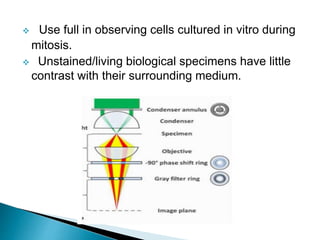
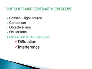
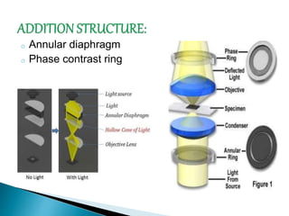
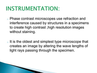
V.MUTHUMEENA
III BSc Biotechnology
Bon Secours College For Women,
Thanjavur.
 Phase contrast microscope was first described
in 1934 by DUTCH physicist FRITZ ZERNIKE.
 He awarded by Nobel prize in physics in 1953.
 Green light used in Phase contrast microscope.
 Application for Phase contrast microscope
equipment range from the study of living
biological specimens , medical applications ,
study of live blood cells and other biological
and science applications.
 Most commonly used to provide contrast of
transparent specimens such as living cells or
small organisms.
 Use full in observing cells cultured in vitro during
mitosis.
 Unstained/living biological specimens have little
contrast with their surrounding medium.
 Posses – light source
 Condenser
 Objective lens
 Ocular lens
 CHARACTERS OF LIGHT(Two types):
Diffraction
Interference
o Annular diaphragm
o Phase contrast ring
o Phase contrast microscopes use refraction and
interference caused by structures in a specimens
to create high contrast ,high resolution images
without staining.
o It is the oldest and simplest type microscope that
creates an image by altering the wave lengths of
light rays passing through the specimen.
o When the wave lengths are out of phase, wave
troughs will cancel out wave peaks, which is called
destructive interference.
 Transparent specimens in living cells and small
organisms.
 Cell cultured in vitro during mitosis.
 Enable visualization of internal cellular
components.
 Study of cytoskeleton.
 Visualize all types of cellular movements.
 Living cells can be examined in their natural state.
 The dynamics of ongoing biological processes
can be observed and recorded in high contrast
with sharp clarity of minute specimen detail.
 It contain special phase contrast objectives and a
special phase contrast condenser.
 Annuli or rings limit the aperture to some extent ,
which decreases resolution.
 Not ideal for thick organisms or particles.
 Images may appear grey or green if white or
green lights are used respectively, resulting in
poor photomicrography.
Phase contrast microscope

Phase contrast microscope

  • 1.
    V.MUTHUMEENA III BSc Biotechnology BonSecours College For Women, Thanjavur.
  • 2.
     Phase contrastmicroscope was first described in 1934 by DUTCH physicist FRITZ ZERNIKE.  He awarded by Nobel prize in physics in 1953.  Green light used in Phase contrast microscope.
  • 3.
     Application forPhase contrast microscope equipment range from the study of living biological specimens , medical applications , study of live blood cells and other biological and science applications.  Most commonly used to provide contrast of transparent specimens such as living cells or small organisms.
  • 4.
     Use fullin observing cells cultured in vitro during mitosis.  Unstained/living biological specimens have little contrast with their surrounding medium.
  • 5.
     Posses –light source  Condenser  Objective lens  Ocular lens  CHARACTERS OF LIGHT(Two types): Diffraction Interference
  • 6.
    o Annular diaphragm oPhase contrast ring
  • 7.
    o Phase contrastmicroscopes use refraction and interference caused by structures in a specimens to create high contrast ,high resolution images without staining. o It is the oldest and simplest type microscope that creates an image by altering the wave lengths of light rays passing through the specimen.
  • 8.
    o When thewave lengths are out of phase, wave troughs will cancel out wave peaks, which is called destructive interference.
  • 9.
     Transparent specimensin living cells and small organisms.  Cell cultured in vitro during mitosis.  Enable visualization of internal cellular components.  Study of cytoskeleton.  Visualize all types of cellular movements.
  • 10.
     Living cellscan be examined in their natural state.  The dynamics of ongoing biological processes can be observed and recorded in high contrast with sharp clarity of minute specimen detail.  It contain special phase contrast objectives and a special phase contrast condenser.
  • 11.
     Annuli orrings limit the aperture to some extent , which decreases resolution.  Not ideal for thick organisms or particles.  Images may appear grey or green if white or green lights are used respectively, resulting in poor photomicrography.