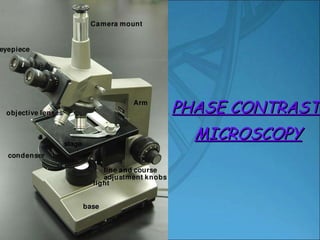
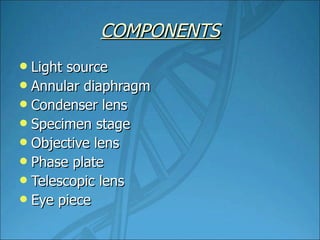
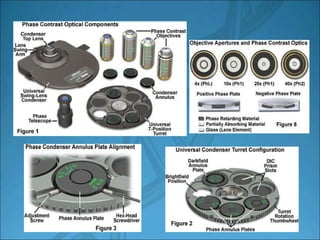
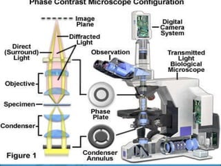
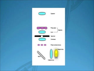
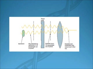
PHASE CONTRAST  MICROSCOPY
Phase contrast microscope is a technique which makes highly transparent objects more visible without killing them. Invented by the Dutch physicist Frederick  Zernike in 1930
PRINCIPLE It converts the different degrees of retardation of light rays passing through a translucent material into amplitude differences to create an image with increased contrast.
COMPONENTS Light source Annular diaphragm Condenser lens Specimen stage Objective lens Phase plate Telescopic lens Eye piece


Cheek cell
Clostridium botulinum,  the bacterium responsible for botulism, with subterminal oval endospores;
Spirillum volutans,  a very large bacterium with flagellar bundles
Paramecium stained to show a large central macronucleus with a small spherical micronucleus at its side
APPLICATIONS Objects with poor intensity can be resolved well. Image of the unstained object can be obtained It is widely used in microbiology and tissue culture research to detect bacteria, cellular organelles and other small entities in living specimens It allows the visualization of living unstained cells and their organelles. For the detection of bacterial components such as endospores and inclusion bodies containing poly β hydroxy butyrate, polymetaphosphate, sulpher and other substances
THANK YOU HARIS P MSC BIOTECHNOLOGY MA COLLEGE  ERNAKULAM

Phase contrst microscope.HRS

  • 1.
  • 2.
    PHASE CONTRAST MICROSCOPY
  • 3.
    Phase contrast microscopeis a technique which makes highly transparent objects more visible without killing them. Invented by the Dutch physicist Frederick Zernike in 1930
  • 4.
    PRINCIPLE It convertsthe different degrees of retardation of light rays passing through a translucent material into amplitude differences to create an image with increased contrast.
  • 5.
    COMPONENTS Light sourceAnnular diaphragm Condenser lens Specimen stage Objective lens Phase plate Telescopic lens Eye piece
  • 6.
  • 7.
  • 8.
  • 9.
  • 10.
  • 11.
    Clostridium botulinum, the bacterium responsible for botulism, with subterminal oval endospores;
  • 12.
    Spirillum volutans, a very large bacterium with flagellar bundles
  • 13.
    Paramecium stained toshow a large central macronucleus with a small spherical micronucleus at its side
  • 14.
    APPLICATIONS Objects withpoor intensity can be resolved well. Image of the unstained object can be obtained It is widely used in microbiology and tissue culture research to detect bacteria, cellular organelles and other small entities in living specimens It allows the visualization of living unstained cells and their organelles. For the detection of bacterial components such as endospores and inclusion bodies containing poly β hydroxy butyrate, polymetaphosphate, sulpher and other substances
  • 15.
    THANK YOU HARISP MSC BIOTECHNOLOGY MA COLLEGE ERNAKULAM