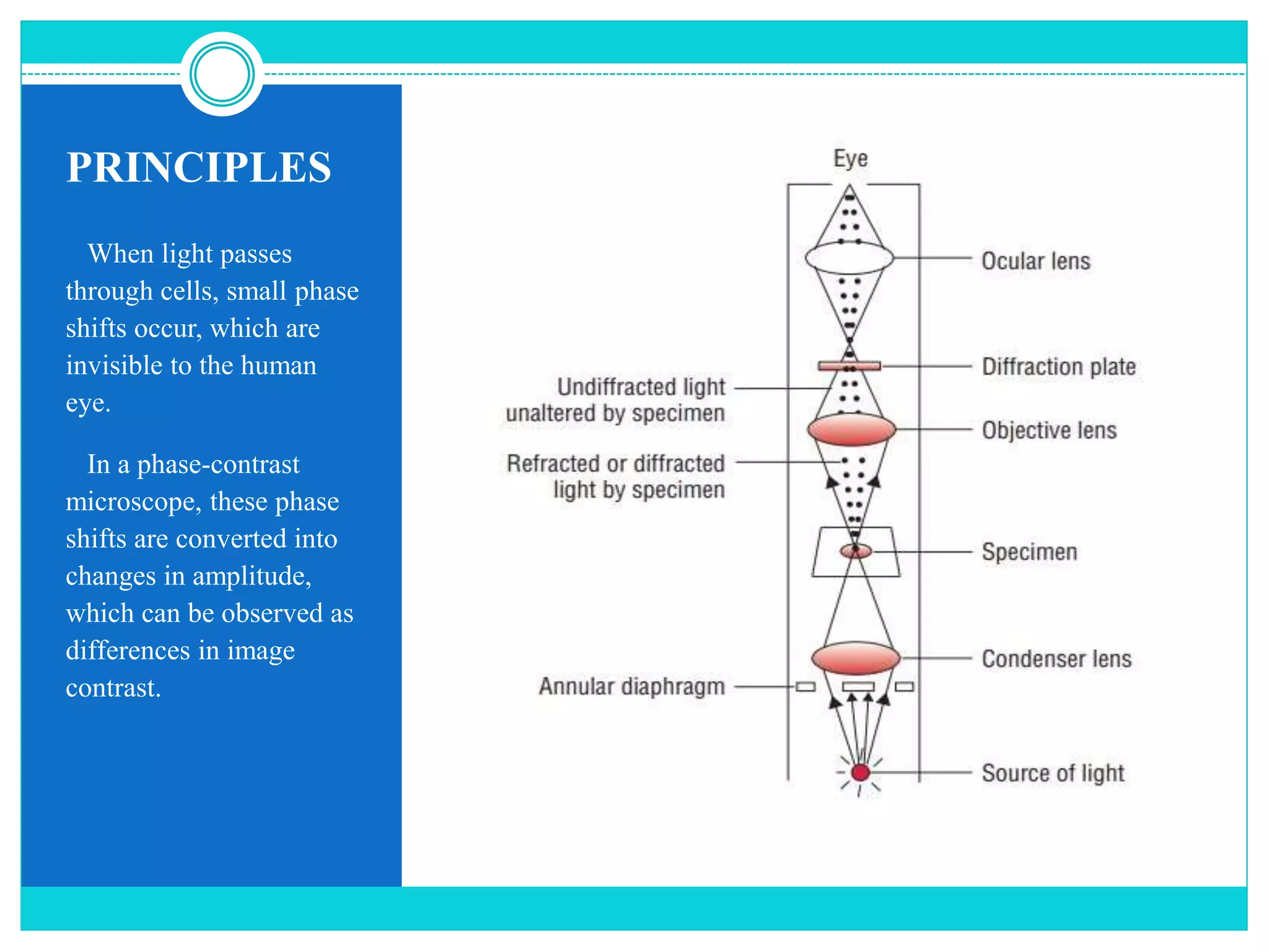
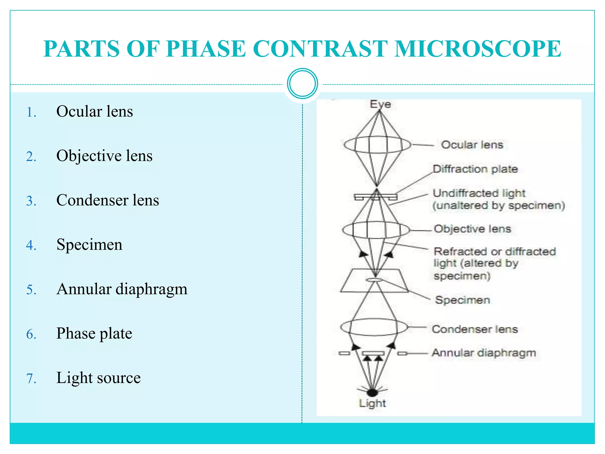
SIVASANGARI SHANMUGAM
PHASE CONTRAST MICROSCOPY
CONTENTS
1. Introduction
2. Principles
3. Parts of Microscope
4. How does it works?
5. Applications
6. Advantages
7. Disadvantages
INTRODUCTION
 Phase contrast microscopy, first described by Dutch physicist Frits Zernike
in 1934.
 It can be utilized to produce high-contrast images of transparent specimens,
such as living cells (usually in culture), microorganisms, thin tissue slices,
fibers, latex dispersions, glass fragments, and subcellular particles (including
nuclei and other organelles).
 It is an optical microscopy technique that converts phase shifts in the light
passing through a transparent specimen to brightness changes in the image.
 A phase-contrast microscope splits a beam of light into 2 types of light, direct
and reflected and brings them together to form an image of the specimen.
 Where the lights are “in-phase” the image is brighter, where the lights are
“out of phase” the image is darker, and by amplifying these differences in
the light, it enhances contrast.
 Phase-contrast microscopy allows for the detailed observation of living
organisms, especially the internal structures.
PRINCIPLES
When light passes
through cells, small phase
shifts occur, which are
invisible to the human
eye.
In a phase-contrast
microscope, these phase
shifts are converted into
changes in amplitude,
which can be observed as
differences in image
contrast.
PARTS OF PHASE CONTRAST MICROSCOPE
1. Ocular lens
2. Objective lens
3. Condenser lens
4. Specimen
5. Annular diaphragm
6. Phase plate
7. Light source
The annular diaphragm
 It is situated below the condenser.
 It is made up of a circular disc having a circular annular groove.
 The light rays are allowed to pass through the annular groove.
 Through the annular groove of the annular diaphragm, the light rays fall on the
specimen or object to be studied.
 At the back focal plane of the objective develops an image.
 The annular phase plate is placed at this back focal plane.
The phase plate
 It is either a negative phase plate having a thick circular area or a positive phase
plate having a thin circular groove.
 This thick or thin area in the phase plate is called the conjugate area.
 The phase plate is a transparent disc.
 With the help of the annular diaphragm and the phase plate, the phase contrast is
obtained in this microscope.
 This is obtained by separating the direct rays from the diffracted rays.
 The direct light rays pass through the annular groove whereas the diffracted light
rays pass through the region outside the groove.
 Depending upon the different refractive indices of different cell components, the
object to be studied shows a different degree of contrast in this microscope.
HOW DOES IT WORKS?
HOW DOES IT WORKS?
 Partially coherent illumination produced from tungsten -halogen lamp is
directed through a collector lens and focused on a specialized annulus
positioned in the sub stage condenser front focal plane.
 Wave fronts passing through the annulus illuminate the specimen and either
pass through undeviated or are diffracted and retarded in phase by structures
and phase gradients present in the specimen.
 Undeviated and diffracted light collected by the objective segregated at the
rear focal plane by a phase plate and focused at the intermediate image plane
to form the final phase- contrast image observed in the eyepiece.
APPLICATIONS
1. To produce high-contrast images of transparent specimens, such as Living
cells (usually in culture), Microorganisms, Thin tissue slices, Lithographic
patterns, Fibers, Subcellular particles (including nuclei and other
organelles).
2. Phase contrast is by far the most frequently used method in biological light
microscopy. It is an established microscopy technique in cell culture and live
cell imaging.
3. When using this inexpensive technique, living cells can be observed in their
natural state without previous fixation or labeling.
ADVANTAGES
1. Living cells can be observed in their natural state without previous fixation or labeling.
2. It makes a highly transparent object more visible.
3. No special preparation of fixation or staining etc. is needed to study an object under a
phase-contrast microscope which saves a lot of time.
4. Examining intracellular components of living cells at relatively high resolution. eg:
The dynamic motility of mitochondria, mitotic chromosomes & vacuoles.
5. It made it possible for biologists to study living cells and how they proliferate through
cell division.
6. Phase-contrast optical components can be added to virtually any brigh-tfield
microscope, provided the specialized phase objectives conform to the tube length
parameters, and the condenser will accept an annular phase ring of the correct size.
DISADVANTAGES
1. Phase-contrast condensers and objective lenses add considerable cost to a
microscope, and so phase contrast is often not used in teaching labs except
perhaps in classes in the health professions.
2. To use phase-contrast the light path must be aligned.
3. Generally, more light is needed for phase contrast than for corresponding
bright-field viewing, since the technique is based on the diminishment of the
brightness of most objects.
REFERENCES
1. https://www.microscopyu.com/techniques/phase-contrast/introduction-to-
phase-contrast-microscopy
2. https://microbenotes.com/phase-contrast-microscopy/
3. https://ibidi.com/content/213-phase-contrast
4. https://www.olympus-lifescience.com/en/microscope-
resource/primer/techniques/phasecontrast/phase/
5. https://bio.libretexts.org/Bookshelves/Microbiology/Book%3A_Microbiolog
y_(Boundless)/3%3A_Microscopy/3.3%3A_Other_Types_of_Microscopy/3
.3B%3A_Phase-Contrast_Microscopy
THANK YOU

Phase contrast microscope

  • 1.
  • 2.
    CONTENTS 1. Introduction 2. Principles 3.Parts of Microscope 4. How does it works? 5. Applications 6. Advantages 7. Disadvantages
  • 3.
    INTRODUCTION  Phase contrastmicroscopy, first described by Dutch physicist Frits Zernike in 1934.  It can be utilized to produce high-contrast images of transparent specimens, such as living cells (usually in culture), microorganisms, thin tissue slices, fibers, latex dispersions, glass fragments, and subcellular particles (including nuclei and other organelles).  It is an optical microscopy technique that converts phase shifts in the light passing through a transparent specimen to brightness changes in the image.  A phase-contrast microscope splits a beam of light into 2 types of light, direct and reflected and brings them together to form an image of the specimen.
  • 4.
     Where thelights are “in-phase” the image is brighter, where the lights are “out of phase” the image is darker, and by amplifying these differences in the light, it enhances contrast.  Phase-contrast microscopy allows for the detailed observation of living organisms, especially the internal structures.
  • 6.
    PRINCIPLES When light passes throughcells, small phase shifts occur, which are invisible to the human eye. In a phase-contrast microscope, these phase shifts are converted into changes in amplitude, which can be observed as differences in image contrast.
  • 7.
    PARTS OF PHASECONTRAST MICROSCOPE 1. Ocular lens 2. Objective lens 3. Condenser lens 4. Specimen 5. Annular diaphragm 6. Phase plate 7. Light source
  • 9.
    The annular diaphragm It is situated below the condenser.  It is made up of a circular disc having a circular annular groove.  The light rays are allowed to pass through the annular groove.  Through the annular groove of the annular diaphragm, the light rays fall on the specimen or object to be studied.  At the back focal plane of the objective develops an image.  The annular phase plate is placed at this back focal plane.
  • 10.
    The phase plate It is either a negative phase plate having a thick circular area or a positive phase plate having a thin circular groove.  This thick or thin area in the phase plate is called the conjugate area.  The phase plate is a transparent disc.  With the help of the annular diaphragm and the phase plate, the phase contrast is obtained in this microscope.  This is obtained by separating the direct rays from the diffracted rays.  The direct light rays pass through the annular groove whereas the diffracted light rays pass through the region outside the groove.  Depending upon the different refractive indices of different cell components, the object to be studied shows a different degree of contrast in this microscope.
  • 11.
  • 12.
    HOW DOES ITWORKS?  Partially coherent illumination produced from tungsten -halogen lamp is directed through a collector lens and focused on a specialized annulus positioned in the sub stage condenser front focal plane.  Wave fronts passing through the annulus illuminate the specimen and either pass through undeviated or are diffracted and retarded in phase by structures and phase gradients present in the specimen.  Undeviated and diffracted light collected by the objective segregated at the rear focal plane by a phase plate and focused at the intermediate image plane to form the final phase- contrast image observed in the eyepiece.
  • 13.
    APPLICATIONS 1. To producehigh-contrast images of transparent specimens, such as Living cells (usually in culture), Microorganisms, Thin tissue slices, Lithographic patterns, Fibers, Subcellular particles (including nuclei and other organelles). 2. Phase contrast is by far the most frequently used method in biological light microscopy. It is an established microscopy technique in cell culture and live cell imaging. 3. When using this inexpensive technique, living cells can be observed in their natural state without previous fixation or labeling.
  • 14.
    ADVANTAGES 1. Living cellscan be observed in their natural state without previous fixation or labeling. 2. It makes a highly transparent object more visible. 3. No special preparation of fixation or staining etc. is needed to study an object under a phase-contrast microscope which saves a lot of time. 4. Examining intracellular components of living cells at relatively high resolution. eg: The dynamic motility of mitochondria, mitotic chromosomes & vacuoles. 5. It made it possible for biologists to study living cells and how they proliferate through cell division. 6. Phase-contrast optical components can be added to virtually any brigh-tfield microscope, provided the specialized phase objectives conform to the tube length parameters, and the condenser will accept an annular phase ring of the correct size.
  • 15.
    DISADVANTAGES 1. Phase-contrast condensersand objective lenses add considerable cost to a microscope, and so phase contrast is often not used in teaching labs except perhaps in classes in the health professions. 2. To use phase-contrast the light path must be aligned. 3. Generally, more light is needed for phase contrast than for corresponding bright-field viewing, since the technique is based on the diminishment of the brightness of most objects.
  • 16.
    REFERENCES 1. https://www.microscopyu.com/techniques/phase-contrast/introduction-to- phase-contrast-microscopy 2. https://microbenotes.com/phase-contrast-microscopy/ 3.https://ibidi.com/content/213-phase-contrast 4. https://www.olympus-lifescience.com/en/microscope- resource/primer/techniques/phasecontrast/phase/ 5. https://bio.libretexts.org/Bookshelves/Microbiology/Book%3A_Microbiolog y_(Boundless)/3%3A_Microscopy/3.3%3A_Other_Types_of_Microscopy/3 .3B%3A_Phase-Contrast_Microscopy
  • 17.