SlideShare a Scribd company logo
1 of 2
Download to read offline
MICROSCOPY 1
PHASE-CONTRAST MICROSCOPE
 The first phase contrast microscope was developed by FRITZ ZERNIKE (FREDRICK
ZERNIKE) in 1933, hence also referred as ZERNIKE MICROSCOPE.
 The phase contrast microscope enables to differentiate transparent, unstained, living
(without killing or altering the living component) structures.
 Phase contrast is an illumination technique provides greater degree differentiation
inside the cells by phase contrast optics.
WORKING PRINCIPLE
Light from the illumination (light) source from the base of the Microscope stand is
aimed at annular diaphragm stop. The annular-diaphragm-stop allows only the hollow
cone of light rays to pass through sub-stage condenser lens. The sub-stage condenser
lens focuses light through slit in the stage onto the sample. The projected light from the
sample is collected by special set of objective lens with phase plate and phase rings
which are placed in the back/rare focal plane of the objective. The direct rays (unaltered
amplitude and phase, but retarded by ¼ wavelength) from transparent sample converge
on the phase ring within the objective and produce phase shift. The most diffracted rays
(altered rays due to difference in density) pass through phase plate by missing phase ring.
The convergence of diffracted and direct rays on the image plane results in image.
ADVANTAGES
 Specimens which have a refractive index similar to their surroundings can be invisible
in Brightfield, but are well defined in Phase Contrast.
 Phase Contrast is normally used to examine unstained biological specimens.
 Living microorganisms and their minute details such as Cilia, flagella can be observed.
MICROSCOPY 2
FLUORESCENT MICROSCOPE
When a substance absorbs light, the electrons present at the outermost orbit absorbs
energy and get excited; on the way back to the ground state, it emits a part of the energy
absorbed. This phenomenon is termed as fluorescence.Fluorescent microscopeinvolves
staining of specimens with special fluorescent dyes (fluorescein, acridine orange, etc).
When a fluorescent dye is applied to a substance, it absorbs a wavelength of light
(excitation wavelength) and emits light of different wavelength (emission wavelength).
WORKING PRINCIPLE
Illumination (light) is provided by a bright mercury vapor lamp (very expensive +
harmful), produces light range of 200-400nm and generates considerable amount of heat.
The heat filter absorbs heat, allows UV rays and visible rays by blocking infrared rays.
The exciter filter ensures high energy - short wavelength - monochromatic light towards
dichroic mirror. Dichroic mirror (beam splitter) eliminates visible light and reflects
excited UV light to the dark-field condenser, which provides high contrast for
fluorescence and also deflects majority of UV light.The excitation light is focused on to
the fluorochrome specimen. The fluorescent labeled specimen absorbs light and emits
excitatory light along with florescent light, which reaches objectivelens .As per the
capacity of objective lens, the specimen would be magnified and are directed towards
barrier filter. The additional barrier filter permits only the fluorescent wavelength and
rejects excitation light. The fluorescent light passing through ocular lens creates the
magnified image, which can also be detected by detector to give a photographic image.
APPLICATIONS
 Imaging the genetic material (DNA & RNA) and other structural components.
 Monitoring the environment for microbial contamination.
 Certain micro-organism can be detected and identified only by this microscopy.

More Related Content

What's hot

Phase contrast & Flurosence microscope
Phase contrast & Flurosence microscopePhase contrast & Flurosence microscope
Phase contrast & Flurosence microscopemicrobiology Notes
 
Confocal laser scanning microscopy
Confocal laser scanning microscopyConfocal laser scanning microscopy
Confocal laser scanning microscopyBaskar Selvam
 
Principles and application of light, phase constrast and fluorescence microscope
Principles and application of light, phase constrast and fluorescence microscopePrinciples and application of light, phase constrast and fluorescence microscope
Principles and application of light, phase constrast and fluorescence microscopeMaitriThakor
 
Phase contrast microscope
Phase contrast microscopePhase contrast microscope
Phase contrast microscopeSuganyaPaulraj
 
Bright Field Microscopy.pptx
Bright Field Microscopy.pptxBright Field Microscopy.pptx
Bright Field Microscopy.pptxARVINDPRAJAPATI41
 
Microscopy
MicroscopyMicroscopy
Microscopykarade
 
Unit – 2 Microscopy
Unit – 2 MicroscopyUnit – 2 Microscopy
Unit – 2 Microscopysanchu yadav
 
Microscopy - Magnification, Resolving power, Principles, Types and Applications
Microscopy - Magnification, Resolving power, Principles, Types and ApplicationsMicroscopy - Magnification, Resolving power, Principles, Types and Applications
Microscopy - Magnification, Resolving power, Principles, Types and ApplicationsNethravathi Siri
 
Principle of transmission electron microscope.
Principle of transmission electron microscope.Principle of transmission electron microscope.
Principle of transmission electron microscope.naren
 
Bright field microscopy, Principle and applications
Bright field microscopy, Principle and applicationsBright field microscopy, Principle and applications
Bright field microscopy, Principle and applicationsKAUSHAL SAHU
 
Light phase contrast and fluorescence microscopy
Light phase contrast and fluorescence microscopyLight phase contrast and fluorescence microscopy
Light phase contrast and fluorescence microscopyKeerthana Chithanathan
 

What's hot (20)

Phase contrast & Flurosence microscope
Phase contrast & Flurosence microscopePhase contrast & Flurosence microscope
Phase contrast & Flurosence microscope
 
Fluorescence Microscopy
Fluorescence MicroscopyFluorescence Microscopy
Fluorescence Microscopy
 
phase contrast microscope
phase contrast microscopephase contrast microscope
phase contrast microscope
 
Confocal laser scanning microscopy
Confocal laser scanning microscopyConfocal laser scanning microscopy
Confocal laser scanning microscopy
 
Dark field microscope
Dark field microscopeDark field microscope
Dark field microscope
 
Light microscope
Light microscopeLight microscope
Light microscope
 
Principles and application of light, phase constrast and fluorescence microscope
Principles and application of light, phase constrast and fluorescence microscopePrinciples and application of light, phase constrast and fluorescence microscope
Principles and application of light, phase constrast and fluorescence microscope
 
Phase contrast microscope
Phase contrast microscopePhase contrast microscope
Phase contrast microscope
 
Bright Field Microscopy.pptx
Bright Field Microscopy.pptxBright Field Microscopy.pptx
Bright Field Microscopy.pptx
 
Microscopy
MicroscopyMicroscopy
Microscopy
 
Unit – 2 Microscopy
Unit – 2 MicroscopyUnit – 2 Microscopy
Unit – 2 Microscopy
 
Microscopy - Magnification, Resolving power, Principles, Types and Applications
Microscopy - Magnification, Resolving power, Principles, Types and ApplicationsMicroscopy - Magnification, Resolving power, Principles, Types and Applications
Microscopy - Magnification, Resolving power, Principles, Types and Applications
 
Principle of transmission electron microscope.
Principle of transmission electron microscope.Principle of transmission electron microscope.
Principle of transmission electron microscope.
 
Simple microscope
Simple microscopeSimple microscope
Simple microscope
 
Phase contrast microscope
Phase contrast microscopePhase contrast microscope
Phase contrast microscope
 
Compound microscope
Compound microscopeCompound microscope
Compound microscope
 
Numerical aperture and limits of resolution of microscope
Numerical aperture and limits of resolution of microscopeNumerical aperture and limits of resolution of microscope
Numerical aperture and limits of resolution of microscope
 
Bright field microscopy, Principle and applications
Bright field microscopy, Principle and applicationsBright field microscopy, Principle and applications
Bright field microscopy, Principle and applications
 
Light microscope
Light microscopeLight microscope
Light microscope
 
Light phase contrast and fluorescence microscopy
Light phase contrast and fluorescence microscopyLight phase contrast and fluorescence microscopy
Light phase contrast and fluorescence microscopy
 

Similar to 4. Microscope - Phase contrast & Fluorescent

FLUOROSCENCE & PHASE CONTRAST MICROSCOPY.pptx
FLUOROSCENCE & PHASE CONTRAST MICROSCOPY.pptxFLUOROSCENCE & PHASE CONTRAST MICROSCOPY.pptx
FLUOROSCENCE & PHASE CONTRAST MICROSCOPY.pptxSagarBhakare1
 
Phase contrast and Fluorescent microscope
Phase contrast and Fluorescent microscopePhase contrast and Fluorescent microscope
Phase contrast and Fluorescent microscopeNithiya Pappuraj
 
Lectures Notes Types of microscopes.pptx
Lectures Notes Types of microscopes.pptxLectures Notes Types of microscopes.pptx
Lectures Notes Types of microscopes.pptxPeninaTabitha
 
DIFFERENT TYPES OF MICROSCOPY.pptx
DIFFERENT TYPES OF MICROSCOPY.pptxDIFFERENT TYPES OF MICROSCOPY.pptx
DIFFERENT TYPES OF MICROSCOPY.pptxISHAgunwal2
 
Different types of microscopes
Different types of microscopesDifferent types of microscopes
Different types of microscopesUTTAM KUMAR DAS
 
microscopy in bacteriology by Tejpal.pptx
microscopy in bacteriology by Tejpal.pptxmicroscopy in bacteriology by Tejpal.pptx
microscopy in bacteriology by Tejpal.pptxVetico
 
Microscope ppt, by jitendra kumar pandey,medical micro,2nd yr, mgm medical co...
Microscope ppt, by jitendra kumar pandey,medical micro,2nd yr, mgm medical co...Microscope ppt, by jitendra kumar pandey,medical micro,2nd yr, mgm medical co...
Microscope ppt, by jitendra kumar pandey,medical micro,2nd yr, mgm medical co...jitendra Pandey
 
Fluorescence microscope by Subhankar Das
Fluorescence microscope by Subhankar DasFluorescence microscope by Subhankar Das
Fluorescence microscope by Subhankar DasSubhankar Das
 
Microscope part 2 BY DR. C. P. ARYA (B.Sc. B.D.S.; M.D.S.; P.M.S.; R.N.T.C.P.)
Microscope part 2 BY DR. C. P. ARYA (B.Sc. B.D.S.; M.D.S.; P.M.S.; R.N.T.C.P.)Microscope part 2 BY DR. C. P. ARYA (B.Sc. B.D.S.; M.D.S.; P.M.S.; R.N.T.C.P.)
Microscope part 2 BY DR. C. P. ARYA (B.Sc. B.D.S.; M.D.S.; P.M.S.; R.N.T.C.P.)DR. C. P. ARYA
 
fluroscence microscopy final- ppt.pptx
fluroscence microscopy final- ppt.pptxfluroscence microscopy final- ppt.pptx
fluroscence microscopy final- ppt.pptxMonalisaMathan1
 
Microscopy and Micrometry
Microscopy and MicrometryMicroscopy and Micrometry
Microscopy and MicrometrySuprakash Das
 
Fluorescence Microscopy
Fluorescence MicroscopyFluorescence Microscopy
Fluorescence MicroscopyAalap Tripathy
 
Fluorescence microscopy
Fluorescence microscopy   Fluorescence microscopy
Fluorescence microscopy Fida Muhammad
 

Similar to 4. Microscope - Phase contrast & Fluorescent (20)

FLUOROSCENCE & PHASE CONTRAST MICROSCOPY.pptx
FLUOROSCENCE & PHASE CONTRAST MICROSCOPY.pptxFLUOROSCENCE & PHASE CONTRAST MICROSCOPY.pptx
FLUOROSCENCE & PHASE CONTRAST MICROSCOPY.pptx
 
Phase contrast and Fluorescent microscope
Phase contrast and Fluorescent microscopePhase contrast and Fluorescent microscope
Phase contrast and Fluorescent microscope
 
Lectures Notes Types of microscopes.pptx
Lectures Notes Types of microscopes.pptxLectures Notes Types of microscopes.pptx
Lectures Notes Types of microscopes.pptx
 
DIFFERENT TYPES OF MICROSCOPY.pptx
DIFFERENT TYPES OF MICROSCOPY.pptxDIFFERENT TYPES OF MICROSCOPY.pptx
DIFFERENT TYPES OF MICROSCOPY.pptx
 
Different types of microscopes
Different types of microscopesDifferent types of microscopes
Different types of microscopes
 
Microscopy
MicroscopyMicroscopy
Microscopy
 
Florescense microscopy
Florescense microscopyFlorescense microscopy
Florescense microscopy
 
Types of microscope
Types of microscopeTypes of microscope
Types of microscope
 
microscopy in bacteriology by Tejpal.pptx
microscopy in bacteriology by Tejpal.pptxmicroscopy in bacteriology by Tejpal.pptx
microscopy in bacteriology by Tejpal.pptx
 
Microscope ppt, by jitendra kumar pandey,medical micro,2nd yr, mgm medical co...
Microscope ppt, by jitendra kumar pandey,medical micro,2nd yr, mgm medical co...Microscope ppt, by jitendra kumar pandey,medical micro,2nd yr, mgm medical co...
Microscope ppt, by jitendra kumar pandey,medical micro,2nd yr, mgm medical co...
 
Ppt f1
Ppt f1Ppt f1
Ppt f1
 
Microscopy - Study
Microscopy - StudyMicroscopy - Study
Microscopy - Study
 
Microscopy - Study
Microscopy - StudyMicroscopy - Study
Microscopy - Study
 
Fluorescence microscope by Subhankar Das
Fluorescence microscope by Subhankar DasFluorescence microscope by Subhankar Das
Fluorescence microscope by Subhankar Das
 
Microscope part 2 BY DR. C. P. ARYA (B.Sc. B.D.S.; M.D.S.; P.M.S.; R.N.T.C.P.)
Microscope part 2 BY DR. C. P. ARYA (B.Sc. B.D.S.; M.D.S.; P.M.S.; R.N.T.C.P.)Microscope part 2 BY DR. C. P. ARYA (B.Sc. B.D.S.; M.D.S.; P.M.S.; R.N.T.C.P.)
Microscope part 2 BY DR. C. P. ARYA (B.Sc. B.D.S.; M.D.S.; P.M.S.; R.N.T.C.P.)
 
fluroscence microscopy final- ppt.pptx
fluroscence microscopy final- ppt.pptxfluroscence microscopy final- ppt.pptx
fluroscence microscopy final- ppt.pptx
 
Microscopy and Micrometry
Microscopy and MicrometryMicroscopy and Micrometry
Microscopy and Micrometry
 
Fluorescence Microscopy
Fluorescence MicroscopyFluorescence Microscopy
Fluorescence Microscopy
 
Fluorescence microscopy
Fluorescence microscopy   Fluorescence microscopy
Fluorescence microscopy
 
microscopy
 microscopy microscopy
microscopy
 

More from Nethravathi Siri

Human genetics and holistic health
Human genetics and holistic healthHuman genetics and holistic health
Human genetics and holistic healthNethravathi Siri
 
QUANTITATIVE INHERITANCE - KERNEL COLOR IN WHEAT
QUANTITATIVE INHERITANCE - KERNEL COLOR IN WHEATQUANTITATIVE INHERITANCE - KERNEL COLOR IN WHEAT
QUANTITATIVE INHERITANCE - KERNEL COLOR IN WHEATNethravathi Siri
 
Evolutionary genetics - Theories,
Evolutionary genetics - Theories, Evolutionary genetics - Theories,
Evolutionary genetics - Theories, Nethravathi Siri
 
Holliday model of crossing over
Holliday model of crossing overHolliday model of crossing over
Holliday model of crossing overNethravathi Siri
 
3. Special chromosome - B chromosome
3. Special chromosome - B chromosome 3. Special chromosome - B chromosome
3. Special chromosome - B chromosome Nethravathi Siri
 
3. Special chromosomes - Lampbrush chromosomes
3. Special chromosomes - Lampbrush chromosomes 3. Special chromosomes - Lampbrush chromosomes
3. Special chromosomes - Lampbrush chromosomes Nethravathi Siri
 
2. Special chromosomes - Polytene chromosomes
2. Special chromosomes - Polytene chromosomes2. Special chromosomes - Polytene chromosomes
2. Special chromosomes - Polytene chromosomesNethravathi Siri
 
1. Special chromosomes - Introduction
1. Special chromosomes - Introduction1. Special chromosomes - Introduction
1. Special chromosomes - IntroductionNethravathi Siri
 
Measurement of Radioactivity - Geiger Muller [GM] Counter & SCINTILLATION COU...
Measurement of Radioactivity - Geiger Muller [GM] Counter & SCINTILLATION COU...Measurement of Radioactivity - Geiger Muller [GM] Counter & SCINTILLATION COU...
Measurement of Radioactivity - Geiger Muller [GM] Counter & SCINTILLATION COU...Nethravathi Siri
 
NUCLEOSOME MODEL OF CHROMOSOME
NUCLEOSOME MODEL OF CHROMOSOMENUCLEOSOME MODEL OF CHROMOSOME
NUCLEOSOME MODEL OF CHROMOSOMENethravathi Siri
 
4. Gene interaction - Epistasis - Dominant & Recessive, Non-epistatsis
4. Gene interaction - Epistasis - Dominant & Recessive, Non-epistatsis4. Gene interaction - Epistasis - Dominant & Recessive, Non-epistatsis
4. Gene interaction - Epistasis - Dominant & Recessive, Non-epistatsisNethravathi Siri
 
3. Gene interaction - supplementary
3. Gene interaction - supplementary3. Gene interaction - supplementary
3. Gene interaction - supplementaryNethravathi Siri
 
2. Gene interaction - complementary
2. Gene interaction - complementary2. Gene interaction - complementary
2. Gene interaction - complementaryNethravathi Siri
 
1. Gene interaction - Introduction
1. Gene interaction - Introduction1. Gene interaction - Introduction
1. Gene interaction - IntroductionNethravathi Siri
 
Comparative account on different types of microscopes
Comparative account on different types of microscopesComparative account on different types of microscopes
Comparative account on different types of microscopesNethravathi Siri
 

More from Nethravathi Siri (20)

Human genetics and holistic health
Human genetics and holistic healthHuman genetics and holistic health
Human genetics and holistic health
 
QUANTITATIVE INHERITANCE - KERNEL COLOR IN WHEAT
QUANTITATIVE INHERITANCE - KERNEL COLOR IN WHEATQUANTITATIVE INHERITANCE - KERNEL COLOR IN WHEAT
QUANTITATIVE INHERITANCE - KERNEL COLOR IN WHEAT
 
Evolutionary genetics - Theories,
Evolutionary genetics - Theories, Evolutionary genetics - Theories,
Evolutionary genetics - Theories,
 
Upstream processing
Upstream processing Upstream processing
Upstream processing
 
Retro copia transposons
Retro copia transposonsRetro copia transposons
Retro copia transposons
 
Eukaryotic transcription
Eukaryotic transcription Eukaryotic transcription
Eukaryotic transcription
 
Holliday model of crossing over
Holliday model of crossing overHolliday model of crossing over
Holliday model of crossing over
 
Vitamins - Basics
Vitamins - BasicsVitamins - Basics
Vitamins - Basics
 
3. Special chromosome - B chromosome
3. Special chromosome - B chromosome 3. Special chromosome - B chromosome
3. Special chromosome - B chromosome
 
3. Special chromosomes - Lampbrush chromosomes
3. Special chromosomes - Lampbrush chromosomes 3. Special chromosomes - Lampbrush chromosomes
3. Special chromosomes - Lampbrush chromosomes
 
2. Special chromosomes - Polytene chromosomes
2. Special chromosomes - Polytene chromosomes2. Special chromosomes - Polytene chromosomes
2. Special chromosomes - Polytene chromosomes
 
1. Special chromosomes - Introduction
1. Special chromosomes - Introduction1. Special chromosomes - Introduction
1. Special chromosomes - Introduction
 
Measurement of Radioactivity - Geiger Muller [GM] Counter & SCINTILLATION COU...
Measurement of Radioactivity - Geiger Muller [GM] Counter & SCINTILLATION COU...Measurement of Radioactivity - Geiger Muller [GM] Counter & SCINTILLATION COU...
Measurement of Radioactivity - Geiger Muller [GM] Counter & SCINTILLATION COU...
 
Crossing over basics
Crossing over   basicsCrossing over   basics
Crossing over basics
 
NUCLEOSOME MODEL OF CHROMOSOME
NUCLEOSOME MODEL OF CHROMOSOMENUCLEOSOME MODEL OF CHROMOSOME
NUCLEOSOME MODEL OF CHROMOSOME
 
4. Gene interaction - Epistasis - Dominant & Recessive, Non-epistatsis
4. Gene interaction - Epistasis - Dominant & Recessive, Non-epistatsis4. Gene interaction - Epistasis - Dominant & Recessive, Non-epistatsis
4. Gene interaction - Epistasis - Dominant & Recessive, Non-epistatsis
 
3. Gene interaction - supplementary
3. Gene interaction - supplementary3. Gene interaction - supplementary
3. Gene interaction - supplementary
 
2. Gene interaction - complementary
2. Gene interaction - complementary2. Gene interaction - complementary
2. Gene interaction - complementary
 
1. Gene interaction - Introduction
1. Gene interaction - Introduction1. Gene interaction - Introduction
1. Gene interaction - Introduction
 
Comparative account on different types of microscopes
Comparative account on different types of microscopesComparative account on different types of microscopes
Comparative account on different types of microscopes
 

Recently uploaded

GFP in rDNA Technology (Biotechnology).pptx
GFP in rDNA Technology (Biotechnology).pptxGFP in rDNA Technology (Biotechnology).pptx
GFP in rDNA Technology (Biotechnology).pptxAleenaTreesaSaji
 
SOLUBLE PATTERN RECOGNITION RECEPTORS.pptx
SOLUBLE PATTERN RECOGNITION RECEPTORS.pptxSOLUBLE PATTERN RECOGNITION RECEPTORS.pptx
SOLUBLE PATTERN RECOGNITION RECEPTORS.pptxkessiyaTpeter
 
Artificial Intelligence In Microbiology by Dr. Prince C P
Artificial Intelligence In Microbiology by Dr. Prince C PArtificial Intelligence In Microbiology by Dr. Prince C P
Artificial Intelligence In Microbiology by Dr. Prince C PPRINCE C P
 
All-domain Anomaly Resolution Office U.S. Department of Defense (U) Case: “Eg...
All-domain Anomaly Resolution Office U.S. Department of Defense (U) Case: “Eg...All-domain Anomaly Resolution Office U.S. Department of Defense (U) Case: “Eg...
All-domain Anomaly Resolution Office U.S. Department of Defense (U) Case: “Eg...Sérgio Sacani
 
Isotopic evidence of long-lived volcanism on Io
Isotopic evidence of long-lived volcanism on IoIsotopic evidence of long-lived volcanism on Io
Isotopic evidence of long-lived volcanism on IoSérgio Sacani
 
Biological Classification BioHack (3).pdf
Biological Classification BioHack (3).pdfBiological Classification BioHack (3).pdf
Biological Classification BioHack (3).pdfmuntazimhurra
 
CALL ON ➥8923113531 🔝Call Girls Kesar Bagh Lucknow best Night Fun service 🪡
CALL ON ➥8923113531 🔝Call Girls Kesar Bagh Lucknow best Night Fun service  🪡CALL ON ➥8923113531 🔝Call Girls Kesar Bagh Lucknow best Night Fun service  🪡
CALL ON ➥8923113531 🔝Call Girls Kesar Bagh Lucknow best Night Fun service 🪡anilsa9823
 
Grafana in space: Monitoring Japan's SLIM moon lander in real time
Grafana in space: Monitoring Japan's SLIM moon lander  in real timeGrafana in space: Monitoring Japan's SLIM moon lander  in real time
Grafana in space: Monitoring Japan's SLIM moon lander in real timeSatoshi NAKAHIRA
 
Analytical Profile of Coleus Forskohlii | Forskolin .pptx
Analytical Profile of Coleus Forskohlii | Forskolin .pptxAnalytical Profile of Coleus Forskohlii | Forskolin .pptx
Analytical Profile of Coleus Forskohlii | Forskolin .pptxSwapnil Therkar
 
PossibleEoarcheanRecordsoftheGeomagneticFieldPreservedintheIsuaSupracrustalBe...
PossibleEoarcheanRecordsoftheGeomagneticFieldPreservedintheIsuaSupracrustalBe...PossibleEoarcheanRecordsoftheGeomagneticFieldPreservedintheIsuaSupracrustalBe...
PossibleEoarcheanRecordsoftheGeomagneticFieldPreservedintheIsuaSupracrustalBe...Sérgio Sacani
 
Call Us ≽ 9953322196 ≼ Call Girls In Mukherjee Nagar(Delhi) |
Call Us ≽ 9953322196 ≼ Call Girls In Mukherjee Nagar(Delhi) |Call Us ≽ 9953322196 ≼ Call Girls In Mukherjee Nagar(Delhi) |
Call Us ≽ 9953322196 ≼ Call Girls In Mukherjee Nagar(Delhi) |aasikanpl
 
Call Girls in Munirka Delhi 💯Call Us 🔝9953322196🔝 💯Escort.
Call Girls in Munirka Delhi 💯Call Us 🔝9953322196🔝 💯Escort.Call Girls in Munirka Delhi 💯Call Us 🔝9953322196🔝 💯Escort.
Call Girls in Munirka Delhi 💯Call Us 🔝9953322196🔝 💯Escort.aasikanpl
 
Hubble Asteroid Hunter III. Physical properties of newly found asteroids
Hubble Asteroid Hunter III. Physical properties of newly found asteroidsHubble Asteroid Hunter III. Physical properties of newly found asteroids
Hubble Asteroid Hunter III. Physical properties of newly found asteroidsSérgio Sacani
 
Recombinant DNA technology (Immunological screening)
Recombinant DNA technology (Immunological screening)Recombinant DNA technology (Immunological screening)
Recombinant DNA technology (Immunological screening)PraveenaKalaiselvan1
 
Physiochemical properties of nanomaterials and its nanotoxicity.pptx
Physiochemical properties of nanomaterials and its nanotoxicity.pptxPhysiochemical properties of nanomaterials and its nanotoxicity.pptx
Physiochemical properties of nanomaterials and its nanotoxicity.pptxAArockiyaNisha
 
Discovery of an Accretion Streamer and a Slow Wide-angle Outflow around FUOri...
Discovery of an Accretion Streamer and a Slow Wide-angle Outflow around FUOri...Discovery of an Accretion Streamer and a Slow Wide-angle Outflow around FUOri...
Discovery of an Accretion Streamer and a Slow Wide-angle Outflow around FUOri...Sérgio Sacani
 
A relative description on Sonoporation.pdf
A relative description on Sonoporation.pdfA relative description on Sonoporation.pdf
A relative description on Sonoporation.pdfnehabiju2046
 
Boyles law module in the grade 10 science
Boyles law module in the grade 10 scienceBoyles law module in the grade 10 science
Boyles law module in the grade 10 sciencefloriejanemacaya1
 
Botany 4th semester file By Sumit Kumar yadav.pdf
Botany 4th semester file By Sumit Kumar yadav.pdfBotany 4th semester file By Sumit Kumar yadav.pdf
Botany 4th semester file By Sumit Kumar yadav.pdfSumit Kumar yadav
 

Recently uploaded (20)

GFP in rDNA Technology (Biotechnology).pptx
GFP in rDNA Technology (Biotechnology).pptxGFP in rDNA Technology (Biotechnology).pptx
GFP in rDNA Technology (Biotechnology).pptx
 
SOLUBLE PATTERN RECOGNITION RECEPTORS.pptx
SOLUBLE PATTERN RECOGNITION RECEPTORS.pptxSOLUBLE PATTERN RECOGNITION RECEPTORS.pptx
SOLUBLE PATTERN RECOGNITION RECEPTORS.pptx
 
Artificial Intelligence In Microbiology by Dr. Prince C P
Artificial Intelligence In Microbiology by Dr. Prince C PArtificial Intelligence In Microbiology by Dr. Prince C P
Artificial Intelligence In Microbiology by Dr. Prince C P
 
All-domain Anomaly Resolution Office U.S. Department of Defense (U) Case: “Eg...
All-domain Anomaly Resolution Office U.S. Department of Defense (U) Case: “Eg...All-domain Anomaly Resolution Office U.S. Department of Defense (U) Case: “Eg...
All-domain Anomaly Resolution Office U.S. Department of Defense (U) Case: “Eg...
 
Isotopic evidence of long-lived volcanism on Io
Isotopic evidence of long-lived volcanism on IoIsotopic evidence of long-lived volcanism on Io
Isotopic evidence of long-lived volcanism on Io
 
Biological Classification BioHack (3).pdf
Biological Classification BioHack (3).pdfBiological Classification BioHack (3).pdf
Biological Classification BioHack (3).pdf
 
CALL ON ➥8923113531 🔝Call Girls Kesar Bagh Lucknow best Night Fun service 🪡
CALL ON ➥8923113531 🔝Call Girls Kesar Bagh Lucknow best Night Fun service  🪡CALL ON ➥8923113531 🔝Call Girls Kesar Bagh Lucknow best Night Fun service  🪡
CALL ON ➥8923113531 🔝Call Girls Kesar Bagh Lucknow best Night Fun service 🪡
 
Grafana in space: Monitoring Japan's SLIM moon lander in real time
Grafana in space: Monitoring Japan's SLIM moon lander  in real timeGrafana in space: Monitoring Japan's SLIM moon lander  in real time
Grafana in space: Monitoring Japan's SLIM moon lander in real time
 
Analytical Profile of Coleus Forskohlii | Forskolin .pptx
Analytical Profile of Coleus Forskohlii | Forskolin .pptxAnalytical Profile of Coleus Forskohlii | Forskolin .pptx
Analytical Profile of Coleus Forskohlii | Forskolin .pptx
 
PossibleEoarcheanRecordsoftheGeomagneticFieldPreservedintheIsuaSupracrustalBe...
PossibleEoarcheanRecordsoftheGeomagneticFieldPreservedintheIsuaSupracrustalBe...PossibleEoarcheanRecordsoftheGeomagneticFieldPreservedintheIsuaSupracrustalBe...
PossibleEoarcheanRecordsoftheGeomagneticFieldPreservedintheIsuaSupracrustalBe...
 
Call Us ≽ 9953322196 ≼ Call Girls In Mukherjee Nagar(Delhi) |
Call Us ≽ 9953322196 ≼ Call Girls In Mukherjee Nagar(Delhi) |Call Us ≽ 9953322196 ≼ Call Girls In Mukherjee Nagar(Delhi) |
Call Us ≽ 9953322196 ≼ Call Girls In Mukherjee Nagar(Delhi) |
 
Call Girls in Munirka Delhi 💯Call Us 🔝9953322196🔝 💯Escort.
Call Girls in Munirka Delhi 💯Call Us 🔝9953322196🔝 💯Escort.Call Girls in Munirka Delhi 💯Call Us 🔝9953322196🔝 💯Escort.
Call Girls in Munirka Delhi 💯Call Us 🔝9953322196🔝 💯Escort.
 
Hubble Asteroid Hunter III. Physical properties of newly found asteroids
Hubble Asteroid Hunter III. Physical properties of newly found asteroidsHubble Asteroid Hunter III. Physical properties of newly found asteroids
Hubble Asteroid Hunter III. Physical properties of newly found asteroids
 
Engler and Prantl system of classification in plant taxonomy
Engler and Prantl system of classification in plant taxonomyEngler and Prantl system of classification in plant taxonomy
Engler and Prantl system of classification in plant taxonomy
 
Recombinant DNA technology (Immunological screening)
Recombinant DNA technology (Immunological screening)Recombinant DNA technology (Immunological screening)
Recombinant DNA technology (Immunological screening)
 
Physiochemical properties of nanomaterials and its nanotoxicity.pptx
Physiochemical properties of nanomaterials and its nanotoxicity.pptxPhysiochemical properties of nanomaterials and its nanotoxicity.pptx
Physiochemical properties of nanomaterials and its nanotoxicity.pptx
 
Discovery of an Accretion Streamer and a Slow Wide-angle Outflow around FUOri...
Discovery of an Accretion Streamer and a Slow Wide-angle Outflow around FUOri...Discovery of an Accretion Streamer and a Slow Wide-angle Outflow around FUOri...
Discovery of an Accretion Streamer and a Slow Wide-angle Outflow around FUOri...
 
A relative description on Sonoporation.pdf
A relative description on Sonoporation.pdfA relative description on Sonoporation.pdf
A relative description on Sonoporation.pdf
 
Boyles law module in the grade 10 science
Boyles law module in the grade 10 scienceBoyles law module in the grade 10 science
Boyles law module in the grade 10 science
 
Botany 4th semester file By Sumit Kumar yadav.pdf
Botany 4th semester file By Sumit Kumar yadav.pdfBotany 4th semester file By Sumit Kumar yadav.pdf
Botany 4th semester file By Sumit Kumar yadav.pdf
 

4. Microscope - Phase contrast & Fluorescent

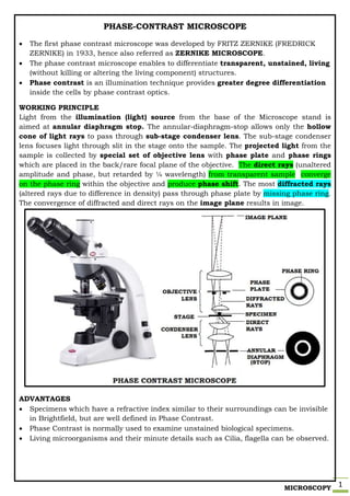
  • 1. MICROSCOPY 1 PHASE-CONTRAST MICROSCOPE  The first phase contrast microscope was developed by FRITZ ZERNIKE (FREDRICK ZERNIKE) in 1933, hence also referred as ZERNIKE MICROSCOPE.  The phase contrast microscope enables to differentiate transparent, unstained, living (without killing or altering the living component) structures.  Phase contrast is an illumination technique provides greater degree differentiation inside the cells by phase contrast optics. WORKING PRINCIPLE Light from the illumination (light) source from the base of the Microscope stand is aimed at annular diaphragm stop. The annular-diaphragm-stop allows only the hollow cone of light rays to pass through sub-stage condenser lens. The sub-stage condenser lens focuses light through slit in the stage onto the sample. The projected light from the sample is collected by special set of objective lens with phase plate and phase rings which are placed in the back/rare focal plane of the objective. The direct rays (unaltered amplitude and phase, but retarded by ¼ wavelength) from transparent sample converge on the phase ring within the objective and produce phase shift. The most diffracted rays (altered rays due to difference in density) pass through phase plate by missing phase ring. The convergence of diffracted and direct rays on the image plane results in image. ADVANTAGES  Specimens which have a refractive index similar to their surroundings can be invisible in Brightfield, but are well defined in Phase Contrast.  Phase Contrast is normally used to examine unstained biological specimens.  Living microorganisms and their minute details such as Cilia, flagella can be observed.
  • 2. MICROSCOPY 2 FLUORESCENT MICROSCOPE When a substance absorbs light, the electrons present at the outermost orbit absorbs energy and get excited; on the way back to the ground state, it emits a part of the energy absorbed. This phenomenon is termed as fluorescence.Fluorescent microscopeinvolves staining of specimens with special fluorescent dyes (fluorescein, acridine orange, etc). When a fluorescent dye is applied to a substance, it absorbs a wavelength of light (excitation wavelength) and emits light of different wavelength (emission wavelength). WORKING PRINCIPLE Illumination (light) is provided by a bright mercury vapor lamp (very expensive + harmful), produces light range of 200-400nm and generates considerable amount of heat. The heat filter absorbs heat, allows UV rays and visible rays by blocking infrared rays. The exciter filter ensures high energy - short wavelength - monochromatic light towards dichroic mirror. Dichroic mirror (beam splitter) eliminates visible light and reflects excited UV light to the dark-field condenser, which provides high contrast for fluorescence and also deflects majority of UV light.The excitation light is focused on to the fluorochrome specimen. The fluorescent labeled specimen absorbs light and emits excitatory light along with florescent light, which reaches objectivelens .As per the capacity of objective lens, the specimen would be magnified and are directed towards barrier filter. The additional barrier filter permits only the fluorescent wavelength and rejects excitation light. The fluorescent light passing through ocular lens creates the magnified image, which can also be detected by detector to give a photographic image. APPLICATIONS  Imaging the genetic material (DNA & RNA) and other structural components.  Monitoring the environment for microbial contamination.  Certain micro-organism can be detected and identified only by this microscopy.