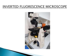
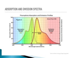
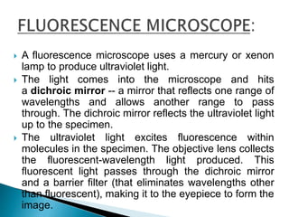
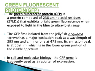
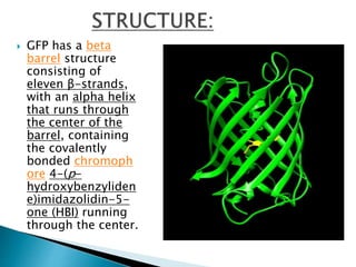
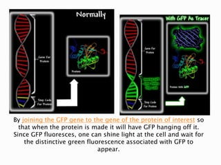
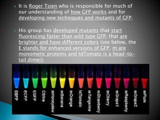
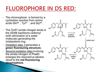
PRESENTED BY: JYOTSNA VERMA
RAHUL VERMA
SRISHTI SHARMA
 A process by which a photon is absorbed at
one wavelength and released at a different
wavelength or energy.
 JABLONSKI
DIAGRAM
 A fluorescence microscope uses a mercury or xenon
lamp to produce ultraviolet light.
 The light comes into the microscope and hits
a dichroic mirror -- a mirror that reflects one range of
wavelengths and allows another range to pass
through. The dichroic mirror reflects the ultraviolet light
up to the specimen.
 The ultraviolet light excites fluorescence within
molecules in the specimen. The objective lens collects
the fluorescent-wavelength light produced. This
fluorescent light passes through the dichroic mirror
and a barrier filter (that eliminates wavelengths other
than fluorescent), making it to the eyepiece to form the
image.
 Fluorescence microscopy requires intense, near-
monochromatic, illumination which some
widespread light sources, like halogen
lamps cannot provide.
 Four main types of light source are used,
including xenon arc lamps or mercury-vapor
lamps .
 Lasers are most widely used for more complex
fluorescence microscopy techniques like confocal
microscopy and total internal reflection
fluorescence microscopy while xenon lamps, and
mercury lamps, and LEDs with
a dichroic excitation filter are commonly used for
widefield epifluorescence microscopes.
 Epifluorescence microscopy is a
method of fluorescence
microscopy that is widely used in
life sciences.
 The excitory light is passed from
above(or,for inverted
microscope,from below),through
the objective lens and then onto
the specimen instead of passing it
first throgh the specimen.
 The fluorescence in the specimen
then gives rise to the emitted
light which is focused to the
detector by the same objective
lens that is used for excitation.
 A fluorophore (or fluorochrome, similarly to a
chromophore) is a fluorescent chemical compound
that can re-emit light upon light excitation.
Fluorophores typically contain several combined
aromatic groups, or plane or cyclic molecules with
several π bonds.
 Types:
 internal: are part of molecule .e.g.Trp amino acid
in proteins.
 external: molecules that lack internal
fluoroscense are linked with other small
fluorescent molecules externally
 Many fluorescent stains have been designed for a
range of biological molecules.
 Some of these are small molecules which are
intrinsically fluorescent and bind a biological
molecule of interest. Major examples of these
are nucleic acid stains like DAPI and
Hoechst (excited by UV wavelength light).
 DAPI (4',6-diamidino-2-phenylindole) is
a fluorescent stain that binds strongly to A-T
rich regions in DNA.
 Hoechst stains are part of a family of
bluefluorescent dyes used to stain DNA.
 A major example of fluorescent stain
is phalloidin which is used to
stain actin fibres in mammalian cells
 There are many fluorescent molecules
called fluorophores or fluorochromes such
as fluorescein, Alexa Fluors or DyLight 488,
which can be chemically linked to a different
molecule which binds the target of interest
within the sample.
1. Immunology: An antibody is first prepared by having a fluorescent chemical
group attached, and the sites (e.g., on a microscopic specimen) where the
antibody has bound can be seen, and even quantified, by the fluorescence.
2. Cell and molecular biology:
• detection of colocalization using fluorescence-labelled.
• Imaging structural components of small specimens, such as cells.
• Conducting viability studies on cell populations (are they alive or dead?)
• Viewing specific cells within a larger population with techniques such as
FISH. Detection and determination of the proteins localization in cell and
tissue
3. Diagnostic of diseases
 The green fluorescent protein (GFP) is
a protein composed of 238 amino acid residues
(27kDa) that exhibits bright green fluorescence when
exposed to light in the blue to ultraviolet range.
 The GFP,first isolated from the jellyfish Aequorea
victoria has a major excitation peak at a wavelength of
395 nm and a minor one at 475 nm. Its emission peak
is at 509 nm, which is in the lower green portion of
the visible spectrum.
 In cell and molecular biology, the GFP gene is
frequently used as a reporter of expression.
 GFP has a beta
barrel structure
consisting of
eleven β-strands,
with an alpha helix
that runs through
the center of the
barrel, containing
the covalently
bonded chromoph
ore 4-(p-
hydroxybenzyliden
e)imidazolidin-5-
one (HBI) running
through the center.
 The chromophore is located in the middle
of the beta-barrel, it is occasionally referred
to as the “light in the can.”
 Five shorter alpha helices form caps on the
ends of the structure.
 The beta barrel structure is a nearly perfect
cylinder, 42Å long and 24Å in
diameter,creating what is referred to as a "β-
can" formation, which is unique to the GFP-
like family.
 HBI, the spontaneously modified form of the
tripeptide Ser65–Tyr66–Gly67, is
nonfluorescent in the absence of the properly
folded GFP scaffold.
 Biological marker :
Fusion of GFP to a protein does not alter the function ,mobility
abilities or location of the protein.
This feature has enabled researchers to use GFP in living systems,
and it has led to GFP’s widespread use in cell dynamics and
development studies.
 Reporter Gene:
- The first applications of GFP were as a reporter
gene -Gene expression level in living cells.
By joining the GFP gene to the gene of the protein of interest so
that when the protein is made it will have GFP hanging off it.
Since GFP fluoresces, one can shine light at the cell and wait for
the distinctive green fluorescence associated with GFP to
appear.
 GFP has the ability to exhibit intrinsic fluorescence through three
amino acids that cyclise (Ser65-Tyr66-Gly67) and then undergo an
oxidation step during a complex maturation process.
The 3 amino acids forming
the chormophore in GFP
undergoes oxidation and
cyclization.
The formed active
chromophore
contains conjugated double
bonds.
These double bonds store
and release the energy from
electrons.
 Topology of Folding
 The above diagram illustrates the topology of the
folding pattern. Beta-strands are shown in green,
alpha-helices in red and connecting loops in black. The
numbers of the residues at the beginning and end of
the secondary structure elements are given as well.
 It is Roger Tsien who is responsible for much of
our understanding of how GFP works and for
developing new techniques and mutants of GFP.
 His group has developed mutants that start
fluorescing faster than wild type GFP, that are
brighter and have different colors (see below, the
E stands for enhanced versions of GFP, m are
monomeric proteins and tdTomato is a head-to-
tail dimer).
 The DsRed is a protein tetramer having the
property of emitting a fluorescence color red .
 From a coral (Discosoma ), this protein is
intrinsically fluorescent.
 Its gene can be fused in vitro to the gene of a
protein that one wishes to study. The
recombinant gene is then reintroduced
into cells or an embryo , which will then
synthesize the then fluorescent fusion protein. It
can then be observed using a fluorescence
microscope, for example. This method makes it
possible to study proteins in their natural
environment: the living cell.
 DsRed has excitation maxima at 558 nm (green
light) and emission maxima wavelength
is 583nm.
 The chromophore is formed by a
cyclization reaction from amino
acids Gln66 , Tyr67 , and Gly68 .
 The Tyr67 amide nitrogen binds to
the Gln66 backbone carbonyl
(with elimination of a water
molecule) generating the
imidazolone ring.
 Oxidation step 1:generates a
green fluorescing structure.
 Second oxidation step:involving
the amide nitrogen of Phe65
enlarges the resonant pi electon
cloud to the red fluorescing
chromophore.
 One of its crippling defects is the slow
processing of the protein as one of the
intermediate forms has a green fluorescence
similar to that of the green fluorescent
protein (GFP).
 Furthermore, DsRed is an obligate tetramer and
can form large protein aggregates in living cells.
 In contrast to the jellyfish fluorescent proteins,
which have been successfully used to tag
hundreds of proteins, DsRed conjugates have
proven much less successful and are often toxic.
 But wild-type DsRed has several drawbacks, including
slow chromophore maturation and poor solubility. To
overcome the slow maturation, we used random and
directed mutagenesis to create DsRed variants that
mature 10–15 times faster than the wild-type
protein.
 An asparagine-to-glutamine substitution at position
42 greatly accelerates the maturation of DsRed, but
also increases the level of green emission.
 Additional amino acid substitutions suppress this
green emission while further accelerating the
maturation.
 To enhance the solubility of DsRed, we reduced the
net charge near the N terminus of the protein. The
optimized DsRed variants yield bright fluorescence
even in rapidly growing organisms such as yeast.
 There are now different variants of DsRed that have been
obtained by modifying it by genetic engineering. These
mutation steps made it possible to obtain dimeric,
monomeric forms, to increase the brightness or to change
the range of wavelengths involved in the excitation and
emission spectra.
 Variants obtained by mutagenesis:
 dimer2 : dimeric variant
 mRFP1 : monomeric variant , slow maturation
 dTomato : dimeric variant
 tdTomato : tetrameric variant
 mOrange : monomeric variant, orange fluorescence
 mBanana : monomeric variant, orange fluorescence
 mHoneydew : monomeric variant, green-orange
fluorescence
 mStrawberry : monomeric variant, red fluorescence
between DsRed and mRFP.
 mCherry : monomeric variant, mRFP-like fluorescence with
faster maturation and better stability
 http://www.chm.bris.ac.uk/motm/GFP/GFPh.htm
 https://www.conncoll.edu/ccacad/zimmer/GFP-
ww/GFP-1.htm
 https://www.microscopyu.com/techniques/fluores
cence/introduction-to-fluorescence-microscopy
 https://www.conncoll.edu/ccacad/zimmer/GFP-
ww/GFP-1.htm
Fluorescence microscopy

Fluorescence microscopy

  • 1.
    PRESENTED BY: JYOTSNAVERMA RAHUL VERMA SRISHTI SHARMA
  • 3.
     A processby which a photon is absorbed at one wavelength and released at a different wavelength or energy.  JABLONSKI DIAGRAM
  • 8.
     A fluorescencemicroscope uses a mercury or xenon lamp to produce ultraviolet light.  The light comes into the microscope and hits a dichroic mirror -- a mirror that reflects one range of wavelengths and allows another range to pass through. The dichroic mirror reflects the ultraviolet light up to the specimen.  The ultraviolet light excites fluorescence within molecules in the specimen. The objective lens collects the fluorescent-wavelength light produced. This fluorescent light passes through the dichroic mirror and a barrier filter (that eliminates wavelengths other than fluorescent), making it to the eyepiece to form the image.
  • 10.
     Fluorescence microscopyrequires intense, near- monochromatic, illumination which some widespread light sources, like halogen lamps cannot provide.  Four main types of light source are used, including xenon arc lamps or mercury-vapor lamps .  Lasers are most widely used for more complex fluorescence microscopy techniques like confocal microscopy and total internal reflection fluorescence microscopy while xenon lamps, and mercury lamps, and LEDs with a dichroic excitation filter are commonly used for widefield epifluorescence microscopes.
  • 13.
     Epifluorescence microscopyis a method of fluorescence microscopy that is widely used in life sciences.  The excitory light is passed from above(or,for inverted microscope,from below),through the objective lens and then onto the specimen instead of passing it first throgh the specimen.  The fluorescence in the specimen then gives rise to the emitted light which is focused to the detector by the same objective lens that is used for excitation.
  • 14.
     A fluorophore(or fluorochrome, similarly to a chromophore) is a fluorescent chemical compound that can re-emit light upon light excitation. Fluorophores typically contain several combined aromatic groups, or plane or cyclic molecules with several π bonds.  Types:  internal: are part of molecule .e.g.Trp amino acid in proteins.  external: molecules that lack internal fluoroscense are linked with other small fluorescent molecules externally
  • 15.
     Many fluorescentstains have been designed for a range of biological molecules.  Some of these are small molecules which are intrinsically fluorescent and bind a biological molecule of interest. Major examples of these are nucleic acid stains like DAPI and Hoechst (excited by UV wavelength light).  DAPI (4',6-diamidino-2-phenylindole) is a fluorescent stain that binds strongly to A-T rich regions in DNA.  Hoechst stains are part of a family of bluefluorescent dyes used to stain DNA.
  • 16.
     A majorexample of fluorescent stain is phalloidin which is used to stain actin fibres in mammalian cells  There are many fluorescent molecules called fluorophores or fluorochromes such as fluorescein, Alexa Fluors or DyLight 488, which can be chemically linked to a different molecule which binds the target of interest within the sample.
  • 17.
    1. Immunology: Anantibody is first prepared by having a fluorescent chemical group attached, and the sites (e.g., on a microscopic specimen) where the antibody has bound can be seen, and even quantified, by the fluorescence. 2. Cell and molecular biology: • detection of colocalization using fluorescence-labelled. • Imaging structural components of small specimens, such as cells. • Conducting viability studies on cell populations (are they alive or dead?) • Viewing specific cells within a larger population with techniques such as FISH. Detection and determination of the proteins localization in cell and tissue 3. Diagnostic of diseases
  • 19.
     The greenfluorescent protein (GFP) is a protein composed of 238 amino acid residues (27kDa) that exhibits bright green fluorescence when exposed to light in the blue to ultraviolet range.  The GFP,first isolated from the jellyfish Aequorea victoria has a major excitation peak at a wavelength of 395 nm and a minor one at 475 nm. Its emission peak is at 509 nm, which is in the lower green portion of the visible spectrum.  In cell and molecular biology, the GFP gene is frequently used as a reporter of expression.
  • 20.
     GFP hasa beta barrel structure consisting of eleven β-strands, with an alpha helix that runs through the center of the barrel, containing the covalently bonded chromoph ore 4-(p- hydroxybenzyliden e)imidazolidin-5- one (HBI) running through the center.
  • 21.
     The chromophoreis located in the middle of the beta-barrel, it is occasionally referred to as the “light in the can.”  Five shorter alpha helices form caps on the ends of the structure.  The beta barrel structure is a nearly perfect cylinder, 42Å long and 24Å in diameter,creating what is referred to as a "β- can" formation, which is unique to the GFP- like family.  HBI, the spontaneously modified form of the tripeptide Ser65–Tyr66–Gly67, is nonfluorescent in the absence of the properly folded GFP scaffold.
  • 22.
     Biological marker: Fusion of GFP to a protein does not alter the function ,mobility abilities or location of the protein. This feature has enabled researchers to use GFP in living systems, and it has led to GFP’s widespread use in cell dynamics and development studies.  Reporter Gene: - The first applications of GFP were as a reporter gene -Gene expression level in living cells.
  • 23.
    By joining theGFP gene to the gene of the protein of interest so that when the protein is made it will have GFP hanging off it. Since GFP fluoresces, one can shine light at the cell and wait for the distinctive green fluorescence associated with GFP to appear.
  • 24.
     GFP hasthe ability to exhibit intrinsic fluorescence through three amino acids that cyclise (Ser65-Tyr66-Gly67) and then undergo an oxidation step during a complex maturation process. The 3 amino acids forming the chormophore in GFP undergoes oxidation and cyclization. The formed active chromophore contains conjugated double bonds. These double bonds store and release the energy from electrons.
  • 25.
     Topology ofFolding  The above diagram illustrates the topology of the folding pattern. Beta-strands are shown in green, alpha-helices in red and connecting loops in black. The numbers of the residues at the beginning and end of the secondary structure elements are given as well.
  • 27.
     It isRoger Tsien who is responsible for much of our understanding of how GFP works and for developing new techniques and mutants of GFP.  His group has developed mutants that start fluorescing faster than wild type GFP, that are brighter and have different colors (see below, the E stands for enhanced versions of GFP, m are monomeric proteins and tdTomato is a head-to- tail dimer).
  • 29.
     The DsRedis a protein tetramer having the property of emitting a fluorescence color red .  From a coral (Discosoma ), this protein is intrinsically fluorescent.  Its gene can be fused in vitro to the gene of a protein that one wishes to study. The recombinant gene is then reintroduced into cells or an embryo , which will then synthesize the then fluorescent fusion protein. It can then be observed using a fluorescence microscope, for example. This method makes it possible to study proteins in their natural environment: the living cell.  DsRed has excitation maxima at 558 nm (green light) and emission maxima wavelength is 583nm.
  • 30.
     The chromophoreis formed by a cyclization reaction from amino acids Gln66 , Tyr67 , and Gly68 .  The Tyr67 amide nitrogen binds to the Gln66 backbone carbonyl (with elimination of a water molecule) generating the imidazolone ring.  Oxidation step 1:generates a green fluorescing structure.  Second oxidation step:involving the amide nitrogen of Phe65 enlarges the resonant pi electon cloud to the red fluorescing chromophore.
  • 31.
     One ofits crippling defects is the slow processing of the protein as one of the intermediate forms has a green fluorescence similar to that of the green fluorescent protein (GFP).  Furthermore, DsRed is an obligate tetramer and can form large protein aggregates in living cells.  In contrast to the jellyfish fluorescent proteins, which have been successfully used to tag hundreds of proteins, DsRed conjugates have proven much less successful and are often toxic.
  • 32.
     But wild-typeDsRed has several drawbacks, including slow chromophore maturation and poor solubility. To overcome the slow maturation, we used random and directed mutagenesis to create DsRed variants that mature 10–15 times faster than the wild-type protein.  An asparagine-to-glutamine substitution at position 42 greatly accelerates the maturation of DsRed, but also increases the level of green emission.  Additional amino acid substitutions suppress this green emission while further accelerating the maturation.  To enhance the solubility of DsRed, we reduced the net charge near the N terminus of the protein. The optimized DsRed variants yield bright fluorescence even in rapidly growing organisms such as yeast.
  • 33.
     There arenow different variants of DsRed that have been obtained by modifying it by genetic engineering. These mutation steps made it possible to obtain dimeric, monomeric forms, to increase the brightness or to change the range of wavelengths involved in the excitation and emission spectra.  Variants obtained by mutagenesis:  dimer2 : dimeric variant  mRFP1 : monomeric variant , slow maturation  dTomato : dimeric variant  tdTomato : tetrameric variant  mOrange : monomeric variant, orange fluorescence  mBanana : monomeric variant, orange fluorescence  mHoneydew : monomeric variant, green-orange fluorescence  mStrawberry : monomeric variant, red fluorescence between DsRed and mRFP.  mCherry : monomeric variant, mRFP-like fluorescence with faster maturation and better stability
  • 34.
     http://www.chm.bris.ac.uk/motm/GFP/GFPh.htm  https://www.conncoll.edu/ccacad/zimmer/GFP- ww/GFP-1.htm https://www.microscopyu.com/techniques/fluores cence/introduction-to-fluorescence-microscopy  https://www.conncoll.edu/ccacad/zimmer/GFP- ww/GFP-1.htm