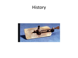
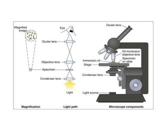
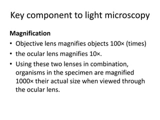
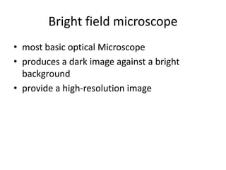
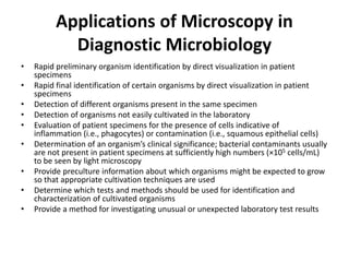
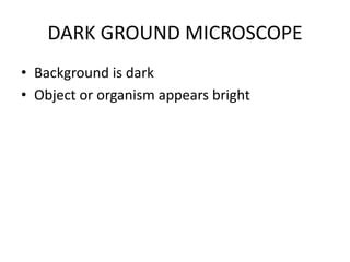
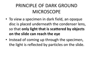
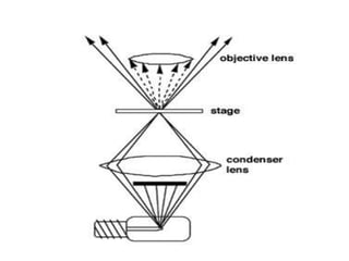
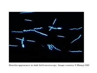
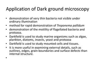
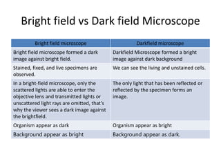
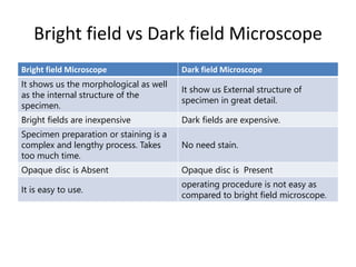
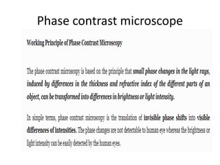
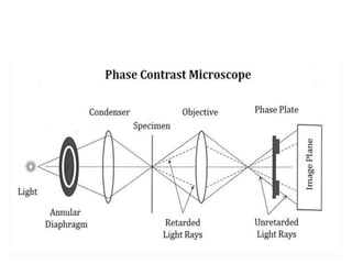
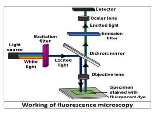
SlideShare a Scribd company logo
Light
microscope
Dr. Rasika Deshmukh
(MBBS, MD)
• The evolution of the Microbiology field put to
perspective the need to identify, view, observe
and understand microorganisms, including
their structural morphologies and
mechanisms.
History
• The first simple microscope was discovered
by two Dutch scientists, Zaccharias Janssen
and his father, Hans who made spectacles,
were the first to experiment with their lenses
by combining lenses in a tube and observed
that the objects that were nearby, appeared
closer and larger.
History
• Antony Van Lewnehoueek an amateur
Microbiologist made the first simple
microscope, that enabled him to observe the
presence of tiny living organisms in pond
water that appeared like dots. His simple
microscope was made up of a double convex
glass lens that was held between two silver
plates.
History
What is a light microscope?
• Instrument or tool, that uses visible light to
detect and magnify very small objects and
enlarge them.
• Use lenses to focus light on the specimen,
magnifying it thus producing an image
Principles of Light Microscopy
• For light microscopy, visible light is passed
through the specimen and then through a
series of lenses that bend the light in a
manner that results in magnification of the
organisms present in the specimen . The total
magnification achieved is the product of the
lenses used.
Key component to light microscopy
Magnification
• Objective lens magnifies objects 100× (times)
• the ocular lens magnifies 10×.
• Using these two lenses in combination,
organisms in the specimen are magnified
1000× their actual size when viewed through
the ocular lens.
Contrast
• Needed to make objects stand out from the
background
• Contrast is most commonly achieved by
staining techniques that highlight organisms
and allow them to be differentiated from one
another and from background material and
debris.
Resolution
• To optimize visualization, other factors besides
magnification must be considered.
• Resolution, defined as the extent to which
detail in the magnified object is maintained, is
also essential.
Types of microscopes
Depending on the number of lenses, there are
two types of microscopes
• Simple light microscope (it has low
magnification because it uses a single lens)
• Compound light microscope (it has a higher
magnification compared to the simple
microscope because it uses at least two sets of
lenses, an objective lens, and an eyepiece).
Light microscope
The modern types of Light Microscopes include:
• Bright field light microscope
• Dark field light microscope
• Phase contrast light microscope
• Fluorescence light microscope
Bright field microscope
• most basic optical Microscope
• produces a dark image against a bright
background
• provide a high-resolution image
Parts of a bright-field microscope
• Objective lens: magnifies the image by 10x, 20x,
40x and 100x
• Ocular lens: magnifies the image by 10x
• Condenser: focuses a beam of light onto the
specimen
• Diaphragm: controls the diameter of the beam of
light that passes through the condenser
• Two focusing knobs: focus on the image and
sharpen the image.
Applications of Microscopy in
Diagnostic Microbiology
• Rapid preliminary organism identification by direct visualization in patient
specimens
• Rapid final identification of certain organisms by direct visualization in patient
specimens
• Detection of different organisms present in the same specimen
• Detection of organisms not easily cultivated in the laboratory
• Evaluation of patient specimens for the presence of cells indicative of
inflammation (i.e., phagocytes) or contamination (i.e., squamous epithelial cells)
• Determination of an organism’s clinical significance; bacterial contaminants usually
are not present in patient specimens at sufficiently high numbers (×105 cells/mL)
to be seen by light microscopy
• Provide preculture information about which organisms might be expected to grow
so that appropriate cultivation techniques are used
• Determine which tests and methods should be used for identification and
characterization of cultivated organisms
• Provide a method for investigating unusual or unexpected laboratory test results
DARK GROUND MICROSCOPE
• Background is dark
• Object or organism appears bright
PRINCIPLE OF DARK GROUND
MICROSCOPE
• To view a specimen in dark field, an opaque
disc is placed underneath the condenser lens,
so that only light that is scattered by objects
on the slide can reach the eye
• Instead of coming up through the specimen,
the light is reflected by particles on the slide.
Application of Dark ground microscopy
• demonstration of very thin bacteria not visible under
ordinary illumination
• method for rapid demonstration of Treponema pallidum
• demonstration of the motility of flagellated bacteria and
protozoa.
• Darkfield is used to study marine organisms such as algae,
plankton, diatoms, insects, yeast and protozoa
• Darkfield is used to study mounted cells and tissues.
• It is more useful in examining external details, such as
outlines, edges, grain boundaries and surface defects than
internal structure.
•
Bright field vs Dark field Microscope
Bright field microscope Darkfield microscope
Bright field microscope formed a dark
image against bright field.
Darkfield Microscope formed a bright
image against dark background
Stained, fixed, and live specimens are
observed.
We can see the living and unstained cells.
In a bright-field microscope, only the
scattered lights are able to enter the
objective lens and transmitted lights or
unscattered light rays are omitted, that’s
why the viewer sees a dark image against
the brightfield.
The only light that has been reflected or
reflected by the specimen forms an
image.
Organism appear as dark Organism appear as bright
Background appear as bright Background appear as dark.
Bright field vs Dark field Microscope
Bright field Microscope Dark field Microscope
It shows us the morphological as well
as the internal structure of the
specimen.
It show us External structure of
specimen in great detail.
Bright fields are inexpensive Dark fields are expensive.
Specimen preparation or staining is a
complex and lengthy process. Takes
too much time.
No need stain.
Opaque disc is Absent Opaque disc is Present
It is easy to use.
operating procedure is not easy as
compared to bright field microscope.
Phase contrast microscope
Application
The Fluorescent Microscope
• dye molecule added to the specimen.
Advantages
• helps in the study of cell behaviour.
• highlights the biomolecule of interest.
• highlight the image of particular structural
components within the microscopic organisms.
• highly sensitive technique that can detect around
50 molecules/µm3.
• analyze or track the physiochemical properties of
multiple biomolecules simultaneously.
• Disadvantages
• It only allows the observation of specific structures
inside a cell tagged with the fluorescent dye.
• The photobleaching due to the electron excitation
during the process of fluorescence may affect reactive
molecules of the fluorescent dyes. As a result, the
reactive dyes might lose their chemical property of
fluorescence emission intensity.
• The cells are susceptible to the phototoxic effect after
staining with fluorescent dyes, as the fluorophore
molecules absorb the high energy photons from the
short-wavelength light.
Thank you

More Related Content

What's hot

Microscope and its types
Microscope and its typesMicroscope and its types
Microscope and its types
Ketan Patil
 
Light microscopy
Light microscopyLight microscopy
Light microscopy
Saurabh Bhargava
 
DARK FIELD MICROSCOPY
DARK FIELD MICROSCOPYDARK FIELD MICROSCOPY
DARK FIELD MICROSCOPY
Sivasangari Shanmugam
 
Microscopy
MicroscopyMicroscopy
Microscopy
Daya Pavitrakar
 
Bright field microscopy, Principle and applications
Bright field microscopy, Principle and applicationsBright field microscopy, Principle and applications
Bright field microscopy, Principle and applications
KAUSHAL SAHU
 
Fluorescence microscope
Fluorescence microscopeFluorescence microscope
Fluorescence microscope
gohil sanjay bhagvanji
 
Phase contrast microscope
Phase contrast microscopePhase contrast microscope
Phase contrast microscope
Ravi Kumar
 
Microscopy
 Microscopy  Microscopy
Microscopy
Baljinder Gill
 
Electron microscope, principle and application
Electron microscope, principle and applicationElectron microscope, principle and application
Electron microscope, principle and application
KAUSHAL SAHU
 
Dark field microscopy
Dark field microscopyDark field microscopy
Dark field microscopy
Abhishek Indurkar
 
Numerical aperture and limits of resolution of microscope
Numerical aperture and limits of resolution of microscopeNumerical aperture and limits of resolution of microscope
Numerical aperture and limits of resolution of microscope
Tahir Ali,Punjab University Lahore
 
3. Microscope simple, compound & stereo - Basics
3. Microscope   simple, compound & stereo - Basics3. Microscope   simple, compound & stereo - Basics
3. Microscope simple, compound & stereo - Basics
Nethravathi Siri
 
Phase contrast & Flurosence microscope
Phase contrast & Flurosence microscopePhase contrast & Flurosence microscope
Phase contrast & Flurosence microscope
microbiology Notes
 
Light microscope
Light microscopeLight microscope
Light microscope
Sivasangari Shanmugam
 
Phase contrast microscope
Phase contrast microscopePhase contrast microscope
Phase contrast microscope
NithyaNandapal
 
Scanning and transmission electron microscopy
Scanning and transmission electron microscopyScanning and transmission electron microscopy
Scanning and transmission electron microscopy
gohil sanjay bhagvanji
 
Microscope
MicroscopeMicroscope
Electron microscope ppt
Electron microscope pptElectron microscope ppt
Electron microscope ppt
NithyaNandapal
 
Lecture 3 Unit-I Bright-field & Dark-field Microscopy.pdf
Lecture 3 Unit-I Bright-field & Dark-field Microscopy.pdfLecture 3 Unit-I Bright-field & Dark-field Microscopy.pdf
Lecture 3 Unit-I Bright-field & Dark-field Microscopy.pdf
SushmitaChoudhary16
 
Compound microscope
Compound microscopeCompound microscope
Compound microscope
NithyaNandapal
 

What's hot (20)

Microscope and its types
Microscope and its typesMicroscope and its types
Microscope and its types
 
Light microscopy
Light microscopyLight microscopy
Light microscopy
 
DARK FIELD MICROSCOPY
DARK FIELD MICROSCOPYDARK FIELD MICROSCOPY
DARK FIELD MICROSCOPY
 
Microscopy
MicroscopyMicroscopy
Microscopy
 
Bright field microscopy, Principle and applications
Bright field microscopy, Principle and applicationsBright field microscopy, Principle and applications
Bright field microscopy, Principle and applications
 
Fluorescence microscope
Fluorescence microscopeFluorescence microscope
Fluorescence microscope
 
Phase contrast microscope
Phase contrast microscopePhase contrast microscope
Phase contrast microscope
 
Microscopy
 Microscopy  Microscopy
Microscopy
 
Electron microscope, principle and application
Electron microscope, principle and applicationElectron microscope, principle and application
Electron microscope, principle and application
 
Dark field microscopy
Dark field microscopyDark field microscopy
Dark field microscopy
 
Numerical aperture and limits of resolution of microscope
Numerical aperture and limits of resolution of microscopeNumerical aperture and limits of resolution of microscope
Numerical aperture and limits of resolution of microscope
 
3. Microscope simple, compound & stereo - Basics
3. Microscope   simple, compound & stereo - Basics3. Microscope   simple, compound & stereo - Basics
3. Microscope simple, compound & stereo - Basics
 
Phase contrast & Flurosence microscope
Phase contrast & Flurosence microscopePhase contrast & Flurosence microscope
Phase contrast & Flurosence microscope
 
Light microscope
Light microscopeLight microscope
Light microscope
 
Phase contrast microscope
Phase contrast microscopePhase contrast microscope
Phase contrast microscope
 
Scanning and transmission electron microscopy
Scanning and transmission electron microscopyScanning and transmission electron microscopy
Scanning and transmission electron microscopy
 
Microscope
MicroscopeMicroscope
Microscope
 
Electron microscope ppt
Electron microscope pptElectron microscope ppt
Electron microscope ppt
 
Lecture 3 Unit-I Bright-field & Dark-field Microscopy.pdf
Lecture 3 Unit-I Bright-field & Dark-field Microscopy.pdfLecture 3 Unit-I Bright-field & Dark-field Microscopy.pdf
Lecture 3 Unit-I Bright-field & Dark-field Microscopy.pdf
 
Compound microscope
Compound microscopeCompound microscope
Compound microscope
 

Similar to Light microscope

Compound microscope
Compound microscopeCompound microscope
Compound microscope
meghashridhar
 
MICROSCOPY.pptx
MICROSCOPY.pptxMICROSCOPY.pptx
MICROSCOPY.pptx
ishtiyakjafarjafar
 
MICROSCOPY.pptx
MICROSCOPY.pptxMICROSCOPY.pptx
MICROSCOPY.pptx
Saraswathi29574
 
B.Sc. Biochemistry II Cellular Biochemistry Unit 4 Basic Techniques in Microb...
B.Sc. Biochemistry II Cellular Biochemistry Unit 4 Basic Techniques in Microb...B.Sc. Biochemistry II Cellular Biochemistry Unit 4 Basic Techniques in Microb...
B.Sc. Biochemistry II Cellular Biochemistry Unit 4 Basic Techniques in Microb...
Rai University
 
B sc micro i btm u 1 microscopy and staining
B sc micro i btm u 1 microscopy and staining B sc micro i btm u 1 microscopy and staining
B sc micro i btm u 1 microscopy and staining
Rai University
 
B sc micro i btm u 1 microscopy and staining
B sc micro i btm u 1 microscopy and staining B sc micro i btm u 1 microscopy and staining
B sc micro i btm u 1 microscopy and staining
Rai University
 
History and types of Microscopy.pptx
History and types of Microscopy.pptxHistory and types of Microscopy.pptx
History and types of Microscopy.pptx
SandeepKaur926499
 
microscope- lecture 2.pptx
microscope- lecture 2.pptxmicroscope- lecture 2.pptx
microscope- lecture 2.pptx
OsmanAli92
 
Microscope
MicroscopeMicroscope
Microscope
MUKESH SINGH
 
Light microscopy.pptx bright dark phase contrast microscope
Light microscopy.pptx bright dark phase contrast microscopeLight microscopy.pptx bright dark phase contrast microscope
Light microscopy.pptx bright dark phase contrast microscope
DarshanN65
 
Presentation1 4
Presentation1 4Presentation1 4
Presentation1 4
jiwaji university gwalior
 
Microscopy 4 anna
Microscopy 4 annaMicroscopy 4 anna
Microscopy 4 anna
Annakurian9
 
microscope.pptx
microscope.pptxmicroscope.pptx
microscope.pptx
Dinraj Kulkarni
 
Types of Microscopes with their applications
Types of Microscopes with their applicationsTypes of Microscopes with their applications
Types of Microscopes with their applications
Microbiology Note
 
Electron Microscope: Definition, Types, Parts, Application, Advantages, Disad...
Electron Microscope: Definition, Types, Parts, Application, Advantages, Disad...Electron Microscope: Definition, Types, Parts, Application, Advantages, Disad...
Electron Microscope: Definition, Types, Parts, Application, Advantages, Disad...
Microbiology Note
 
History of microscopy i
History of microscopy iHistory of microscopy i
History of microscopy i
Pharmacy Universe
 
agri microbiology for Micro Hort PPT.pptx
agri microbiology for Micro Hort PPT.pptxagri microbiology for Micro Hort PPT.pptx
agri microbiology for Micro Hort PPT.pptx
DawitGetahun6
 
Microscopy.ppt Microscopy.pptvMicroscopy.pptMicroscopy.pptMicroscopy.pptMicro...
Microscopy.ppt Microscopy.pptvMicroscopy.pptMicroscopy.pptMicroscopy.pptMicro...Microscopy.ppt Microscopy.pptvMicroscopy.pptMicroscopy.pptMicroscopy.pptMicro...
Microscopy.ppt Microscopy.pptvMicroscopy.pptMicroscopy.pptMicroscopy.pptMicro...
KAOUKAAlaedin
 
Histo tool.pptx
Histo tool.pptxHisto tool.pptx
Histo tool.pptx
DeanParamedics
 
Histological tools
Histological toolsHistological tools
Histological tools
Navneet Randhawa
 

Similar to Light microscope (20)

Compound microscope
Compound microscopeCompound microscope
Compound microscope
 
MICROSCOPY.pptx
MICROSCOPY.pptxMICROSCOPY.pptx
MICROSCOPY.pptx
 
MICROSCOPY.pptx
MICROSCOPY.pptxMICROSCOPY.pptx
MICROSCOPY.pptx
 
B.Sc. Biochemistry II Cellular Biochemistry Unit 4 Basic Techniques in Microb...
B.Sc. Biochemistry II Cellular Biochemistry Unit 4 Basic Techniques in Microb...B.Sc. Biochemistry II Cellular Biochemistry Unit 4 Basic Techniques in Microb...
B.Sc. Biochemistry II Cellular Biochemistry Unit 4 Basic Techniques in Microb...
 
B sc micro i btm u 1 microscopy and staining
B sc micro i btm u 1 microscopy and staining B sc micro i btm u 1 microscopy and staining
B sc micro i btm u 1 microscopy and staining
 
B sc micro i btm u 1 microscopy and staining
B sc micro i btm u 1 microscopy and staining B sc micro i btm u 1 microscopy and staining
B sc micro i btm u 1 microscopy and staining
 
History and types of Microscopy.pptx
History and types of Microscopy.pptxHistory and types of Microscopy.pptx
History and types of Microscopy.pptx
 
microscope- lecture 2.pptx
microscope- lecture 2.pptxmicroscope- lecture 2.pptx
microscope- lecture 2.pptx
 
Microscope
MicroscopeMicroscope
Microscope
 
Light microscopy.pptx bright dark phase contrast microscope
Light microscopy.pptx bright dark phase contrast microscopeLight microscopy.pptx bright dark phase contrast microscope
Light microscopy.pptx bright dark phase contrast microscope
 
Presentation1 4
Presentation1 4Presentation1 4
Presentation1 4
 
Microscopy 4 anna
Microscopy 4 annaMicroscopy 4 anna
Microscopy 4 anna
 
microscope.pptx
microscope.pptxmicroscope.pptx
microscope.pptx
 
Types of Microscopes with their applications
Types of Microscopes with their applicationsTypes of Microscopes with their applications
Types of Microscopes with their applications
 
Electron Microscope: Definition, Types, Parts, Application, Advantages, Disad...
Electron Microscope: Definition, Types, Parts, Application, Advantages, Disad...Electron Microscope: Definition, Types, Parts, Application, Advantages, Disad...
Electron Microscope: Definition, Types, Parts, Application, Advantages, Disad...
 
History of microscopy i
History of microscopy iHistory of microscopy i
History of microscopy i
 
agri microbiology for Micro Hort PPT.pptx
agri microbiology for Micro Hort PPT.pptxagri microbiology for Micro Hort PPT.pptx
agri microbiology for Micro Hort PPT.pptx
 
Microscopy.ppt Microscopy.pptvMicroscopy.pptMicroscopy.pptMicroscopy.pptMicro...
Microscopy.ppt Microscopy.pptvMicroscopy.pptMicroscopy.pptMicroscopy.pptMicro...Microscopy.ppt Microscopy.pptvMicroscopy.pptMicroscopy.pptMicroscopy.pptMicro...
Microscopy.ppt Microscopy.pptvMicroscopy.pptMicroscopy.pptMicroscopy.pptMicro...
 
Histo tool.pptx
Histo tool.pptxHisto tool.pptx
Histo tool.pptx
 
Histological tools
Histological toolsHistological tools
Histological tools
 

More from Rasika Deshmukh

Malaria
MalariaMalaria
HHV 8.pptx
HHV 8.pptxHHV 8.pptx
HHV 8.pptx
Rasika Deshmukh
 
Amp C (1).pptx
Amp C (1).pptxAmp C (1).pptx
Amp C (1).pptx
Rasika Deshmukh
 
atypical pneumonia.pptx
atypical pneumonia.pptxatypical pneumonia.pptx
atypical pneumonia.pptx
Rasika Deshmukh
 
malaria.pptx
malaria.pptxmalaria.pptx
malaria.pptx
Rasika Deshmukh
 
systems for processing blood cultures.pptx
systems for processing blood cultures.pptxsystems for processing blood cultures.pptx
systems for processing blood cultures.pptx
Rasika Deshmukh
 
Electron microscope
Electron microscopeElectron microscope
Electron microscope
Rasika Deshmukh
 
Mechanism of immunoevasion in parasites 2018 06-17
Mechanism of immunoevasion in parasites  2018 06-17Mechanism of immunoevasion in parasites  2018 06-17
Mechanism of immunoevasion in parasites 2018 06-17
Rasika Deshmukh
 

More from Rasika Deshmukh (8)

Malaria
MalariaMalaria
Malaria
 
HHV 8.pptx
HHV 8.pptxHHV 8.pptx
HHV 8.pptx
 
Amp C (1).pptx
Amp C (1).pptxAmp C (1).pptx
Amp C (1).pptx
 
atypical pneumonia.pptx
atypical pneumonia.pptxatypical pneumonia.pptx
atypical pneumonia.pptx
 
malaria.pptx
malaria.pptxmalaria.pptx
malaria.pptx
 
systems for processing blood cultures.pptx
systems for processing blood cultures.pptxsystems for processing blood cultures.pptx
systems for processing blood cultures.pptx
 
Electron microscope
Electron microscopeElectron microscope
Electron microscope
 
Mechanism of immunoevasion in parasites 2018 06-17
Mechanism of immunoevasion in parasites  2018 06-17Mechanism of immunoevasion in parasites  2018 06-17
Mechanism of immunoevasion in parasites 2018 06-17
 

Recently uploaded

Preventing Pickleball Injuries & Treatment
Preventing Pickleball Injuries & TreatmentPreventing Pickleball Injuries & Treatment
Preventing Pickleball Injuries & Treatment
LAB Sports Therapy
 
Antibiotic Stewardship by Anushri Srivastava.pptx
Antibiotic Stewardship by Anushri Srivastava.pptxAntibiotic Stewardship by Anushri Srivastava.pptx
Antibiotic Stewardship by Anushri Srivastava.pptx
AnushriSrivastav
 
GURGAON Call Girls ❤8901183002❤ #ℂALL# #gIRLS# In GURGAON ₹,2500 Cash Payment...
GURGAON Call Girls ❤8901183002❤ #ℂALL# #gIRLS# In GURGAON ₹,2500 Cash Payment...GURGAON Call Girls ❤8901183002❤ #ℂALL# #gIRLS# In GURGAON ₹,2500 Cash Payment...
GURGAON Call Girls ❤8901183002❤ #ℂALL# #gIRLS# In GURGAON ₹,2500 Cash Payment...
ranishasharma67
 
Surgery-Mini-OSCE-All-Past-Years-Questions-Modified.
Surgery-Mini-OSCE-All-Past-Years-Questions-Modified.Surgery-Mini-OSCE-All-Past-Years-Questions-Modified.
Surgery-Mini-OSCE-All-Past-Years-Questions-Modified.
preciousstephanie75
 
Introduction to Forensic Pathology course
Introduction to Forensic Pathology courseIntroduction to Forensic Pathology course
Introduction to Forensic Pathology course
fprxsqvnz5
 
Anatomy and Physiology Chapter-16_Digestive-System.pptx
Anatomy and Physiology Chapter-16_Digestive-System.pptxAnatomy and Physiology Chapter-16_Digestive-System.pptx
Anatomy and Physiology Chapter-16_Digestive-System.pptx
shanicedivinagracia2
 
A Community health , health for prisoners
A Community health  , health for prisonersA Community health  , health for prisoners
A Community health , health for prisoners
Ahmed Elmi
 
GENERAL PHARMACOLOGY - INTRODUCTION DENTAL.ppt
GENERAL PHARMACOLOGY - INTRODUCTION DENTAL.pptGENERAL PHARMACOLOGY - INTRODUCTION DENTAL.ppt
GENERAL PHARMACOLOGY - INTRODUCTION DENTAL.ppt
Mangaiarkkarasi
 
💘Ludhiana ℂall Girls 📞]][89011★83002][[ 📱 ❤ESCORTS service in Ludhiana💃💦Ludhi...
💘Ludhiana ℂall Girls 📞]][89011★83002][[ 📱 ❤ESCORTS service in Ludhiana💃💦Ludhi...💘Ludhiana ℂall Girls 📞]][89011★83002][[ 📱 ❤ESCORTS service in Ludhiana💃💦Ludhi...
💘Ludhiana ℂall Girls 📞]][89011★83002][[ 📱 ❤ESCORTS service in Ludhiana💃💦Ludhi...
ranishasharma67
 
CHAPTER 1 SEMESTER V PREVENTIVE-PEDIATRICS.pdf
CHAPTER 1 SEMESTER V PREVENTIVE-PEDIATRICS.pdfCHAPTER 1 SEMESTER V PREVENTIVE-PEDIATRICS.pdf
CHAPTER 1 SEMESTER V PREVENTIVE-PEDIATRICS.pdf
Sachin Sharma
 
The Docs PPG - 30.05.2024.pptx..........
The Docs PPG - 30.05.2024.pptx..........The Docs PPG - 30.05.2024.pptx..........
The Docs PPG - 30.05.2024.pptx..........
TheDocs
 
Navigating the Health Insurance Market_ Understanding Trends and Options.pdf
Navigating the Health Insurance Market_ Understanding Trends and Options.pdfNavigating the Health Insurance Market_ Understanding Trends and Options.pdf
Navigating the Health Insurance Market_ Understanding Trends and Options.pdf
Enterprise Wired
 
HEAT WAVE presented by priya bhojwani..pptx
HEAT WAVE presented by priya bhojwani..pptxHEAT WAVE presented by priya bhojwani..pptx
HEAT WAVE presented by priya bhojwani..pptx
priyabhojwani1200
 
Contact Now 89011**83002 Dehradun ℂall Girls By Full Service ℂall Girl In De...
Contact Now  89011**83002 Dehradun ℂall Girls By Full Service ℂall Girl In De...Contact Now  89011**83002 Dehradun ℂall Girls By Full Service ℂall Girl In De...
Contact Now 89011**83002 Dehradun ℂall Girls By Full Service ℂall Girl In De...
aunty1x2
 
一比一原版纽约大学毕业证(NYU毕业证)成绩单留信认证
一比一原版纽约大学毕业证(NYU毕业证)成绩单留信认证一比一原版纽约大学毕业证(NYU毕业证)成绩单留信认证
一比一原版纽约大学毕业证(NYU毕业证)成绩单留信认证
o6ov5dqmf
 
India Clinical Trials Market: Industry Size and Growth Trends [2030] Analyzed...
India Clinical Trials Market: Industry Size and Growth Trends [2030] Analyzed...India Clinical Trials Market: Industry Size and Growth Trends [2030] Analyzed...
India Clinical Trials Market: Industry Size and Growth Trends [2030] Analyzed...
Kumar Satyam
 
Artificial Intelligence to Optimize Cardiovascular Therapy
Artificial Intelligence to Optimize Cardiovascular TherapyArtificial Intelligence to Optimize Cardiovascular Therapy
Artificial Intelligence to Optimize Cardiovascular Therapy
Iris Thiele Isip-Tan
 
Nursing Care of Client With Acute And Chronic Renal Failure.ppt
Nursing Care of Client With Acute And Chronic Renal Failure.pptNursing Care of Client With Acute And Chronic Renal Failure.ppt
Nursing Care of Client With Acute And Chronic Renal Failure.ppt
Rommel Luis III Israel
 
VERIFICATION AND VALIDATION TOOLKIT Determining Performance Characteristics o...
VERIFICATION AND VALIDATION TOOLKIT Determining Performance Characteristics o...VERIFICATION AND VALIDATION TOOLKIT Determining Performance Characteristics o...
VERIFICATION AND VALIDATION TOOLKIT Determining Performance Characteristics o...
Nguyễn Thị Vân Anh
 
Dimensions of Healthcare Quality
Dimensions of Healthcare QualityDimensions of Healthcare Quality
Dimensions of Healthcare Quality
Naeemshahzad51
 

Recently uploaded (20)

Preventing Pickleball Injuries & Treatment
Preventing Pickleball Injuries & TreatmentPreventing Pickleball Injuries & Treatment
Preventing Pickleball Injuries & Treatment
 
Antibiotic Stewardship by Anushri Srivastava.pptx
Antibiotic Stewardship by Anushri Srivastava.pptxAntibiotic Stewardship by Anushri Srivastava.pptx
Antibiotic Stewardship by Anushri Srivastava.pptx
 
GURGAON Call Girls ❤8901183002❤ #ℂALL# #gIRLS# In GURGAON ₹,2500 Cash Payment...
GURGAON Call Girls ❤8901183002❤ #ℂALL# #gIRLS# In GURGAON ₹,2500 Cash Payment...GURGAON Call Girls ❤8901183002❤ #ℂALL# #gIRLS# In GURGAON ₹,2500 Cash Payment...
GURGAON Call Girls ❤8901183002❤ #ℂALL# #gIRLS# In GURGAON ₹,2500 Cash Payment...
 
Surgery-Mini-OSCE-All-Past-Years-Questions-Modified.
Surgery-Mini-OSCE-All-Past-Years-Questions-Modified.Surgery-Mini-OSCE-All-Past-Years-Questions-Modified.
Surgery-Mini-OSCE-All-Past-Years-Questions-Modified.
 
Introduction to Forensic Pathology course
Introduction to Forensic Pathology courseIntroduction to Forensic Pathology course
Introduction to Forensic Pathology course
 
Anatomy and Physiology Chapter-16_Digestive-System.pptx
Anatomy and Physiology Chapter-16_Digestive-System.pptxAnatomy and Physiology Chapter-16_Digestive-System.pptx
Anatomy and Physiology Chapter-16_Digestive-System.pptx
 
A Community health , health for prisoners
A Community health  , health for prisonersA Community health  , health for prisoners
A Community health , health for prisoners
 
GENERAL PHARMACOLOGY - INTRODUCTION DENTAL.ppt
GENERAL PHARMACOLOGY - INTRODUCTION DENTAL.pptGENERAL PHARMACOLOGY - INTRODUCTION DENTAL.ppt
GENERAL PHARMACOLOGY - INTRODUCTION DENTAL.ppt
 
💘Ludhiana ℂall Girls 📞]][89011★83002][[ 📱 ❤ESCORTS service in Ludhiana💃💦Ludhi...
💘Ludhiana ℂall Girls 📞]][89011★83002][[ 📱 ❤ESCORTS service in Ludhiana💃💦Ludhi...💘Ludhiana ℂall Girls 📞]][89011★83002][[ 📱 ❤ESCORTS service in Ludhiana💃💦Ludhi...
💘Ludhiana ℂall Girls 📞]][89011★83002][[ 📱 ❤ESCORTS service in Ludhiana💃💦Ludhi...
 
CHAPTER 1 SEMESTER V PREVENTIVE-PEDIATRICS.pdf
CHAPTER 1 SEMESTER V PREVENTIVE-PEDIATRICS.pdfCHAPTER 1 SEMESTER V PREVENTIVE-PEDIATRICS.pdf
CHAPTER 1 SEMESTER V PREVENTIVE-PEDIATRICS.pdf
 
The Docs PPG - 30.05.2024.pptx..........
The Docs PPG - 30.05.2024.pptx..........The Docs PPG - 30.05.2024.pptx..........
The Docs PPG - 30.05.2024.pptx..........
 
Navigating the Health Insurance Market_ Understanding Trends and Options.pdf
Navigating the Health Insurance Market_ Understanding Trends and Options.pdfNavigating the Health Insurance Market_ Understanding Trends and Options.pdf
Navigating the Health Insurance Market_ Understanding Trends and Options.pdf
 
HEAT WAVE presented by priya bhojwani..pptx
HEAT WAVE presented by priya bhojwani..pptxHEAT WAVE presented by priya bhojwani..pptx
HEAT WAVE presented by priya bhojwani..pptx
 
Contact Now 89011**83002 Dehradun ℂall Girls By Full Service ℂall Girl In De...
Contact Now  89011**83002 Dehradun ℂall Girls By Full Service ℂall Girl In De...Contact Now  89011**83002 Dehradun ℂall Girls By Full Service ℂall Girl In De...
Contact Now 89011**83002 Dehradun ℂall Girls By Full Service ℂall Girl In De...
 
一比一原版纽约大学毕业证(NYU毕业证)成绩单留信认证
一比一原版纽约大学毕业证(NYU毕业证)成绩单留信认证一比一原版纽约大学毕业证(NYU毕业证)成绩单留信认证
一比一原版纽约大学毕业证(NYU毕业证)成绩单留信认证
 
India Clinical Trials Market: Industry Size and Growth Trends [2030] Analyzed...
India Clinical Trials Market: Industry Size and Growth Trends [2030] Analyzed...India Clinical Trials Market: Industry Size and Growth Trends [2030] Analyzed...
India Clinical Trials Market: Industry Size and Growth Trends [2030] Analyzed...
 
Artificial Intelligence to Optimize Cardiovascular Therapy
Artificial Intelligence to Optimize Cardiovascular TherapyArtificial Intelligence to Optimize Cardiovascular Therapy
Artificial Intelligence to Optimize Cardiovascular Therapy
 
Nursing Care of Client With Acute And Chronic Renal Failure.ppt
Nursing Care of Client With Acute And Chronic Renal Failure.pptNursing Care of Client With Acute And Chronic Renal Failure.ppt
Nursing Care of Client With Acute And Chronic Renal Failure.ppt
 
VERIFICATION AND VALIDATION TOOLKIT Determining Performance Characteristics o...
VERIFICATION AND VALIDATION TOOLKIT Determining Performance Characteristics o...VERIFICATION AND VALIDATION TOOLKIT Determining Performance Characteristics o...
VERIFICATION AND VALIDATION TOOLKIT Determining Performance Characteristics o...
 
Dimensions of Healthcare Quality
Dimensions of Healthcare QualityDimensions of Healthcare Quality
Dimensions of Healthcare Quality
 

Light microscope

  • 2. • The evolution of the Microbiology field put to perspective the need to identify, view, observe and understand microorganisms, including their structural morphologies and mechanisms.
  • 3. History • The first simple microscope was discovered by two Dutch scientists, Zaccharias Janssen and his father, Hans who made spectacles, were the first to experiment with their lenses by combining lenses in a tube and observed that the objects that were nearby, appeared closer and larger.
  • 4. History • Antony Van Lewnehoueek an amateur Microbiologist made the first simple microscope, that enabled him to observe the presence of tiny living organisms in pond water that appeared like dots. His simple microscope was made up of a double convex glass lens that was held between two silver plates.
  • 6. What is a light microscope? • Instrument or tool, that uses visible light to detect and magnify very small objects and enlarge them. • Use lenses to focus light on the specimen, magnifying it thus producing an image
  • 7. Principles of Light Microscopy • For light microscopy, visible light is passed through the specimen and then through a series of lenses that bend the light in a manner that results in magnification of the organisms present in the specimen . The total magnification achieved is the product of the lenses used.
  • 8.
  • 9. Key component to light microscopy Magnification • Objective lens magnifies objects 100× (times) • the ocular lens magnifies 10×. • Using these two lenses in combination, organisms in the specimen are magnified 1000× their actual size when viewed through the ocular lens.
  • 10. Contrast • Needed to make objects stand out from the background • Contrast is most commonly achieved by staining techniques that highlight organisms and allow them to be differentiated from one another and from background material and debris.
  • 11. Resolution • To optimize visualization, other factors besides magnification must be considered. • Resolution, defined as the extent to which detail in the magnified object is maintained, is also essential.
  • 12. Types of microscopes Depending on the number of lenses, there are two types of microscopes • Simple light microscope (it has low magnification because it uses a single lens) • Compound light microscope (it has a higher magnification compared to the simple microscope because it uses at least two sets of lenses, an objective lens, and an eyepiece).
  • 13. Light microscope The modern types of Light Microscopes include: • Bright field light microscope • Dark field light microscope • Phase contrast light microscope • Fluorescence light microscope
  • 14. Bright field microscope • most basic optical Microscope • produces a dark image against a bright background • provide a high-resolution image
  • 15. Parts of a bright-field microscope
  • 16. • Objective lens: magnifies the image by 10x, 20x, 40x and 100x • Ocular lens: magnifies the image by 10x • Condenser: focuses a beam of light onto the specimen • Diaphragm: controls the diameter of the beam of light that passes through the condenser • Two focusing knobs: focus on the image and sharpen the image.
  • 17.
  • 18. Applications of Microscopy in Diagnostic Microbiology • Rapid preliminary organism identification by direct visualization in patient specimens • Rapid final identification of certain organisms by direct visualization in patient specimens • Detection of different organisms present in the same specimen • Detection of organisms not easily cultivated in the laboratory • Evaluation of patient specimens for the presence of cells indicative of inflammation (i.e., phagocytes) or contamination (i.e., squamous epithelial cells) • Determination of an organism’s clinical significance; bacterial contaminants usually are not present in patient specimens at sufficiently high numbers (×105 cells/mL) to be seen by light microscopy • Provide preculture information about which organisms might be expected to grow so that appropriate cultivation techniques are used • Determine which tests and methods should be used for identification and characterization of cultivated organisms • Provide a method for investigating unusual or unexpected laboratory test results
  • 19. DARK GROUND MICROSCOPE • Background is dark • Object or organism appears bright
  • 20. PRINCIPLE OF DARK GROUND MICROSCOPE • To view a specimen in dark field, an opaque disc is placed underneath the condenser lens, so that only light that is scattered by objects on the slide can reach the eye • Instead of coming up through the specimen, the light is reflected by particles on the slide.
  • 21.
  • 22.
  • 23. Application of Dark ground microscopy • demonstration of very thin bacteria not visible under ordinary illumination • method for rapid demonstration of Treponema pallidum • demonstration of the motility of flagellated bacteria and protozoa. • Darkfield is used to study marine organisms such as algae, plankton, diatoms, insects, yeast and protozoa • Darkfield is used to study mounted cells and tissues. • It is more useful in examining external details, such as outlines, edges, grain boundaries and surface defects than internal structure. •
  • 24. Bright field vs Dark field Microscope Bright field microscope Darkfield microscope Bright field microscope formed a dark image against bright field. Darkfield Microscope formed a bright image against dark background Stained, fixed, and live specimens are observed. We can see the living and unstained cells. In a bright-field microscope, only the scattered lights are able to enter the objective lens and transmitted lights or unscattered light rays are omitted, that’s why the viewer sees a dark image against the brightfield. The only light that has been reflected or reflected by the specimen forms an image. Organism appear as dark Organism appear as bright Background appear as bright Background appear as dark.
  • 25. Bright field vs Dark field Microscope Bright field Microscope Dark field Microscope It shows us the morphological as well as the internal structure of the specimen. It show us External structure of specimen in great detail. Bright fields are inexpensive Dark fields are expensive. Specimen preparation or staining is a complex and lengthy process. Takes too much time. No need stain. Opaque disc is Absent Opaque disc is Present It is easy to use. operating procedure is not easy as compared to bright field microscope.
  • 27.
  • 28.
  • 29.
  • 31. The Fluorescent Microscope • dye molecule added to the specimen.
  • 32.
  • 33.
  • 34.
  • 35.
  • 36. Advantages • helps in the study of cell behaviour. • highlights the biomolecule of interest. • highlight the image of particular structural components within the microscopic organisms. • highly sensitive technique that can detect around 50 molecules/µm3. • analyze or track the physiochemical properties of multiple biomolecules simultaneously.
  • 37. • Disadvantages • It only allows the observation of specific structures inside a cell tagged with the fluorescent dye. • The photobleaching due to the electron excitation during the process of fluorescence may affect reactive molecules of the fluorescent dyes. As a result, the reactive dyes might lose their chemical property of fluorescence emission intensity. • The cells are susceptible to the phototoxic effect after staining with fluorescent dyes, as the fluorophore molecules absorb the high energy photons from the short-wavelength light.

Editor's Notes

  1. Microbiology’s scope is to study organisms and minute agents that can only be examined and observed with a microscope.
  2. Objective lenses of lower magnification are available so that those of 10×, 20×, and 40× magnification power can provide total magnifications of 100×, 200×, and 400×, respectively. Magnification of 1000× allows for the visualization of fungi, most parasites, and most bacteria, but it is not sufficient for observing viruses, which require magnification of 100,000× or more (see Electron Microscopy in this chapter).
  3. . Because microorganisms are essentially transparent, owing to their microscopic dimensions and high water content, they cannot be easily detected among the background materials and debris in patient specimens. In the absence of staining, the simplest way to improve contrast is to reduce the diameter of the microscope aperture diaphragm increasing contrast at the expense of the resolution