Downloaded 31 times
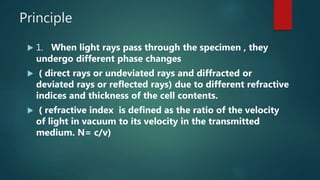

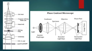
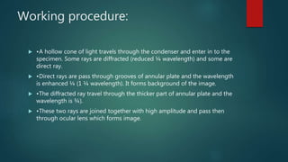
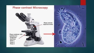
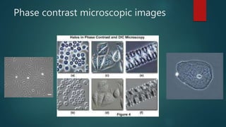
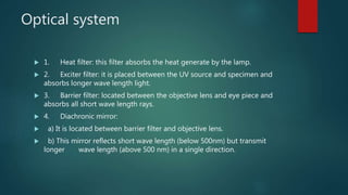
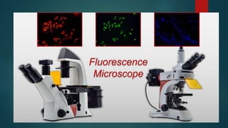
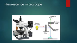


Phase contrast and fluorescence microscopes allow viewing of unstained live samples. Phase contrast microscopy uses interference of light waves passing through a sample to create contrast between structures of different refractive indices. Fluorescence microscopy employs fluorophores and fluorescent dyes excited by UV or blue light to emit visible light, allowing specific structures to be viewed with a dark background. Both techniques have advanced biological and medical research by enabling observation of otherwise transparent live cells and structures.


