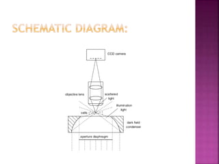
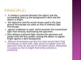
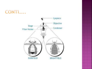
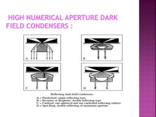
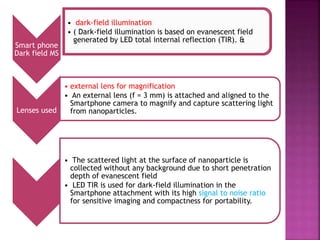
Presented by:
Rafia Altaf
 What is dark field microscopy?
 Principle and working
 Applications
 Advantages & disadvantages
Definition:
Dark field microscopy produces an image
with a dark background.
 Dark field microscopy is a technique used to
observe unstained samples causing them to
appear brightly lit against a dark, almost purely
black, background.
 used to examine live microorganisms that are
not visible with light microscope and cannot
stained, or distorted by staining and cannot be
identified.
 Instead of normal condenser, dark
field microscope uses dark field condenser
that contain a opaque disc. This disk blocks
light that would enter the lens directly, only
the light is reflected off the specimen enter
objective lens.
 As there is no background light, the
specimen appears light the against black
background the dark field.
 it creates a contrast between the object and the
surrounding field e.g the background is dark and the
object is bright.
 the objective and the ocular lenses used in the dark
ground microscope are same as that of ordinary light
microscope.
 special condenser is used, which prevents the transmitted
light from directly illuminating the specimen.
 Only oblique scattered light reaches the specimen and
passes onto the lens system causing the object to appear
bright against a dark background.
 Oblique illumination was the first step toward dark field
microscopy. In dark field, light seems to radiate from the
specimen while all the rest of the field is black. Resolution
is as good as that in bright field while contrast is
enhanced.
High Numerical Aperture Dark Field
Condensers :
 The first condenser made specifically for
dark field was produced by Francis H.
Wenham and George Shadbolt in 1855.
 This condenser used a parabolic glass
reflector to create a hollow cone of light.
Unlike a refracting condenser, a reflecting
condenser does not produce chromatic
aberrations and a parabolic shape minimizes
spherical aberrations. The result is a more
finely focused spot of light.
 A low magnification dark field condenser can
be nothing more than an ordinary bright field
condenser with an opaque disk of the proper
diameter placed in its front focal plane.
 The diameter of the opaque disk must be
just large enough to prevent any direct light
from entering the objective.
 Very thin histological sections can be used if
unstained or if only certain components are
stained, as in silver stains.
 Biological fluids from animals and plants, cell
cultures, microbes, foods, fibers, crystals,
colloids, and sub-microscopic particles are
all suitable for dark field microscopy.
 Viewing blood cell
 viewing bacteria
 Viewing different type of algae
 Viewing hair line metal fracture
 Viewing diamonds and other precious stones
Recently, fluorescence-based
nanoparticle detection was reported
using Smartphone adding
nanotechnology (NT) to Smartphone-
based imaging methods
Smart phone
Dark field MS
• dark-field illumination
• ( Dark-field illumination is based on evanescent field
generated by LED total internal reflection (TIR). &
Lenses used
• external lens for magnification
• An external lens (f = 3 mm) is attached and aligned to the
Smartphone camera to magnify and capture scattering light
from nanoparticles.
• The scattered light at the surface of nanoparticle is
collected without any background due to short penetration
depth of evanescent field
• LED TIR is used for dark-field illumination in the
Smartphone attachment with its high signal to noise ratio
for sensitive imaging and compactness for portability.
 It is ideal for viewing unstained objects,
transparent and absorb little or no light.
 These specimens often have similar refractive
indices as their surrounding, making them hard
to distinguish with other illumination techniques.
 Used in research of live bacterium, as well as
mounted cells and tissues.
 Useful in examining external details, edges and
surface defects.
 Dark field has regained its popularity when
combined with other illumination techniques,
such as fluorescence, which widens its possible
employment in certain field.
 not good for internal structure
 -Dust on sample, optics, bubbles in oil are
not tolerated with this technique
 If oil or water on the condenser or slide, air
bubble will produce.
 Q. Wei, H. Qi, W. Luo, D. Tseng, S. J. Ki, Z.
Wan, Z. Göröcs, L. A. Bentolila, T. Wu, R.
Sun, and A. Ozcan, “Fluorescent Imaging of
Single Nanoparticles and Viruses on a Smart
Phone,” ACS Nano, 7, (10), pp 9147-9155,
2013.
 http://www.microscormaster.com.dark-
field-miroscopy,html
 http://public.wse.edu/omoto/papers/darkfi
eld.html
 http://www,microscopeworld.com/t-
darkfield-microscopy
Dark Field microscopy

Dark Field microscopy

  • 2.
  • 3.
     What isdark field microscopy?  Principle and working  Applications  Advantages & disadvantages
  • 4.
    Definition: Dark field microscopyproduces an image with a dark background.  Dark field microscopy is a technique used to observe unstained samples causing them to appear brightly lit against a dark, almost purely black, background.  used to examine live microorganisms that are not visible with light microscope and cannot stained, or distorted by staining and cannot be identified.
  • 5.
     Instead ofnormal condenser, dark field microscope uses dark field condenser that contain a opaque disc. This disk blocks light that would enter the lens directly, only the light is reflected off the specimen enter objective lens.  As there is no background light, the specimen appears light the against black background the dark field.
  • 7.
     it createsa contrast between the object and the surrounding field e.g the background is dark and the object is bright.  the objective and the ocular lenses used in the dark ground microscope are same as that of ordinary light microscope.  special condenser is used, which prevents the transmitted light from directly illuminating the specimen.  Only oblique scattered light reaches the specimen and passes onto the lens system causing the object to appear bright against a dark background.  Oblique illumination was the first step toward dark field microscopy. In dark field, light seems to radiate from the specimen while all the rest of the field is black. Resolution is as good as that in bright field while contrast is enhanced.
  • 10.
    High Numerical ApertureDark Field Condensers :  The first condenser made specifically for dark field was produced by Francis H. Wenham and George Shadbolt in 1855.  This condenser used a parabolic glass reflector to create a hollow cone of light. Unlike a refracting condenser, a reflecting condenser does not produce chromatic aberrations and a parabolic shape minimizes spherical aberrations. The result is a more finely focused spot of light.
  • 12.
     A lowmagnification dark field condenser can be nothing more than an ordinary bright field condenser with an opaque disk of the proper diameter placed in its front focal plane.  The diameter of the opaque disk must be just large enough to prevent any direct light from entering the objective.
  • 13.
     Very thinhistological sections can be used if unstained or if only certain components are stained, as in silver stains.  Biological fluids from animals and plants, cell cultures, microbes, foods, fibers, crystals, colloids, and sub-microscopic particles are all suitable for dark field microscopy.
  • 14.
     Viewing bloodcell  viewing bacteria  Viewing different type of algae  Viewing hair line metal fracture  Viewing diamonds and other precious stones
  • 15.
    Recently, fluorescence-based nanoparticle detectionwas reported using Smartphone adding nanotechnology (NT) to Smartphone- based imaging methods
  • 16.
    Smart phone Dark fieldMS • dark-field illumination • ( Dark-field illumination is based on evanescent field generated by LED total internal reflection (TIR). & Lenses used • external lens for magnification • An external lens (f = 3 mm) is attached and aligned to the Smartphone camera to magnify and capture scattering light from nanoparticles. • The scattered light at the surface of nanoparticle is collected without any background due to short penetration depth of evanescent field • LED TIR is used for dark-field illumination in the Smartphone attachment with its high signal to noise ratio for sensitive imaging and compactness for portability.
  • 18.
     It isideal for viewing unstained objects, transparent and absorb little or no light.  These specimens often have similar refractive indices as their surrounding, making them hard to distinguish with other illumination techniques.  Used in research of live bacterium, as well as mounted cells and tissues.  Useful in examining external details, edges and surface defects.  Dark field has regained its popularity when combined with other illumination techniques, such as fluorescence, which widens its possible employment in certain field.
  • 19.
     not goodfor internal structure  -Dust on sample, optics, bubbles in oil are not tolerated with this technique  If oil or water on the condenser or slide, air bubble will produce.
  • 20.
     Q. Wei,H. Qi, W. Luo, D. Tseng, S. J. Ki, Z. Wan, Z. Göröcs, L. A. Bentolila, T. Wu, R. Sun, and A. Ozcan, “Fluorescent Imaging of Single Nanoparticles and Viruses on a Smart Phone,” ACS Nano, 7, (10), pp 9147-9155, 2013.  http://www.microscormaster.com.dark- field-miroscopy,html  http://public.wse.edu/omoto/papers/darkfi eld.html  http://www,microscopeworld.com/t- darkfield-microscopy