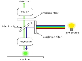
A fluorescence microscope uses fluorescence to enhance its capabilities beyond a regular light microscope. It illuminates samples tagged with fluorescent dyes with high-energy light, which causes the dyes to emit lower-energy light, producing a magnified image. This allows visualization of cell structures and live/dead cell assays. Advanced fluorescence microscopes like confocal microscopes can generate high-resolution 3D images of sample depths using lasers and image reconstruction software. Key applications include imaging cellular components, viability studies, and fluorescence in situ hybridization.